Movie
Movie Controller

[English] 日本語
 Yorodumi
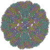
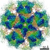
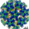
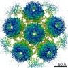
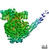
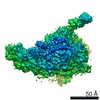
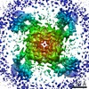
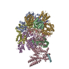
- PDB-7k25: Murine polyomavirus hexavalent capsomer, subparticle reconstruction -
+ Open data

- Basic information
| Entry | Database: PDB / ID: 7k25 | |||||||||||||||||||||
|---|---|---|---|---|---|---|---|---|---|---|---|---|---|---|---|---|---|---|---|---|---|---|
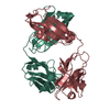
| Title | Murine polyomavirus hexavalent capsomer, subparticle reconstruction | |||||||||||||||||||||
 Components Components | Capsid protein VP1 | |||||||||||||||||||||
 Keywords Keywords | VIRAL PROTEIN / polyomavirus / capsomer | |||||||||||||||||||||
| Function / homology |  Function and homology information Function and homology informationcaveolin-mediated endocytosis of virus by host cell / T=7 icosahedral viral capsid / endocytosis involved in viral entry into host cell / virion attachment to host cell / host cell nucleus / structural molecule activity Similarity search - Function | |||||||||||||||||||||
| Biological species |  Mus musculus polyomavirus 1 Mus musculus polyomavirus 1 | |||||||||||||||||||||
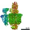
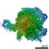
| Method | ELECTRON MICROSCOPY / single particle reconstruction / cryo EM / Resolution: 2.9 Å | |||||||||||||||||||||
 Authors Authors | Goetschius, D.J. / Hafenstein, S.L. | |||||||||||||||||||||
| Funding support |  United States, 6items United States, 6items
| |||||||||||||||||||||
 Citation Citation | Journal: Elife / Year: 2020 Title: Antibody escape by polyomavirus capsid mutation facilitates neurovirulence. Authors: Matthew D Lauver / Daniel J Goetschius / Colleen S Netherby-Winslow / Katelyn N Ayers / Ge Jin / Daniel G Haas / Elizabeth L Frost / Sung Hyun Cho / Carol M Bator / Stephanie M Bywaters / ...Authors: Matthew D Lauver / Daniel J Goetschius / Colleen S Netherby-Winslow / Katelyn N Ayers / Ge Jin / Daniel G Haas / Elizabeth L Frost / Sung Hyun Cho / Carol M Bator / Stephanie M Bywaters / Neil D Christensen / Susan L Hafenstein / Aron E Lukacher / Abstract: JCPyV polyomavirus, a member of the human virome, causes progressive multifocal leukoencephalopathy (PML), an oft-fatal demyelinating brain disease in individuals receiving immunomodulatory therapies. ...JCPyV polyomavirus, a member of the human virome, causes progressive multifocal leukoencephalopathy (PML), an oft-fatal demyelinating brain disease in individuals receiving immunomodulatory therapies. Mutations in the major viral capsid protein, VP1, are common in JCPyV from PML patients (JCPyV-PML) but whether they confer neurovirulence or escape from virus-neutralizing antibody (nAb) in vivo is unknown. A mouse polyomavirus (MuPyV) with a sequence-equivalent JCPyV-PML VP1 mutation replicated poorly in the kidney, a major reservoir for JCPyV persistence, but retained the CNS infectivity, cell tropism, and neuropathology of the parental virus. This mutation rendered MuPyV resistant to a monoclonal Ab (mAb), whose specificity overlapped the endogenous anti-VP1 response. Using cryo-EM and a custom sub-particle refinement approach, we resolved an MuPyV:Fab complex map to 3.2 Å resolution. The structure revealed the mechanism of mAb evasion. Our findings demonstrate convergence between nAb evasion and CNS neurovirulence in vivo by a frequent JCPyV-PML VP1 mutation. | |||||||||||||||||||||
| History |
|
- Structure visualization
| Movie |
Movie viewer |
|---|---|
| Structure viewer | Molecule: MolmilJmol/JSmol |
- Downloads & links
-Download
| PDBx/mmCIF format | 7k25.cif.gz | 309.1 KB | Display | PDBx/mmCIF format |
|---|---|---|---|---|
| PDB format | pdb7k25.ent.gz | 252.3 KB | Display | PDB format |
| PDBx/mmJSON format | 7k25.json.gz | Tree view | PDBx/mmJSON format | |
| Others |  Other downloads Other downloads |
-Validation report
| Arichive directory | https://data.pdbj.org/pub/pdb/validation_reports/k2/7k25ftp://data.pdbj.org/pub/pdb/validation_reports/k2/7k25 | HTTPS FTP |
|---|
-Related structure data
| Related structure data | 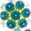 22643MC 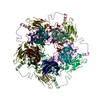 7k22C 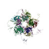 7k23C  7k24C M: map data used to model this data C: citing same article ( |
|---|---|
| Similar structure data |
-Links
 PDBj
PDBj


- Assembly
| Deposited unit | 
|
|---|---|
| 1 |
|
-Components
| #1: Protein | Mass: 42493.172 Da / Num. of mol.: 5 Source method: isolated from a genetically manipulated source Source: (gene. exp.) Mus musculus polyomavirus 1 / Production host:  Has protein modification | Y | |
|---|
-Experimental details
-Experiment
| Experiment | Method: ELECTRON MICROSCOPY |
|---|---|
| EM experiment | Aggregation state: PARTICLE / 3D reconstruction method: single particle reconstruction |
- Sample preparation
| Component | Name: Murine polyomavirus strain A2 / Type: VIRUS / Entity ID: all / Source: RECOMBINANT | ||||||||||||||||||||
|---|---|---|---|---|---|---|---|---|---|---|---|---|---|---|---|---|---|---|---|---|---|
| Molecular weight | Experimental value: NO | ||||||||||||||||||||
| Source (natural) | Organism: Murine polyomavirus strain A2 | ||||||||||||||||||||
| Source (recombinant) | Organism: | ||||||||||||||||||||
| Details of virus | Empty: NO / Enveloped: NO / Isolate: STRAIN / Type: VIRION | ||||||||||||||||||||
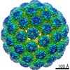
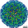
| Virus shell | Diameter: 450 nm / Triangulation number (T number): 7 | ||||||||||||||||||||
| Buffer solution | pH: 7.9 Details: 10 mM HEPES pH 7.9, 1 mM CaCl2, 1 mM MgCl2, 5 mM KCl | ||||||||||||||||||||
| Buffer component |
| ||||||||||||||||||||
| Specimen | Conc.: 2.8 mg/ml / Embedding applied: NO / Shadowing applied: NO / Staining applied: NO / Vitrification applied: YES / Details: MuPyV (2.8 mg/mL) | ||||||||||||||||||||
| Vitrification | Cryogen name: ETHANE / Humidity: 95 % |
- Electron microscopy imaging
| Experimental equipment |  Model: Titan Krios / Image courtesy: FEI Company |
|---|---|
| Microscopy | Model: FEI TITAN KRIOS |
| Electron gun | Electron source:  FIELD EMISSION GUN / Accelerating voltage: 300 kV / Illumination mode: OTHER FIELD EMISSION GUN / Accelerating voltage: 300 kV / Illumination mode: OTHER |
| Electron lens | Mode: BRIGHT FIELD |
| Image recording | Electron dose: 45 e/Å2 / Film or detector model: FEI FALCON III (4k x 4k) |
- Processing
| Software |
| ||||||||||||||||||||||||||||||||
|---|---|---|---|---|---|---|---|---|---|---|---|---|---|---|---|---|---|---|---|---|---|---|---|---|---|---|---|---|---|---|---|---|---|
| EM software |
| ||||||||||||||||||||||||||||||||
| CTF correction | Type: PHASE FLIPPING AND AMPLITUDE CORRECTION | ||||||||||||||||||||||||||||||||
| 3D reconstruction | Resolution: 2.9 Å / Resolution method: FSC 0.143 CUT-OFF / Num. of particles: 929940 / Symmetry type: POINT | ||||||||||||||||||||||||||||||||
| Atomic model building | Space: REAL Details: Homology model for Fab was generated using SwissModel. Initial models were docked into density in Chimera. Fab CDR loops were manually rebuilt in Coot. Iterative rounds of real space ...Details: Homology model for Fab was generated using SwissModel. Initial models were docked into density in Chimera. Fab CDR loops were manually rebuilt in Coot. Iterative rounds of real space refinements (PHENIX) and manual adjustment (coot) were conducted to improve fit to density. | ||||||||||||||||||||||||||||||||
| Atomic model building |
| ||||||||||||||||||||||||||||||||
| Refinement | Cross valid method: NONE Stereochemistry target values: GeoStd + Monomer Library + CDL v1.2 | ||||||||||||||||||||||||||||||||
| Displacement parameters | Biso mean: 27.23 Å2 | ||||||||||||||||||||||||||||||||
| Refine LS restraints |
|