 Movie
Movie Controller

[English] 日本語
 Yorodumi
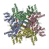
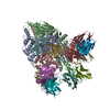
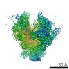
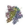
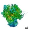
- PDB-7k22: Murine polyomavirus pentavalent capsomer with 8A7H5 Fab, subparti... -
+ Open data

- Basic information
| Entry | Database: PDB / ID: 7k22 | |||||||||||||||||||||||||||||||||||||||
|---|---|---|---|---|---|---|---|---|---|---|---|---|---|---|---|---|---|---|---|---|---|---|---|---|---|---|---|---|---|---|---|---|---|---|---|---|---|---|---|---|
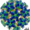
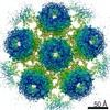
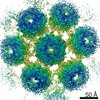
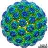
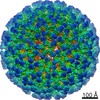
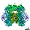
| Title | Murine polyomavirus pentavalent capsomer with 8A7H5 Fab, subparticle reconstruction | |||||||||||||||||||||||||||||||||||||||
 Components Components |
| |||||||||||||||||||||||||||||||||||||||
 Keywords Keywords | VIRAL PROTEIN / polyomavirus / capsomer | |||||||||||||||||||||||||||||||||||||||
| Function / homology |  Function and homology information Function and homology informationT=7 icosahedral viral capsid / endocytosis involved in viral entry into host cell / virion attachment to host cell / host cell nucleus / structural molecule activity Similarity search - Function | |||||||||||||||||||||||||||||||||||||||
| Biological species |  Mus musculus polyomavirus 1 Mus musculus polyomavirus 1 | |||||||||||||||||||||||||||||||||||||||
| Method | ELECTRON MICROSCOPY / single particle reconstruction / cryo EM / Resolution: 3.2 Å | |||||||||||||||||||||||||||||||||||||||
 Authors Authors | Goetschius, D.J. / Hafenstein, S.L. | |||||||||||||||||||||||||||||||||||||||
| Funding support |  United States, 6items United States, 6items
| |||||||||||||||||||||||||||||||||||||||
 Citation Citation | Journal: Elife / Year: 2020 Title: Antibody escape by polyomavirus capsid mutation facilitates neurovirulence. Authors: Matthew D Lauver / Daniel J Goetschius / Colleen S Netherby-Winslow / Katelyn N Ayers / Ge Jin / Daniel G Haas / Elizabeth L Frost / Sung Hyun Cho / Carol M Bator / Stephanie M Bywaters / ...Authors: Matthew D Lauver / Daniel J Goetschius / Colleen S Netherby-Winslow / Katelyn N Ayers / Ge Jin / Daniel G Haas / Elizabeth L Frost / Sung Hyun Cho / Carol M Bator / Stephanie M Bywaters / Neil D Christensen / Susan L Hafenstein / Aron E Lukacher / Abstract: JCPyV polyomavirus, a member of the human virome, causes progressive multifocal leukoencephalopathy (PML), an oft-fatal demyelinating brain disease in individuals receiving immunomodulatory therapies. ...JCPyV polyomavirus, a member of the human virome, causes progressive multifocal leukoencephalopathy (PML), an oft-fatal demyelinating brain disease in individuals receiving immunomodulatory therapies. Mutations in the major viral capsid protein, VP1, are common in JCPyV from PML patients (JCPyV-PML) but whether they confer neurovirulence or escape from virus-neutralizing antibody (nAb) in vivo is unknown. A mouse polyomavirus (MuPyV) with a sequence-equivalent JCPyV-PML VP1 mutation replicated poorly in the kidney, a major reservoir for JCPyV persistence, but retained the CNS infectivity, cell tropism, and neuropathology of the parental virus. This mutation rendered MuPyV resistant to a monoclonal Ab (mAb), whose specificity overlapped the endogenous anti-VP1 response. Using cryo-EM and a custom sub-particle refinement approach, we resolved an MuPyV:Fab complex map to 3.2 Å resolution. The structure revealed the mechanism of mAb evasion. Our findings demonstrate convergence between nAb evasion and CNS neurovirulence in vivo by a frequent JCPyV-PML VP1 mutation. | |||||||||||||||||||||||||||||||||||||||
| History |
|
- Structure visualization
| Movie |
Movie viewer |
|---|---|
| Structure viewer | Molecule: MolmilJmol/JSmol |
- Downloads & links
-Download
| PDBx/mmCIF format | 7k22.cif.gz | 471.2 KB | Display | PDBx/mmCIF format |
|---|---|---|---|---|
| PDB format | pdb7k22.ent.gz | Display | PDB format | |
| PDBx/mmJSON format | 7k22.json.gz | Tree view | PDBx/mmJSON format | |
| Others |  Other downloads Other downloads |
-Validation report
| Arichive directory | https://data.pdbj.org/pub/pdb/validation_reports/k2/7k22ftp://data.pdbj.org/pub/pdb/validation_reports/k2/7k22 | HTTPS FTP |
|---|
-Related structure data
| Related structure data | 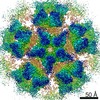 22640MC  7k23C  7k24C  7k25C M: map data used to model this data C: citing same article ( |
|---|---|
| Similar structure data |
-Links
 PDBj
PDBj


- Assembly
| Deposited unit | 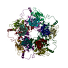
|
|---|---|
| 1 |
|
-Components
| #1: Antibody | Mass: 12086.437 Da / Num. of mol.: 5 / Source method: isolated from a natural source / Source: (natural) #2: Antibody | Mass: 13217.701 Da / Num. of mol.: 5 / Source method: isolated from a natural source / Source: (natural) #3: Protein | Mass: 42493.172 Da / Num. of mol.: 5 Source method: isolated from a genetically manipulated source Source: (gene. exp.) Mus musculus polyomavirus 1 / Production host: Has protein modification | Y | |
|---|
-Experimental details
-Experiment
| Experiment | Method: ELECTRON MICROSCOPY |
|---|---|
| EM experiment | Aggregation state: PARTICLE / 3D reconstruction method: single particle reconstruction |
- Sample preparation
| Component |
| ||||||||||||||||||||||||||||
|---|---|---|---|---|---|---|---|---|---|---|---|---|---|---|---|---|---|---|---|---|---|---|---|---|---|---|---|---|---|
| Molecular weight | Experimental value: NO | ||||||||||||||||||||||||||||
| Source (natural) |
| ||||||||||||||||||||||||||||
| Source (recombinant) | Organism: | ||||||||||||||||||||||||||||
| Details of virus | Empty: NO / Enveloped: NO / Isolate: STRAIN / Type: VIRION | ||||||||||||||||||||||||||||
| Virus shell | Diameter: 450 nm / Triangulation number (T number): 7 | ||||||||||||||||||||||||||||
| Buffer solution | pH: 7.9 Details: 10 mM HEPES pH 7.9, 1 mM CaCl2, 1 mM MgCl2, 5 mM KCl | ||||||||||||||||||||||||||||
| Specimen | Conc.: 2.8 mg/ml / Embedding applied: NO / Shadowing applied: NO / Staining applied: NO / Vitrification applied: YES Details: MuPyV (2.8 mg/mL) was incubated with 8A7H5 Fab (1.1 mg/mL) for 30 m at room temperature | ||||||||||||||||||||||||||||
| Vitrification | Cryogen name: ETHANE |
- Electron microscopy imaging
| Experimental equipment |  Model: Titan Krios / Image courtesy: FEI Company |
|---|---|
| Microscopy | Model: FEI TITAN KRIOS |
| Electron gun | Electron source:  FIELD EMISSION GUN / Accelerating voltage: 300 kV / Illumination mode: OTHER FIELD EMISSION GUN / Accelerating voltage: 300 kV / Illumination mode: OTHER |
| Electron lens | Mode: BRIGHT FIELD |
| Image recording | Electron dose: 45 e/Å2 / Film or detector model: FEI FALCON III (4k x 4k) |
- Processing
| Software | Name: PHENIX / Version: 1.17.1-3660_3260: / Classification: refinement | ||||||||||||||||||||||||
|---|---|---|---|---|---|---|---|---|---|---|---|---|---|---|---|---|---|---|---|---|---|---|---|---|---|
| EM software |
| ||||||||||||||||||||||||
| CTF correction | Type: PHASE FLIPPING AND AMPLITUDE CORRECTION | ||||||||||||||||||||||||
| 3D reconstruction | Resolution: 3.2 Å / Resolution method: FSC 0.143 CUT-OFF / Num. of particles: 109752 / Symmetry type: POINT | ||||||||||||||||||||||||
| Refine LS restraints |
|
