 Movie
Movie Controller

[English] 日本語
 Yorodumi
- EMDB-22640: Murine polyomavirus pentavalent capsomer with 8A7H5 Fab, subparti... -
+ Open data

- Basic information
| Entry | Database: EMDB / ID: EMD-22640 | |||||||||||||||||||||
|---|---|---|---|---|---|---|---|---|---|---|---|---|---|---|---|---|---|---|---|---|---|---|
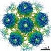
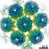
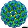
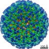
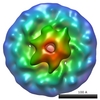
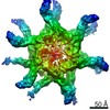
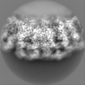
| Title | Murine polyomavirus pentavalent capsomer with 8A7H5 Fab, subparticle reconstruction | |||||||||||||||||||||
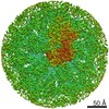
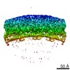
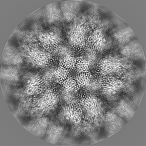
 Map data Map data | Postprocessed map of locally-refined pentavalent capsomer w/ Fab | |||||||||||||||||||||
 Sample Sample |
| |||||||||||||||||||||
 Keywords Keywords | polyomavirus / capsomer / VIRAL PROTEIN | |||||||||||||||||||||
| Function / homology |  Function and homology information Function and homology informationT=7 icosahedral viral capsid / endocytosis involved in viral entry into host cell / virion attachment to host cell / host cell nucleus / structural molecule activity Similarity search - Function | |||||||||||||||||||||
| Biological species |   Murine polyomavirus strain A2 / Mus musculus polyomavirus 1 Murine polyomavirus strain A2 / Mus musculus polyomavirus 1 | |||||||||||||||||||||
| Method | single particle reconstruction / cryo EM / Resolution: 3.2 Å | |||||||||||||||||||||
 Authors Authors | Goetschius DJ / Hafenstein SL | |||||||||||||||||||||
| Funding support |  United States, 6 items United States, 6 items
| |||||||||||||||||||||
 Citation Citation | Journal: Elife / Year: 2020 Title: Antibody escape by polyomavirus capsid mutation facilitates neurovirulence. Authors: Matthew D Lauver / Daniel J Goetschius / Colleen S Netherby-Winslow / Katelyn N Ayers / Ge Jin / Daniel G Haas / Elizabeth L Frost / Sung Hyun Cho / Carol M Bator / Stephanie M Bywaters / ...Authors: Matthew D Lauver / Daniel J Goetschius / Colleen S Netherby-Winslow / Katelyn N Ayers / Ge Jin / Daniel G Haas / Elizabeth L Frost / Sung Hyun Cho / Carol M Bator / Stephanie M Bywaters / Neil D Christensen / Susan L Hafenstein / Aron E Lukacher / Abstract: JCPyV polyomavirus, a member of the human virome, causes progressive multifocal leukoencephalopathy (PML), an oft-fatal demyelinating brain disease in individuals receiving immunomodulatory therapies. ...JCPyV polyomavirus, a member of the human virome, causes progressive multifocal leukoencephalopathy (PML), an oft-fatal demyelinating brain disease in individuals receiving immunomodulatory therapies. Mutations in the major viral capsid protein, VP1, are common in JCPyV from PML patients (JCPyV-PML) but whether they confer neurovirulence or escape from virus-neutralizing antibody (nAb) in vivo is unknown. A mouse polyomavirus (MuPyV) with a sequence-equivalent JCPyV-PML VP1 mutation replicated poorly in the kidney, a major reservoir for JCPyV persistence, but retained the CNS infectivity, cell tropism, and neuropathology of the parental virus. This mutation rendered MuPyV resistant to a monoclonal Ab (mAb), whose specificity overlapped the endogenous anti-VP1 response. Using cryo-EM and a custom sub-particle refinement approach, we resolved an MuPyV:Fab complex map to 3.2 Å resolution. The structure revealed the mechanism of mAb evasion. Our findings demonstrate convergence between nAb evasion and CNS neurovirulence in vivo by a frequent JCPyV-PML VP1 mutation. | |||||||||||||||||||||
| History |
|
- Structure visualization
| Movie |
Movie viewer |
|---|---|
| Structure viewer | EM map: SurfViewMolmilJmol/JSmol |
| Supplemental images |

- Downloads & links
-EMDB archive
| Map data | emd_22640.map.gz | 96.5 MB | EMDB map data format | |
|---|---|---|---|---|
| Header (meta data) | emd-22640-v30.xmlemd-22640.xml | 21.5 KB 21.5 KB | Display Display | EMDB header |
| FSC (resolution estimation) | emd_22640_fsc.xml | 10.7 KB | Display | FSC data file |
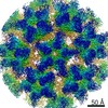
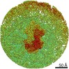
| Images | 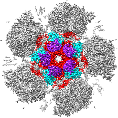 emd_22640.png emd_22640.png | 240.9 KB | ||
| Filedesc metadata | emd-22640.cif.gz | 6.8 KB | ||
| Others | emd_22640_half_map_1.map.gzemd_22640_half_map_2.map.gz | 80.2 MB 80.2 MB | ||
| Archive directory |  http://ftp.pdbj.org/pub/emdb/structures/EMD-22640ftp://ftp.pdbj.org/pub/emdb/structures/EMD-22640 http://ftp.pdbj.org/pub/emdb/structures/EMD-22640ftp://ftp.pdbj.org/pub/emdb/structures/EMD-22640 | HTTPS FTP |
-Related structure data
| Related structure data |  7k22MC  7k23C  7k24C  7k25C M: atomic model generated by this map C: citing same article ( |
|---|---|
| Similar structure data |

-Links
| EMDB pages | EMDB (EBI/PDBe) / EMDataResource |
|---|---|
| Related items in Molecule of the Month |


-Map
| File | Download / File: emd_22640.map.gz / Format: CCP4 / Size: 103 MB / Type: IMAGE STORED AS FLOATING POINT NUMBER (4 BYTES) | ||||||||||||||||||||||||||||||||||||||||||||||||||||||||||||||||||||
|---|---|---|---|---|---|---|---|---|---|---|---|---|---|---|---|---|---|---|---|---|---|---|---|---|---|---|---|---|---|---|---|---|---|---|---|---|---|---|---|---|---|---|---|---|---|---|---|---|---|---|---|---|---|---|---|---|---|---|---|---|---|---|---|---|---|---|---|---|---|
| Annotation | Postprocessed map of locally-refined pentavalent capsomer w/ Fab | ||||||||||||||||||||||||||||||||||||||||||||||||||||||||||||||||||||
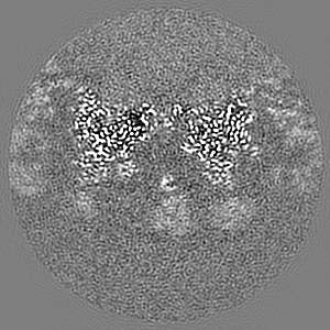
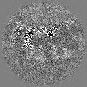
| Projections & slices | Image control
Images are generated by Spider. | ||||||||||||||||||||||||||||||||||||||||||||||||||||||||||||||||||||
| Voxel size | X=Y=Z: 1.1 Å | ||||||||||||||||||||||||||||||||||||||||||||||||||||||||||||||||||||
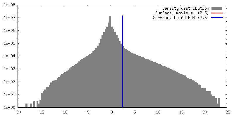
| Density |
| ||||||||||||||||||||||||||||||||||||||||||||||||||||||||||||||||||||
| Symmetry | Space group: 1 | ||||||||||||||||||||||||||||||||||||||||||||||||||||||||||||||||||||
| Details | EMDB XML:
CCP4 map header:
| ||||||||||||||||||||||||||||||||||||||||||||||||||||||||||||||||||||
 Z (Sec.)
 Y (Row.)
 X (Col.)


-Supplemental data
-Half map: Unfiltered halfmap
| File | emd_22640_half_map_1.map | ||||||||||||
|---|---|---|---|---|---|---|---|---|---|---|---|---|---|
| Annotation | Unfiltered halfmap | ||||||||||||
| Projections & Slices |
| ||||||||||||
| Density Histograms |


-Half map: Unfiltered halfmap
| File | emd_22640_half_map_2.map | ||||||||||||
|---|---|---|---|---|---|---|---|---|---|---|---|---|---|
| Annotation | Unfiltered halfmap | ||||||||||||
| Projections & Slices |
| ||||||||||||
| Density Histograms |


- Sample components
-Entire : 8A7H5 Fab light chain, 8A7H5 Fab heavy chain, Capsid protein VP1
| Entire | Name: 8A7H5 Fab light chain, 8A7H5 Fab heavy chain, Capsid protein VP1 |
|---|---|
| Components |
|
-Supramolecule #1: 8A7H5 Fab light chain, 8A7H5 Fab heavy chain, Capsid protein VP1
| Supramolecule | Name: 8A7H5 Fab light chain, 8A7H5 Fab heavy chain, Capsid protein VP1 type: complex / ID: 1 / Parent: 0 / Macromolecule list: all / Details: Fab fragment generated from rat IgG |
|---|
-Supramolecule #2: 8A7H5 Fab light chain, 8A7H5 Fab heavy chain
| Supramolecule | Name: 8A7H5 Fab light chain, 8A7H5 Fab heavy chain / type: complex / ID: 2 / Parent: 1 / Macromolecule list: #1-#2 |
|---|---|
| Source (natural) | Organism: |
-Supramolecule #3: Capsid protein VP1
| Supramolecule | Name: Capsid protein VP1 / type: complex / ID: 3 / Parent: 1 / Macromolecule list: #3 |
|---|---|
| Source (natural) | Organism: Murine polyomavirus strain A2 |
-Macromolecule #1: 8A7H5 Fab light chain
| Macromolecule | Name: 8A7H5 Fab light chain / type: protein_or_peptide / ID: 1 / Number of copies: 5 / Enantiomer: LEVO |
|---|---|
| Source (natural) | Organism: |
| Molecular weight | Theoretical: 12.086437 KDa |
| Sequence | String: DIVMTQSPTS MSISVGDRVT MNCRASQNVY SNVDWYQQKT GQSPKLVIYK ASNRYTGVPD RFTGSGSGTY FTLTITNIQT EDLAVYYCL QSNAFPFTFG SGTKLETTRA |
-Macromolecule #2: 8A7H5 Fab heavy chain
| Macromolecule | Name: 8A7H5 Fab heavy chain / type: protein_or_peptide / ID: 2 / Number of copies: 5 / Enantiomer: LEVO |
|---|---|
| Source (natural) | Organism: |
| Molecular weight | Theoretical: 13.217701 KDa |
| Sequence | String: EESGGGLVQP GKSLKLSCSA SGFTFSSYGM HWIRQVPGKG LDWVAYISSA SDTFYADAVK ERFTISRDNA KNTLYLRLNS LKSEDTAIY YCARTRYPTD HFYDWFPYWG QGTLVTVS |
-Macromolecule #3: Capsid protein VP1
| Macromolecule | Name: Capsid protein VP1 / type: protein_or_peptide / ID: 3 / Number of copies: 5 / Enantiomer: LEVO |
|---|---|
| Source (natural) | Organism: Mus musculus polyomavirus 1 |
| Molecular weight | Theoretical: 42.493172 KDa |
| Recombinant expression | Organism: |
| Sequence | String: APKRKSGVSK CETKCTKACP RPAPVPKLLI KGGMEVLDLV TGPDSVTEIE AFLNPRMGQP PTPESLTEGG QYYGWSRGIN LATSDTEDS PENNTLPTWS MAKLQLPMLN EDLTCDTLQM WEAVSVKTEV VGSGSLLDVH GFNKPTDTVN TKGISTPVEG S QYHVFAVG ...String: APKRKSGVSK CETKCTKACP RPAPVPKLLI KGGMEVLDLV TGPDSVTEIE AFLNPRMGQP PTPESLTEGG QYYGWSRGIN LATSDTEDS PENNTLPTWS MAKLQLPMLN EDLTCDTLQM WEAVSVKTEV VGSGSLLDVH GFNKPTDTVN TKGISTPVEG S QYHVFAVG GEPLDLQGLV TDARTKYKEE GVVTIKTITK KDMVNKDQVL NPISKAKLDK DGMYPVEIWH PDPAKNENTR YF GNYTGGT TTPPVLQFTN TLTTVLLDEN GVGPLCKGEG LYLSCVDIMG WRVTRNYDVH HWRGLPRYFK ITLRKRWVKN PYP MASLIS SLFNNMLPQV QGQPMEGENT QVEEVRVYDG TEPVPGDPDM TRYVDRFGKT KTVFPGN UniProtKB: Capsid protein VP1 |
-Experimental details
-Structure determination
| Method | cryo EM |
|---|---|
 Processing Processing | single particle reconstruction |
| Aggregation state | particle |
-Sample preparation
| Concentration | 2.8 mg/mL |
|---|---|
| Buffer | pH: 7.9 Details: 10 mM HEPES pH 7.9, 1 mM CaCl2, 1 mM MgCl2, 5 mM KCl |
| Vitrification | Cryogen name: ETHANE |
| Details | MuPyV (2.8 mg/mL) was incubated with 8A7H5 Fab (1.1 mg/mL) for 30 m at room temperature |
- Electron microscopy
| Microscope | FEI TITAN KRIOS |
|---|---|
| Image recording | Film or detector model: FEI FALCON III (4k x 4k) / Average electron dose: 45.0 e/Å2 |
| Electron beam | Acceleration voltage: 300 kV / Electron source:  FIELD EMISSION GUN FIELD EMISSION GUN |
| Electron optics | Illumination mode: OTHER / Imaging mode: BRIGHT FIELD |
| Experimental equipment |  Model: Titan Krios / Image courtesy: FEI Company |

