Movie
Movie Controller

[English] 日本語
 Yorodumi
- PDB-3iyi: P22 expanded head coat protein structures reveal a novel mechanis... -
+ Open data

- Basic information
| Entry | Database: PDB / ID: 3iyi | ||||||
|---|---|---|---|---|---|---|---|
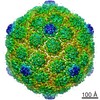
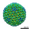
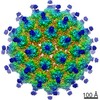
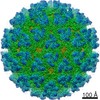
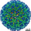
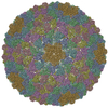
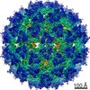
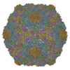
| Title | P22 expanded head coat protein structures reveal a novel mechanism for capsid maturation: Stability without auxiliary proteins or chemical cross-links | ||||||
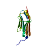
 Components Components | P22 coat protein in procapsid shells | ||||||
 Keywords Keywords | VIRUS / HK97-like fold | ||||||
| Function / homology | Major capsid protein Gp5 / P22 coat protein - gene protein 5 / viral procapsid / viral procapsid maturation / T=7 icosahedral viral capsid / viral capsid / identical protein binding / Major capsid protein Function and homology information Function and homology information | ||||||
| Biological species |  Enterobacteria phage P22 (virus) Enterobacteria phage P22 (virus) | ||||||
| Method | ELECTRON MICROSCOPY / single particle reconstruction / cryo EM / Resolution: 9.1 Å | ||||||
 Authors Authors | Parent, K.N. / Khayat, R. / Tu, L.H. / Suhanovsky, M.M. / Cortines, J.R. / Teschke, C.M. / Johnson, J.E. / Baker, T.S. | ||||||
 Citation Citation | Journal: Structure / Year: 2010 Title: P22 coat protein structures reveal a novel mechanism for capsid maturation: stability without auxiliary proteins or chemical crosslinks. Authors: Kristin N Parent / Reza Khayat / Long H Tu / Margaret M Suhanovsky / Juliana R Cortines / Carolyn M Teschke / John E Johnson / Timothy S Baker /  Abstract: Viral capsid assembly and stability in tailed, dsDNA phage and Herpesviridae are achieved by various means including chemical crosslinks (unique to HK97), or auxiliary proteins (lambda, T4, phi29, ...Viral capsid assembly and stability in tailed, dsDNA phage and Herpesviridae are achieved by various means including chemical crosslinks (unique to HK97), or auxiliary proteins (lambda, T4, phi29, and herpesviruses). All these viruses have coat proteins (CP) with a conserved, HK97-like core structure. We used a combination of trypsin digestion, gold labeling, cryo-electron microscopy, 3D image reconstruction, and comparative modeling to derive two independent, pseudoatomic models of bacteriophage P22 CP: before and after maturation. P22 capsid stabilization results from intersubunit interactions among N-terminal helices and an extensive "P loop," which obviate the need for crosslinks or auxiliary proteins. P22 CP also has a telokin-like Ig domain that likely stabilizes the monomer fold so that assembly may proceed via individual subunit addition rather than via preformed capsomers as occurs in HK97. Hence, the P22 CP structure may be a paradigm for understanding how monomers assemble in viruses like phi29 and HSV-1. | ||||||
| History |
|
- Structure visualization
| Movie |
Movie viewer |
|---|---|
| Structure viewer | Molecule: MolmilJmol/JSmol |
- Downloads & links
-Download
| PDBx/mmCIF format | 3iyi.cif.gz | 88 KB | Display | PDBx/mmCIF format |
|---|---|---|---|---|
| PDB format | pdb3iyi.ent.gz | 54.8 KB | Display | PDB format |
| PDBx/mmJSON format | 3iyi.json.gz | Tree view | PDBx/mmJSON format | |
| Others |  Other downloads Other downloads |
-Validation report
| Arichive directory | https://data.pdbj.org/pub/pdb/validation_reports/iy/3iyiftp://data.pdbj.org/pub/pdb/validation_reports/iy/3iyi | HTTPS FTP |
|---|
-Related structure data
| Related structure data | 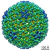 5149MC  5150C  3iyhC M: map data used to model this data C: citing same article ( |
|---|---|
| Similar structure data |
-Links
 PDBj
PDBj
- Assembly
| Deposited unit | 
|
|---|---|
| 1 | x 60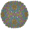
|
| 2 |
|
| 3 | x 5
|
| 4 | x 6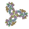
|
| 5 | 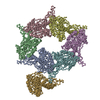
|
| Symmetry | Point symmetry: (Schoenflies symbol: I (icosahedral)) |
-Components
| #1: Protein | Mass: 46795.613 Da / Num. of mol.: 7 / Source method: isolated from a natural source / Source: (natural) Enterobacteria phage P22 (virus) / References: UniProt: P26747 |
|---|
-Experimental details
-Experiment
| Experiment | Method: ELECTRON MICROSCOPY |
|---|---|
| EM experiment | Aggregation state: PARTICLE / 3D reconstruction method: single particle reconstruction |
- Sample preparation
| Component | Name: P22 Procapsid Shell / Type: VIRUS Details: Icosahedral. Scaffolding protein, portal and ejection proteins removed by 0.5 M GuHCl |
|---|---|
| Details of virus | Empty: YES / Enveloped: NO / Host category: BACTERIA(EUBACTERIA) / Isolate: STRAIN / Type: VIRUS-LIKE PARTICLE |
| Natural host | Organism: Salmonella typhimurium |
| Buffer solution | Name: 20 mM sodium phosphate buffer, pH = 7.6 / pH: 7.6 / Details: 20 mM sodium phosphate buffer, pH = 7.6 |
| Specimen | Conc.: 8 mg/ml / Embedding applied: NO / Shadowing applied: NO / Staining applied: NO / Vitrification applied: YES / Details: 20 mM sodium phosphate |
| Specimen support | Details: Quantifoil |
| Vitrification | Instrument: HOMEMADE PLUNGER / Cryogen name: ETHANE / Temp: 89 K / Humidity: 100 % / Method: Blot for 2-3 seconds before plunging |
- Electron microscopy imaging
| Experimental equipment |  Model: Tecnai Polara / Image courtesy: FEI Company |
|---|---|
| Microscopy | Model: FEI POLARA 300 / Date: Jan 1, 2009 |
| Electron gun | Electron source:  FIELD EMISSION GUN / Accelerating voltage: 200 kV / Illumination mode: FLOOD BEAM FIELD EMISSION GUN / Accelerating voltage: 200 kV / Illumination mode: FLOOD BEAM |
| Electron lens | Mode: BRIGHT FIELD / Nominal magnification: 39000 X / Nominal defocus max: 3650 nm / Nominal defocus min: 410 nm / Cs: 2.3 mm / Astigmatism: at working magnification / Camera length: 0 mm |
| Specimen holder | Specimen holder model: OTHER / Specimen holder type: Polrar Multi Specimen Holder / Temperature: 90 K / Temperature (max): 90 K / Temperature (min): 90 K / Tilt angle max: -9999 ° / Tilt angle min: -9999 ° |
| Image recording | Electron dose: 8 e/Å2 / Film or detector model: KODAK SO-163 FILM |
- Processing
| EM software |
| ||||||||||||||||||
|---|---|---|---|---|---|---|---|---|---|---|---|---|---|---|---|---|---|---|---|
| CTF correction | Details: ROBEM | ||||||||||||||||||
| Symmetry | Point symmetry: I (icosahedral) | ||||||||||||||||||
| 3D reconstruction | Method: iterative model-based procedure / Resolution: 9.1 Å / Resolution method: FSC 0.5 CUT-OFF / Num. of particles: 11997 / Nominal pixel size: 1.62 Å / Actual pixel size: 1.62 Å Details: ( Details about the particle: data were collected on film ) Symmetry type: POINT | ||||||||||||||||||
| Atomic model building |
| ||||||||||||||||||
| Atomic model building | Source name: PDB / Type: experimental model
| ||||||||||||||||||
| Refinement step | Cycle: LAST
|