[English] 日本語
Yorodumi
- EMDB-13590: Cryo-EM structure of the dimeric Rhodobacter sphaeroides RC-LH1 c... -

+
Open data


ID or keywords:

Loading...

-
Basic information

Entry
Database: EMDB / ID: EMD-13590
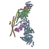
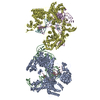
TitleCryo-EM structure of the dimeric Rhodobacter sphaeroides RC-LH1 core complex at 2.9 A: the structural basis for dimerisation
Map datalight harvesting core complex
Sample
  • Complex: Light harvesting core complex
    • Protein or peptide: x 8 types
  • Ligand: x 11 types
Function / homology
Function and homology information


plasma membrane-derived chromatophore membrane / plasma membrane light-harvesting complex / bacteriochlorophyll binding / electron transporter, transferring electrons within the cyclic electron transport pathway of photosynthesis activity / photosynthetic electron transport in photosystem II / metal ion binding
Similarity search - Function
Photosynthetic reaction centre, M subunit / Photosynthetic reaction centre, L/M / Photosystem II protein D1/D2 superfamily / Photosynthetic reaction centre protein / Photosynthetic reaction center proteins signature.
Similarity search - Domain/homology
Reaction center protein M chain
Similarity search - Component
Biological speciesCereibacter sphaeroides 2.4.1 (bacteria)
Methodsingle particle reconstruction / cryo EM / Resolution: 2.9 Å
AuthorsQian P / Hunter CN
Funding support United Kingdom, 3 items
OrganizationGrant numberCountry
Biotechnology and Biological Sciences Research Council (BBSRC)BB/M000265/1 United Kingdom
European Research Council (ERC)845126 United Kingdom
Wellcome Trust209407/Z/17/Z United Kingdom
Citation
Journal: Biochem J / Year: 2021
Title: Cryo-EM structure of the dimeric Rhodobacter sphaeroides RC-LH1 core complex at 2.9 Å: the structural basis for dimerisation.
Authors: Pu Qian / Tristan I Croll / Andrew Hitchcock / Philip J Jackson / Jack H Salisbury / Pablo Castro-Hartmann / Kasim Sader / David J K Swainsbury / C Neil Hunter /
Abstract: The dimeric reaction centre light-harvesting 1 (RC-LH1) core complex of Rhodobacter sphaeroides converts absorbed light energy to a charge separation, and then it reduces a quinone electron and ...The dimeric reaction centre light-harvesting 1 (RC-LH1) core complex of Rhodobacter sphaeroides converts absorbed light energy to a charge separation, and then it reduces a quinone electron and proton acceptor to a quinol. The angle between the two monomers imposes a bent configuration on the dimer complex, which exerts a major influence on the curvature of the membrane vesicles, known as chromatophores, where the light-driven photosynthetic reactions take place. To investigate the dimerisation interface between two RC-LH1 monomers, we determined the cryogenic electron microscopy structure of the dimeric complex at 2.9 Å resolution. The structure shows that each monomer consists of a central RC partly enclosed by a 14-subunit LH1 ring held in an open state by PufX and protein-Y polypeptides, thus enabling quinones to enter and leave the complex. Two monomers are brought together through N-terminal interactions between PufX polypeptides on the cytoplasmic side of the complex, augmented by two novel transmembrane polypeptides, designated protein-Z, that bind to the outer faces of the two central LH1 β polypeptides. The precise fit at the dimer interface, enabled by PufX and protein-Z, by C-terminal interactions between opposing LH1 αβ subunits, and by a series of interactions with a bound sulfoquinovosyl diacylglycerol lipid, bring together each monomer creating an S-shaped array of 28 bacteriochlorophylls. The seamless join between the two sets of LH1 bacteriochlorophylls provides a path for excitation energy absorbed by one half of the complex to migrate across the dimer interface to the other half.
#1: Journal: Acta Crystallogr., Sect. D: Biol. Crystallogr. / Year: 2018
Title: Real-space refinement in PHENIX for cryo-EM and crystallography
Authors: Qian P / Hunter CN
History
DepositionSep 17, 2021-
Header (metadata) releaseNov 24, 2021-
Map releaseNov 24, 2021-
UpdateDec 7, 2022-
Current statusDec 7, 2022Processing site: PDBe / Status: Released

-
Structure visualization

Movie
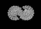
  • Surface view with section colored by density value
  • Surface level: 0.03
  • Imaged by UCSF Chimera
  • Download
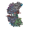
  • Surface view colored by height
  • Surface level: 0.03
  • Imaged by UCSF Chimera
  • Download
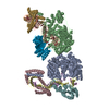
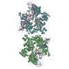
  • Surface view with fitted model
  • Atomic models: PDB-7pqd
  • Surface level: 0.03
  • Imaged by UCSF Chimera
  • Download
Movie viewer
Structure viewerEM map:
SurfViewMolmilJmol/JSmol
Supplemental images

Downloads & links

-
Map

FileDownload / File: emd_13590.map.gz / Format: CCP4 / Size: 512 MB / Type: IMAGE STORED AS FLOATING POINT NUMBER (4 BYTES)
Annotationlight harvesting core complex
Voxel sizeX=Y=Z: 0.65 Å
Density
Contour LevelBy AUTHOR: 0.0222 / Movie #1: 0.03
Minimum - Maximum-0.11344796 - 0.21005328
Average (Standard dev.)0.00032892064 (±0.004754529)
SymmetrySpace group: 1
Details

EMDB XML:

Map geometry
Axis orderXYZ
Origin000
Dimensions512512512
Spacing512512512
CellA=B=C: 332.8 Å
α=β=γ: 90.0 °

CCP4 map header:

modeImage stored as Reals
Å/pix. X/Y/Z0.650.650.65
M x/y/z512512512
origin x/y/z0.0000.0000.000
length x/y/z332.800332.800332.800
α/β/γ90.00090.00090.000
start NX/NY/NZ000
NX/NY/NZ260260260
MAP C/R/S123
start NC/NR/NS000
NC/NR/NS512512512
D min/max/mean-0.1130.2100.000

-
Supplemental data

-
Sample components

+
Entire : Light harvesting core complex

EntireName: Light harvesting core complex
Components
  • Complex: Light harvesting core complex
    • Protein or peptide: LH1-alpha
    • Protein or peptide: LH1-beta
    • Protein or peptide: RC-H
    • Protein or peptide: RC-L
    • Protein or peptide: Reaction center protein M chainPhotosynthetic reaction centre
    • Protein or peptide: PufZ
    • Protein or peptide: PufY
    • Protein or peptide: PufX
  • Ligand: BACTERIOCHLOROPHYLL ABacteriochlorophyll
  • Ligand: 3,4-DIHYDROSPHEROIDENE
  • Ligand: 1,2-Distearoyl-sn-glycerophosphoethanolamine
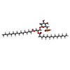
  • Ligand: (2R,5R,11R,14R)-5,8,11-trihydroxy-5,11-dioxido-17-oxo-2,14-bis(tetradecanoyloxy)-4,6,10,12,16-pentaoxa-5,11-diphosphatriacont-1-yl tetradecanoate
  • Ligand: UBIQUINONE-1Coenzyme Q10
  • Ligand: UBIQUINONE-10Coenzyme Q10
  • Ligand: BACTERIOPHEOPHYTIN A
  • Ligand: 1,2-DI-O-ACYL-3-O-[6-DEOXY-6-SULFO-ALPHA-D-GLUCOPYRANOSYL]-SN-GLYCEROL
  • Ligand: DODECYL-BETA-D-MALTOSIDE
  • Ligand: FE (III) ION
  • Ligand: water

+
Supramolecule #1: Light harvesting core complex

SupramoleculeName: Light harvesting core complex / type: complex / ID: 1 / Parent: 0 / Macromolecule list: #1-#8
Source (natural)Organism: Cereibacter sphaeroides 2.4.1 (bacteria)

+
Macromolecule #1: LH1-alpha

MacromoleculeName: LH1-alpha / type: protein_or_peptide / ID: 1 / Number of copies: 28 / Enantiomer: LEVO
Source (natural)Organism: Cereibacter sphaeroides 2.4.1 (bacteria)
Molecular weightTheoretical: 6.84418 KDa
SequenceString:
(FME)SKFYKIWMI FDPRRVFVAQ GVFLFLLAVM IHLILLSTPS YNWLEISAAK YNRVAVAE

+
Macromolecule #2: LH1-beta

MacromoleculeName: LH1-beta / type: protein_or_peptide / ID: 2 / Number of copies: 28 / Enantiomer: LEVO
Source (natural)Organism: Cereibacter sphaeroides 2.4.1 (bacteria)
Molecular weightTheoretical: 5.592361 KDa
SequenceString:
MADKSDLGYT GLTDEQAQEL HSVYMSGLWL FSAVAIVAHL AVYIWRPWF

+
Macromolecule #3: RC-H

MacromoleculeName: RC-H / type: protein_or_peptide / ID: 3 / Number of copies: 2 / Enantiomer: LEVO
Source (natural)Organism: Cereibacter sphaeroides 2.4.1 (bacteria)
Molecular weightTheoretical: 26.544471 KDa
SequenceString: MVGVTAFGNF DLASLAIYSF WIFLAGLIYY LQTENMREGY PLENEDGTPA ANQGPFPLPK PKTFILPHGR GTLTVPGPES EDRPIALAR TAVSEGFPHA PTGDPMKDGV GPASWVARRD LPELDGHGHN KIKPMKAAAG FHVSAGKNPI GLPVRGCDLE I AGKVVDIW ...String:
MVGVTAFGNF DLASLAIYSF WIFLAGLIYY LQTENMREGY PLENEDGTPA ANQGPFPLPK PKTFILPHGR GTLTVPGPES EDRPIALAR TAVSEGFPHA PTGDPMKDGV GPASWVARRD LPELDGHGHN KIKPMKAAAG FHVSAGKNPI GLPVRGCDLE I AGKVVDIW VDIPEQMARF LEVELKDGST RLLPMQMVKV QSNRVHVNAL SSDLFAGIPT IKSPTEVTLL EEDKICGYVA GG LMYAAP

+
Macromolecule #4: RC-L

MacromoleculeName: RC-L / type: protein_or_peptide / ID: 4 / Number of copies: 2 / Enantiomer: LEVO
Source (natural)Organism: Cereibacter sphaeroides 2.4.1 (bacteria)
Molecular weightTheoretical: 31.346389 KDa
SequenceString: ALLSFERKYR VPGGTLVGGN LFDFWVGPFY VGFFGVATFF FAALGIILIA WSAVLQGTWN PQLISVYPPA LEYGLGGAPL AKGGLWQII TICATGAFVS WALREVEICR KLGIGYHIPF AFAFAILAYL TLVLFRPVMM GAWGYAFPYG IWTHLDWVSN T GYTYGNFH ...String:
ALLSFERKYR VPGGTLVGGN LFDFWVGPFY VGFFGVATFF FAALGIILIA WSAVLQGTWN PQLISVYPPA LEYGLGGAPL AKGGLWQII TICATGAFVS WALREVEICR KLGIGYHIPF AFAFAILAYL TLVLFRPVMM GAWGYAFPYG IWTHLDWVSN T GYTYGNFH YNPAHMIAIS FFFTNALALA LHGALVLSAA NPEKGKEMRT PDHEDTFFRD LVGYSIGTLG IHRLGLLLSL SA VFFSALC MIITGTIWFD QWVDWWQWWV KLPWWANIPG GING

+
Macromolecule #5: Reaction center protein M chain

MacromoleculeName: Reaction center protein M chain / type: protein_or_peptide / ID: 5 / Number of copies: 2 / Enantiomer: LEVO
Source (natural)Organism: Cereibacter sphaeroides 2.4.1 (bacteria)
Molecular weightTheoretical: 34.398543 KDa
SequenceString: AEYQNIFSQV QVRGPADLGM TEDVNLANRS GVGPFSTLLG WFGNAQLGPI YLGSLGVLSL FSGLMWFFTI GIWFWYQAGW NPAVFLRDL FFFSLEPPAP EYGLSFAAPL KEGGLWLIAS FFMFVAVWSW WGRTYLRAQA LGMGKHTAWA FLSAIWLWMV L GFIRPILM ...String:
AEYQNIFSQV QVRGPADLGM TEDVNLANRS GVGPFSTLLG WFGNAQLGPI YLGSLGVLSL FSGLMWFFTI GIWFWYQAGW NPAVFLRDL FFFSLEPPAP EYGLSFAAPL KEGGLWLIAS FFMFVAVWSW WGRTYLRAQA LGMGKHTAWA FLSAIWLWMV L GFIRPILM GSWSEAVPYG IFSHLDWTNN FSLVHGNLFY NPFHGLSIAF LYGSALLFAM HGATILAVSR FGGERELEQI AD RGTAAER AALFWRWTMG FNATMEGIHR WAIWMAVLVT LTGGIGILLS GTVVDNWYVW GQNHGMAPLN

+
Macromolecule #6: PufZ

MacromoleculeName: PufZ / type: protein_or_peptide / ID: 6 / Number of copies: 4 / Enantiomer: LEVO
Source (natural)Organism: Cereibacter sphaeroides 2.4.1 (bacteria)
Molecular weightTheoretical: 3.517323 KDa
SequenceString:
MAYMFGIIVF LAMLAVCWFG FMAAERQAGR L

+
Macromolecule #7: PufY

MacromoleculeName: PufY / type: protein_or_peptide / ID: 7 / Number of copies: 2 / Enantiomer: LEVO
Source (natural)Organism: Cereibacter sphaeroides 2.4.1 (bacteria)
Molecular weightTheoretical: 5.126067 KDa
SequenceString:
EVSEFAFRLM MAAVIFVGVG IMFAFAGGHW FVGLVVGGLV AAFFAATPN

+
Macromolecule #8: PufX

MacromoleculeName: PufX / type: protein_or_peptide / ID: 8 / Number of copies: 2 / Enantiomer: LEVO
Source (natural)Organism: Cereibacter sphaeroides 2.4.1 (bacteria)
Molecular weightTheoretical: 6.024157 KDa
SequenceString:
PKTNLRLWVA FQMMKGAGWA GGVFFGTLLL IGFFRVVGRM LPIQENQAPA PNITG

+
Macromolecule #9: BACTERIOCHLOROPHYLL A

MacromoleculeName: BACTERIOCHLOROPHYLL A / type: ligand / ID: 9 / Number of copies: 64 / Formula: BCL
Molecular weightTheoretical: 911.504 Da
Chemical component information

ChemComp-BCL:
BACTERIOCHLOROPHYLL A / Bacteriochlorophyll

+
Macromolecule #10: 3,4-DIHYDROSPHEROIDENE

MacromoleculeName: 3,4-DIHYDROSPHEROIDENE / type: ligand / ID: 10 / Number of copies: 54 / Formula: SP2
Molecular weightTheoretical: 570.93 Da
Chemical component information

ChemComp-SP2:
3,4-DIHYDROSPHEROIDENE

+
Macromolecule #11: 1,2-Distearoyl-sn-glycerophosphoethanolamine

MacromoleculeName: 1,2-Distearoyl-sn-glycerophosphoethanolamine / type: ligand / ID: 11 / Number of copies: 4 / Formula: 3PE
Molecular weightTheoretical: 748.065 Da
Chemical component information

ChemComp-3PE:
1,2-Distearoyl-sn-glycerophosphoethanolamine / phospholipid*YM / Phosphatidylethanolamine

+
Macromolecule #12: (2R,5R,11R,14R)-5,8,11-trihydroxy-5,11-dioxido-17-oxo-2,14-bis(te...

MacromoleculeName: (2R,5R,11R,14R)-5,8,11-trihydroxy-5,11-dioxido-17-oxo-2,14-bis(tetradecanoyloxy)-4,6,10,12,16-pentaoxa-5,11-diphosphatriacont-1-yl tetradecanoate
type: ligand / ID: 12 / Number of copies: 4 / Formula: CD4
Molecular weightTheoretical: 1.241633 KDa
Chemical component information

ChemComp-CD4:
(2R,5R,11R,14R)-5,8,11-trihydroxy-5,11-dioxido-17-oxo-2,14-bis(tetradecanoyloxy)-4,6,10,12,16-pentaoxa-5,11-diphosphatriacont-1-yl tetradecanoate

+
Macromolecule #13: UBIQUINONE-1

MacromoleculeName: UBIQUINONE-1 / type: ligand / ID: 13 / Number of copies: 2 / Formula: UQ1
Molecular weightTheoretical: 250.29 Da
Chemical component information

ChemComp-UQ1:
UBIQUINONE-1

+
Macromolecule #14: UBIQUINONE-10

MacromoleculeName: UBIQUINONE-10 / type: ligand / ID: 14 / Number of copies: 4 / Formula: U10
Molecular weightTheoretical: 863.343 Da
Chemical component information

ChemComp-U10:
UBIQUINONE-10 / Coenzyme Q10

+
Macromolecule #15: BACTERIOPHEOPHYTIN A

MacromoleculeName: BACTERIOPHEOPHYTIN A / type: ligand / ID: 15 / Number of copies: 4 / Formula: BPH
Molecular weightTheoretical: 889.215 Da
Chemical component information

ChemComp-BPH:
BACTERIOPHEOPHYTIN A / Pheophytin

+
Macromolecule #16: 1,2-DI-O-ACYL-3-O-[6-DEOXY-6-SULFO-ALPHA-D-GLUCOPYRANOSYL]-SN-GLYCEROL

MacromoleculeName: 1,2-DI-O-ACYL-3-O-[6-DEOXY-6-SULFO-ALPHA-D-GLUCOPYRANOSYL]-SN-GLYCEROL
type: ligand / ID: 16 / Number of copies: 2 / Formula: SQD
Molecular weightTheoretical: 795.116 Da
Chemical component information

ChemComp-SQD:
1,2-DI-O-ACYL-3-O-[6-DEOXY-6-SULFO-ALPHA-D-GLUCOPYRANOSYL]-SN-GLYCEROL

+
Macromolecule #17: DODECYL-BETA-D-MALTOSIDE

MacromoleculeName: DODECYL-BETA-D-MALTOSIDE / type: ligand / ID: 17 / Number of copies: 2 / Formula: LMT
Molecular weightTheoretical: 510.615 Da
Chemical component information

ChemComp-LMT:
DODECYL-BETA-D-MALTOSIDE / detergent*YM

+
Macromolecule #18: FE (III) ION

MacromoleculeName: FE (III) ION / type: ligand / ID: 18 / Number of copies: 2 / Formula: FE
Molecular weightTheoretical: 55.845 Da

+
Macromolecule #19: water

MacromoleculeName: water / type: ligand / ID: 19 / Number of copies: 144 / Formula: HOH
Molecular weightTheoretical: 18.015 Da
Chemical component information

ChemComp-HOH:
WATER / Water

-
Experimental details

-
Structure determination

Methodcryo EM
Processingsingle particle reconstruction
Aggregation stateparticle

-
Sample preparation

Concentration4.0 mg/mL
BufferpH: 7.8 / Component - Concentration: 20.0 mMol / Component - Formula: HEPES / Details: 20 mM HEPES, pH 7.8, 0.03% beta-DDM
GridModel: Quantifoil R1.2/1.3 / Material: COPPER / Mesh: 300 / Pretreatment - Type: GLOW DISCHARGE / Pretreatment - Time: 60 sec. / Pretreatment - Atmosphere: AIR
VitrificationCryogen name: ETHANE / Chamber humidity: 99 % / Chamber temperature: 277 K / Details: QF R1.2/1.3 300 mesh Cu grid.
DetailsIn 20 mM HEPES, pH 7.8, 0.03% beta-DDM buffer solution

-
Electron microscopy

MicroscopeFEI TITAN KRIOS
Electron beamAcceleration voltage: 300 kV / Electron source: FIELD EMISSION GUN
Electron opticsC2 aperture diameter: 50.0 µm / Illumination mode: FLOOD BEAM / Imaging mode: BRIGHT FIELDBright-field microscopy / Cs: 2.7 mm / Nominal defocus max: 2.2 µm / Nominal defocus min: 0.8 µm / Nominal magnification: 120000
Sample stageSpecimen holder model: FEI TITAN KRIOS AUTOGRID HOLDER / Cooling holder cryogen: NITROGEN
TemperatureMax: 80.0 K
Image recordingFilm or detector model: FEI FALCON IV (4k x 4k) / Number grids imaged: 1 / Number real images: 5058 / Average exposure time: 12.21 sec. / Average electron dose: 45.36 e/Å2
Experimental equipment
Model: Titan Krios / Image courtesy: FEI Company

-
Image processing

Particle selectionNumber selected: 223786
Startup modelType of model: PDB ENTRY
PDB model - PDB ID:
Initial angle assignmentType: MAXIMUM LIKELIHOOD / Software - Name: RELION (ver. 3.1)
Final 3D classificationNumber classes: 4 / Software - Name: RELION (ver. 3.1)
Final angle assignmentType: MAXIMUM LIKELIHOOD / Software - Name: RELION (ver. 3.1)
Final reconstructionNumber classes used: 1 / Applied symmetry - Point group: C2 (2 fold cyclic) / Algorithm: BACK PROJECTION / Resolution.type: BY AUTHOR / Resolution: 2.9 Å / Resolution method: FSC 0.143 CUT-OFF / Software - Name: RELION (ver. 3.1) / Number images used: 58945
FSC plot (resolution estimation)

+
About Yorodumi

-
News

-
Feb 9, 2022. New format data for meta-information of EMDB entries

New format data for meta-information of EMDB entries

  • Version 3 of the EMDB header file is now the official format.
  • The previous official version 1.9 will be removed from the archive.

Related info.:EMDB header

External links:wwPDB to switch to version 3 of the EMDB data model

-
Aug 12, 2020. Covid-19 info

Covid-19 info

URL: https://pdbj.org/emnavi/covid19.php

New page: Covid-19 featured information page in EM Navigator.

Related info.:Covid-19 info / Mar 5, 2020. Novel coronavirus structure data

+
Mar 5, 2020. Novel coronavirus structure data

Novel coronavirus structure data

Related info.:Yorodumi Speices / Aug 12, 2020. Covid-19 info

External links:COVID-19 featured content - PDBj / Molecule of the Month (242):Coronavirus Proteases

+
Jan 31, 2019. EMDB accession codes are about to change! (news from PDBe EMDB page)

EMDB accession codes are about to change! (news from PDBe EMDB page)

  • The allocation of 4 digits for EMDB accession codes will soon come to an end. Whilst these codes will remain in use, new EMDB accession codes will include an additional digit and will expand incrementally as the available range of codes is exhausted. The current 4-digit format prefixed with “EMD-” (i.e. EMD-XXXX) will advance to a 5-digit format (i.e. EMD-XXXXX), and so on. It is currently estimated that the 4-digit codes will be depleted around Spring 2019, at which point the 5-digit format will come into force.
  • The EM Navigator/Yorodumi systems omit the EMD- prefix.

Related info.:Q: What is EMD? / ID/Accession-code notation in Yorodumi/EM Navigator

External links:EMDB Accession Codes are Changing Soon! / Contact to PDBj

+
Jul 12, 2017. Major update of PDB

Major update of PDB

  • wwPDB released updated PDB data conforming to the new PDBx/mmCIF dictionary.
  • This is a major update changing the version number from 4 to 5, and with Remediation, in which all the entries are updated.
  • In this update, many items about electron microscopy experimental information are reorganized (e.g. em_software).
  • Now, EM Navigator and Yorodumi are based on the updated data.

External links:wwPDB Remediation / Enriched Model Files Conforming to OneDep Data Standards Now Available in the PDB FTP Archive

-
Yorodumi

Thousand views of thousand structures

  • Yorodumi is a browser for structure data from EMDB, PDB, SASBDB, etc.
  • This page is also the successor to EM Navigator detail page, and also detail information page/front-end page for Omokage search.
  • The word "yorodu" (or yorozu) is an old Japanese word meaning "ten thousand". "mi" (miru) is to see.

Related info.:EMDB / PDB / SASBDB / Comparison of 3 databanks / Yorodumi Search / Aug 31, 2016. New EM Navigator & Yorodumi / Yorodumi Papers / Jmol/JSmol / Function and homology information / Changes in new EM Navigator and Yorodumi

Read more