 Movie
Movie Controller

[English] 日本語
 Yorodumi
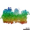
- PDB-7pqd: Cryo-EM structure of the dimeric Rhodobacter sphaeroides RC-LH1 c... -
+ Open data

- Basic information
| Entry | Database: PDB / ID: 7pqd | ||||||||||||
|---|---|---|---|---|---|---|---|---|---|---|---|---|---|
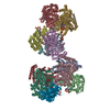
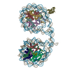
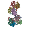
| Title | Cryo-EM structure of the dimeric Rhodobacter sphaeroides RC-LH1 core complex at 2.9 A: the structural basis for dimerisation | ||||||||||||
 Components Components |
| ||||||||||||
 Keywords Keywords | PHOTOSYNTHESIS / light harvesting complex / Cryo-EM / purple bacteria / RC-LH1 / RC-LH1-PufXYZ / dimer / dimeric core complex | ||||||||||||
| Function / homology |  Function and homology information Function and homology informationplasma membrane-derived chromatophore membrane / plasma membrane light-harvesting complex / bacteriochlorophyll binding / photosynthetic electron transport in photosystem II / metal ion binding Similarity search - Function | ||||||||||||
| Biological species |  Cereibacter sphaeroides 2.4.1 (bacteria) Cereibacter sphaeroides 2.4.1 (bacteria) | ||||||||||||
| Method | ELECTRON MICROSCOPY / single particle reconstruction / cryo EM / Resolution: 2.9 Å | ||||||||||||
 Authors Authors | Qian, P. / Hunter, C.N. | ||||||||||||
| Funding support |  United Kingdom, 3items United Kingdom, 3items
| ||||||||||||
 Citation Citation | Journal: Biochem J / Year: 2021 Title: Cryo-EM structure of the dimeric Rhodobacter sphaeroides RC-LH1 core complex at 2.9 Å: the structural basis for dimerisation. Authors: Pu Qian / Tristan I Croll / Andrew Hitchcock / Philip J Jackson / Jack H Salisbury / Pablo Castro-Hartmann / Kasim Sader / David J K Swainsbury / C Neil Hunter /  Abstract: The dimeric reaction centre light-harvesting 1 (RC-LH1) core complex of Rhodobacter sphaeroides converts absorbed light energy to a charge separation, and then it reduces a quinone electron and ...The dimeric reaction centre light-harvesting 1 (RC-LH1) core complex of Rhodobacter sphaeroides converts absorbed light energy to a charge separation, and then it reduces a quinone electron and proton acceptor to a quinol. The angle between the two monomers imposes a bent configuration on the dimer complex, which exerts a major influence on the curvature of the membrane vesicles, known as chromatophores, where the light-driven photosynthetic reactions take place. To investigate the dimerisation interface between two RC-LH1 monomers, we determined the cryogenic electron microscopy structure of the dimeric complex at 2.9 Å resolution. The structure shows that each monomer consists of a central RC partly enclosed by a 14-subunit LH1 ring held in an open state by PufX and protein-Y polypeptides, thus enabling quinones to enter and leave the complex. Two monomers are brought together through N-terminal interactions between PufX polypeptides on the cytoplasmic side of the complex, augmented by two novel transmembrane polypeptides, designated protein-Z, that bind to the outer faces of the two central LH1 β polypeptides. The precise fit at the dimer interface, enabled by PufX and protein-Z, by C-terminal interactions between opposing LH1 αβ subunits, and by a series of interactions with a bound sulfoquinovosyl diacylglycerol lipid, bring together each monomer creating an S-shaped array of 28 bacteriochlorophylls. The seamless join between the two sets of LH1 bacteriochlorophylls provides a path for excitation energy absorbed by one half of the complex to migrate across the dimer interface to the other half. #1: Journal: Acta Crystallogr., Sect. D: Biol. Crystallogr. / Year: 2018Title: Real-space refinement in PHENIX for cryo-EM and crystallography Authors: Qian, P. / Hunter, C.N. | ||||||||||||
| History |
|
- Structure visualization
| Movie |
Movie viewer |
|---|---|
| Structure viewer | Molecule: MolmilJmol/JSmol |
- Downloads & links
-Download
| PDBx/mmCIF format | 7pqd.cif.gz | 1.3 MB | Display | PDBx/mmCIF format |
|---|---|---|---|---|
| PDB format | pdb7pqd.ent.gz | Display | PDB format | |
| PDBx/mmJSON format | 7pqd.json.gz | Tree view | PDBx/mmJSON format | |
| Others |  Other downloads Other downloads |
-Validation report
| Arichive directory | https://data.pdbj.org/pub/pdb/validation_reports/pq/7pqdftp://data.pdbj.org/pub/pdb/validation_reports/pq/7pqd | HTTPS FTP |
|---|
-Related structure data
| Related structure data | 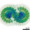 13590MC M: map data used to model this data C: citing same article ( |
|---|---|
| Similar structure data |

-Links
 PDBj
PDBj


- Assembly
| Deposited unit | 
|
|---|---|
| 1 |
|
-Components
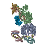
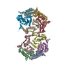
-Protein , 5 types, 36 molecules AAABACADAEAFAGAHAIAJAKALAMANaaabacadaeafagahaiajakalamanHh...
| #1: Protein | Mass: 6844.180 Da / Num. of mol.: 28 / Source method: isolated from a natural source / Source: (natural) Cereibacter sphaeroides 2.4.1 (bacteria)#3: Protein | Mass: 26544.471 Da / Num. of mol.: 2 / Source method: isolated from a natural source / Source: (natural) Cereibacter sphaeroides 2.4.1 (bacteria)#4: Protein | Mass: 31346.389 Da / Num. of mol.: 2 / Source method: isolated from a natural source / Source: (natural) Cereibacter sphaeroides 2.4.1 (bacteria)#5: Protein | Mass: 34398.543 Da / Num. of mol.: 2 / Source method: isolated from a natural source / Source: (natural) Cereibacter sphaeroides 2.4.1 (bacteria) / References: UniProt: Q3J1A6#8: Protein | Mass: 6024.157 Da / Num. of mol.: 2 / Source method: isolated from a natural source / Source: (natural) Cereibacter sphaeroides 2.4.1 (bacteria) |
|---|
-Protein/peptide , 3 types, 34 molecules BABBBCBDBEBFBGBHBIBJBKBLBMBNbabbbcbdbebfbgbhbibjbkblbmbnUAUB...
| #2: Protein/peptide | Mass: 5592.361 Da / Num. of mol.: 28 / Source method: isolated from a natural source / Source: (natural) Cereibacter sphaeroides 2.4.1 (bacteria)#6: Protein/peptide | Mass: 3517.323 Da / Num. of mol.: 4 / Source method: isolated from a natural source / Source: (natural) Cereibacter sphaeroides 2.4.1 (bacteria)#7: Protein/peptide | Mass: 5126.067 Da / Num. of mol.: 2 / Source method: isolated from a natural source / Source: (natural) Cereibacter sphaeroides 2.4.1 (bacteria) |
|---|
-Sugars , 1 types, 2 molecules 
| #17: Sugar |  Type: D-saccharide / Mass: 510.615 Da / Num. of mol.: 2 / Source method: obtained synthetically / Formula: C24H46O11 / Comment: detergent*YM Type: D-saccharide / Mass: 510.615 Da / Num. of mol.: 2 / Source method: obtained synthetically / Formula: C24H46O11 / Comment: detergent*YM |
|---|
-Non-polymers , 10 types, 284 molecules 


| #9: Chemical | ChemComp-BCL /  Mass: 911.504 Da / Num. of mol.: 64 / Source method: obtained synthetically / Formula: C55H74MgN4O6 / Feature type: SUBJECT OF INVESTIGATION Mass: 911.504 Da / Num. of mol.: 64 / Source method: obtained synthetically / Formula: C55H74MgN4O6 / Feature type: SUBJECT OF INVESTIGATION#10: Chemical | ChemComp-SP2 /  Mass: 570.930 Da / Num. of mol.: 54 / Source method: obtained synthetically / Formula: C41H62O Mass: 570.930 Da / Num. of mol.: 54 / Source method: obtained synthetically / Formula: C41H62O#11: Chemical | ChemComp-3PE /  Mass: 748.065 Da / Num. of mol.: 4 / Source method: obtained synthetically / Formula: C41H82NO8P / Comment: phospholipid*YM Mass: 748.065 Da / Num. of mol.: 4 / Source method: obtained synthetically / Formula: C41H82NO8P / Comment: phospholipid*YM#12: Chemical | ChemComp-CD4 / (  Mass: 1241.633 Da / Num. of mol.: 4 / Source method: obtained synthetically / Formula: C65H126O17P2 Mass: 1241.633 Da / Num. of mol.: 4 / Source method: obtained synthetically / Formula: C65H126O17P2#13: Chemical |  Mass: 250.290 Da / Num. of mol.: 2 / Source method: obtained synthetically / Formula: C14H18O4 Mass: 250.290 Da / Num. of mol.: 2 / Source method: obtained synthetically / Formula: C14H18O4#14: Chemical | ChemComp-U10 /  Mass: 863.343 Da / Num. of mol.: 4 / Source method: obtained synthetically / Formula: C59H90O4 / Feature type: SUBJECT OF INVESTIGATION Mass: 863.343 Da / Num. of mol.: 4 / Source method: obtained synthetically / Formula: C59H90O4 / Feature type: SUBJECT OF INVESTIGATION#15: Chemical | ChemComp-BPH /  Mass: 889.215 Da / Num. of mol.: 4 / Source method: obtained synthetically / Formula: C55H76N4O6 / Feature type: SUBJECT OF INVESTIGATION Mass: 889.215 Da / Num. of mol.: 4 / Source method: obtained synthetically / Formula: C55H76N4O6 / Feature type: SUBJECT OF INVESTIGATION#16: Chemical |  Mass: 795.116 Da / Num. of mol.: 2 / Source method: obtained synthetically / Formula: C41H78O12S / Feature type: SUBJECT OF INVESTIGATION Mass: 795.116 Da / Num. of mol.: 2 / Source method: obtained synthetically / Formula: C41H78O12S / Feature type: SUBJECT OF INVESTIGATION#18: Chemical |  Mass: 55.845 Da / Num. of mol.: 2 / Source method: obtained synthetically / Formula: Fe Mass: 55.845 Da / Num. of mol.: 2 / Source method: obtained synthetically / Formula: Fe#19: Water | ChemComp-HOH / | Mass: 18.015 Da / Num. of mol.: 144 / Source method: isolated from a natural source / Formula: H2O |
|---|
-Details
| Has ligand of interest | Y |
|---|---|
| Has protein modification | Y |
-Experimental details
-Experiment
| Experiment | Method: ELECTRON MICROSCOPY |
|---|---|
| EM experiment | Aggregation state: PARTICLE / 3D reconstruction method: single particle reconstruction |
- Sample preparation
| Component | Name: Light harvesting core complex / Type: COMPLEX / Entity ID: #1-#8 / Source: NATURAL |
|---|---|
| Source (natural) | Organism: Cereibacter sphaeroides 2.4.1 (bacteria) |
| Buffer solution | pH: 7.8 / Details: 20 mM HEPES, pH 7.8, 0.03% beta-DDM |
| Buffer component | Conc.: 20 mMol / Formula: HEPES |
| Specimen | Conc.: 4 mg/ml / Embedding applied: NO / Shadowing applied: NO / Staining applied: NO / Vitrification applied: YES Details: In 20 mM HEPES, pH 7.8, 0.03% beta-DDM buffer solution |
| Specimen support | Grid material: COPPER / Grid mesh size: 300 divisions/in. / Grid type: Quantifoil R1.2/1.3 |
| Vitrification | Cryogen name: ETHANE / Humidity: 99 % / Chamber temperature: 277 K / Details: QF R1.2/1.3 300 mesh Cu grid |
- Electron microscopy imaging
| Experimental equipment |  Model: Titan Krios / Image courtesy: FEI Company |
|---|---|
| Microscopy | Model: FEI TITAN KRIOS |
| Electron gun | Electron source:  FIELD EMISSION GUN / Accelerating voltage: 300 kV / Illumination mode: FLOOD BEAM FIELD EMISSION GUN / Accelerating voltage: 300 kV / Illumination mode: FLOOD BEAM |
| Electron lens | Mode: BRIGHT FIELD / Nominal magnification: 120000 X / Nominal defocus max: 2200 nm / Nominal defocus min: 800 nm / Cs: 2.7 mm / C2 aperture diameter: 50 µm / Alignment procedure: COMA FREE |
| Specimen holder | Cryogen: NITROGEN / Specimen holder model: FEI TITAN KRIOS AUTOGRID HOLDER / Temperature (max): 80 K |
| Image recording | Average exposure time: 12.21 sec. / Electron dose: 45.36 e/Å2 / Film or detector model: FEI FALCON IV (4k x 4k) / Num. of grids imaged: 1 / Num. of real images: 5058 |
- Processing
| Software |
| ||||||||||||||||||||||||||||
|---|---|---|---|---|---|---|---|---|---|---|---|---|---|---|---|---|---|---|---|---|---|---|---|---|---|---|---|---|---|
| EM software |
| ||||||||||||||||||||||||||||
| CTF correction | Type: PHASE FLIPPING AND AMPLITUDE CORRECTION | ||||||||||||||||||||||||||||
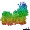
| Particle selection | Num. of particles selected: 223786 | ||||||||||||||||||||||||||||
| Symmetry | Point symmetry: C2 (2 fold cyclic) | ||||||||||||||||||||||||||||
| 3D reconstruction | Resolution: 2.9 Å / Resolution method: FSC 0.143 CUT-OFF / Num. of particles: 58945 / Algorithm: BACK PROJECTION / Num. of class averages: 1 / Symmetry type: POINT |
