 Movie
Movie Controller

[English] 日本語
 Yorodumi
- PDB-5kah: Crystal structure of a dioxygenase in the Crotonase superfamily i... -
+ Open data

- Basic information
| Entry | Database: PDB / ID: 5kah | ||||||
|---|---|---|---|---|---|---|---|
| Title | Crystal structure of a dioxygenase in the Crotonase superfamily in P21, V425T mutant | ||||||
 Components Components | (3,5-dihydroxyphenyl)acetyl-CoA 1,2-dioxygenase | ||||||
 Keywords Keywords | OXIDOREDUCTASE / dioxygenase / DpgC | ||||||
| Function / homology |  Function and homology information Function and homology information(3,5-dihydroxyphenyl)acetyl-CoA 1,2-dioxygenase / oxidoreductase activity, acting on single donors with incorporation of molecular oxygen, incorporation of two atoms of oxygen / fatty acid beta-oxidation / antibiotic biosynthetic process / identical protein binding Similarity search - Function | ||||||
| Biological species |  Streptomyces toyocaensis (bacteria) Streptomyces toyocaensis (bacteria) | ||||||
| Method |  X-RAY DIFFRACTION / SYNCHROTRON / MOLECULAR REPLACEMENT / Resolution: 2.779 Å X-RAY DIFFRACTION / SYNCHROTRON / MOLECULAR REPLACEMENT / Resolution: 2.779 Å | ||||||
 Authors Authors | Li, K. / Fielding, E.N. / Condurso, H.L. / Bruner, S.D. | ||||||
| Funding support |  United States, 1items United States, 1items
| ||||||
 Citation Citation | Journal: Acta Crystallogr D Struct Biol / Year: 2017 Title: Probing the structural basis of oxygen binding in a cofactor-independent dioxygenase. Authors: Li, K. / Fielding, E.N. / Condurso, H.L. / Bruner, S.D. | ||||||
| History |
|
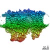
- Structure visualization
| Structure viewer | Molecule: MolmilJmol/JSmol |
|---|
- Downloads & links
-Download
| PDBx/mmCIF format | 5kah.cif.gz | 968.6 KB | Display | PDBx/mmCIF format |
|---|---|---|---|---|
| PDB format | pdb5kah.ent.gz | 807 KB | Display | PDB format |
| PDBx/mmJSON format | 5kah.json.gz | Tree view | PDBx/mmJSON format | |
| Others |  Other downloads Other downloads |
-Validation report
| Arichive directory | https://data.pdbj.org/pub/pdb/validation_reports/ka/5kahftp://data.pdbj.org/pub/pdb/validation_reports/ka/5kah | HTTPS FTP |
|---|
-Related structure data
| Related structure data |  5kagC 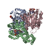 5kajC 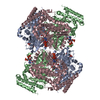 2np9S C: citing same article ( S: Starting model for refinement |
|---|---|
| Similar structure data |


-Links
 PDBj
PDBj- Assembly
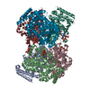
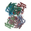
| Deposited unit | 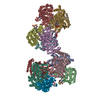
| ||||||||
|---|---|---|---|---|---|---|---|---|---|
| 1 | 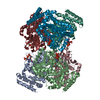
| ||||||||
| 2 | 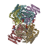
| ||||||||
| Unit cell |
|
-Components
| #1: Protein | ( Mass: 48243.105 Da / Num. of mol.: 12 / Mutation: V425T Source method: isolated from a genetically manipulated source Source: (gene. exp.) Streptomyces toyocaensis (bacteria) / Gene: BU52_01220 / Production host: References: UniProt: Q8KLK7, (3,5-dihydroxyphenyl)acetyl-CoA 1,2-dioxygenase #2: Chemical | ChemComp-YE1 / [(   Mass: 900.615 Da / Num. of mol.: 12 / Source method: isolated from a natural source / Formula: C29H43N8O19P3 Mass: 900.615 Da / Num. of mol.: 12 / Source method: isolated from a natural source / Formula: C29H43N8O19P3#3: Water | ChemComp-HOH / |  Mass: 18.015 Da / Num. of mol.: 738 / Source method: isolated from a natural source / Formula: H2O Mass: 18.015 Da / Num. of mol.: 738 / Source method: isolated from a natural source / Formula: H2O |
|---|
-Experimental details
-Experiment
| Experiment | Method: X-RAY DIFFRACTION / Number of used crystals: 1 |
|---|
- Sample preparation
| Crystal | Density Matthews: 3.37 Å3/Da / Density % sol: 63.55 % |
|---|---|
| Crystal grow | Temperature: 298 K / Method: vapor diffusion, hanging drop / pH: 5.6 Details: 140 mM ammonium acetate, 14% w/v PEG 4,000, and 100 mM sodium citrate pH 5.6 |
-Data collection
| Diffraction | Mean temperature: 100 K |
|---|---|
| Diffraction source | Source: SYNCHROTRON / Site: NSLS / Beamline: X12C / Wavelength: 1.1 Å |
| Detector | Type: ADSC QUANTUM 210 / Detector: CCD / Date: Mar 15, 2007 |
| Radiation | Protocol: SINGLE WAVELENGTH / Monochromatic (M) / Laue (L): M / Scattering type: x-ray |
| Radiation wavelength | Wavelength: 1.1 Å / Relative weight: 1 |
| Reflection | Resolution: 2.779→39.028 Å / Num. obs: 184721 / % possible obs: 99.9 % / Redundancy: 4.9 % / Biso Wilson estimate: 17.9 Å2 / Rmerge(I) obs: 0.11 / Rsym value: 0.14 / Net I/σ(I): 11.8 |
| Reflection shell | Resolution: 2.779→2.83 Å / Redundancy: 4.7 % / Rmerge(I) obs: 0.54 / Mean I/σ(I) obs: 2.6 / % possible all: 89.7 |
- Processing
| Software |
| |||||||||||||||||||||||||||||||||||||||||||||||||||||||||||||||||||||||||||||||||||||||||||||||||||||||||||||||||||||||||||||||||||||||||||||||||||
|---|---|---|---|---|---|---|---|---|---|---|---|---|---|---|---|---|---|---|---|---|---|---|---|---|---|---|---|---|---|---|---|---|---|---|---|---|---|---|---|---|---|---|---|---|---|---|---|---|---|---|---|---|---|---|---|---|---|---|---|---|---|---|---|---|---|---|---|---|---|---|---|---|---|---|---|---|---|---|---|---|---|---|---|---|---|---|---|---|---|---|---|---|---|---|---|---|---|---|---|---|---|---|---|---|---|---|---|---|---|---|---|---|---|---|---|---|---|---|---|---|---|---|---|---|---|---|---|---|---|---|---|---|---|---|---|---|---|---|---|---|---|---|---|---|---|---|---|---|
| Refinement | Method to determine structure: MOLECULAR REPLACEMENT Starting model: 2NP9 Resolution: 2.779→39.028 Å / Cross valid method: FREE R-VALUE / σ(F): 1.36 / Phase error: 27.75 / Stereochemistry target values: TWIN_LSQ_F
| |||||||||||||||||||||||||||||||||||||||||||||||||||||||||||||||||||||||||||||||||||||||||||||||||||||||||||||||||||||||||||||||||||||||||||||||||||
| Solvent computation | Shrinkage radii: 0.9 Å / VDW probe radii: 1.11 Å / Solvent model: FLAT BULK SOLVENT MODEL | |||||||||||||||||||||||||||||||||||||||||||||||||||||||||||||||||||||||||||||||||||||||||||||||||||||||||||||||||||||||||||||||||||||||||||||||||||
| Refinement step | Cycle: LAST / Resolution: 2.779→39.028 Å
| |||||||||||||||||||||||||||||||||||||||||||||||||||||||||||||||||||||||||||||||||||||||||||||||||||||||||||||||||||||||||||||||||||||||||||||||||||
| Refine LS restraints |
| |||||||||||||||||||||||||||||||||||||||||||||||||||||||||||||||||||||||||||||||||||||||||||||||||||||||||||||||||||||||||||||||||||||||||||||||||||
| LS refinement shell |
|
