 Movie
Movie Controller

[English] 日本語
 Yorodumi
- PDB-6k61: Cryo-EM structure of the tetrameric photosystem I from a heterocy... -
+ Open data

- Basic information
| Entry | Database: PDB / ID: 6k61 | |||||||||
|---|---|---|---|---|---|---|---|---|---|---|
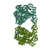
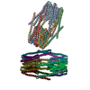
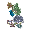
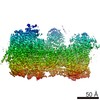
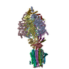
| Title | Cryo-EM structure of the tetrameric photosystem I from a heterocyst-forming cyanobacterium Anabaena sp. PCC7120 | |||||||||
 Components Components | (Photosystem I ...) x 12 | |||||||||
 Keywords Keywords | PHOTOSYNTHESIS / Photosystem I / PSI / PCC7120 | |||||||||
| Function / homology |  Function and homology information Function and homology informationphotosystem I reaction center / photosystem I / photosynthetic electron transport in photosystem I / photosystem I / plasma membrane-derived thylakoid membrane / chlorophyll binding / photosynthesis / 4 iron, 4 sulfur cluster binding / electron transfer activity / oxidoreductase activity ...photosystem I reaction center / photosystem I / photosynthetic electron transport in photosystem I / photosystem I / plasma membrane-derived thylakoid membrane / chlorophyll binding / photosynthesis / 4 iron, 4 sulfur cluster binding / electron transfer activity / oxidoreductase activity / magnesium ion binding / metal ion binding Similarity search - Function | |||||||||
| Biological species |  Nostoc sp. PCC 7120 (bacteria) Nostoc sp. PCC 7120 (bacteria) | |||||||||
| Method | ELECTRON MICROSCOPY / single particle reconstruction / cryo EM / Resolution: 2.37 Å | |||||||||
 Authors Authors | Zheng, L. / Li, Y. / Li, X. / Zhong, Q. / Li, N. / Zhang, K. / Zhang, Y. / Chu, H. / Ma, C. / Li, G. ...Zheng, L. / Li, Y. / Li, X. / Zhong, Q. / Li, N. / Zhang, K. / Zhang, Y. / Chu, H. / Ma, C. / Li, G. / Zhao, J. / Gao, N. | |||||||||
| Funding support | 1items
| |||||||||
 Citation Citation | Journal: Nat Plants / Year: 2019 Title: Structural and functional insights into the tetrameric photosystem I from heterocyst-forming cyanobacteria. Authors: Lvqin Zheng / Yanbing Li / Xiying Li / Qinglu Zhong / Ningning Li / Kun Zhang / Yuebin Zhang / Huiying Chu / Chengying Ma / Guohui Li / Jindong Zhao / Ning Gao /  Abstract: Two large protein-cofactor complexes, photosystem I and photosystem II, are the central components of photosynthesis in the thylakoid membranes. Here, we report the 2.37-Å structure of a tetrameric ...Two large protein-cofactor complexes, photosystem I and photosystem II, are the central components of photosynthesis in the thylakoid membranes. Here, we report the 2.37-Å structure of a tetrameric photosystem I complex from a heterocyst-forming cyanobacterium Anabaena sp. PCC 7120. Four photosystem I monomers, organized in a dimer of dimer, form two distinct interfaces that are largely mediated by specifically orientated polar lipids, such as sulfoquinovosyl diacylglycerol. The structure depicts a more closely connected network of chlorophylls across monomer interfaces than those seen in trimeric PSI from thermophilic cyanobacteria, possibly allowing a more efficient energy transfer between monomers. Our physiological data also revealed a functional link of photosystem I oligomerization to cyclic electron flow and thylakoid membrane organization. | |||||||||
| History |
|
- Structure visualization
| Movie |
Movie viewer |
|---|---|
| Structure viewer | Molecule: MolmilJmol/JSmol |
- Downloads & links
-Download
| PDBx/mmCIF format | 6k61.cif.gz | 1.1 MB | Display | PDBx/mmCIF format |
|---|---|---|---|---|
| PDB format | pdb6k61.ent.gz | 976.9 KB | Display | PDB format |
| PDBx/mmJSON format | 6k61.json.gz | Tree view | PDBx/mmJSON format | |
| Others |  Other downloads Other downloads |
-Validation report
| Arichive directory | https://data.pdbj.org/pub/pdb/validation_reports/k6/6k61ftp://data.pdbj.org/pub/pdb/validation_reports/k6/6k61 | HTTPS FTP |
|---|
-Related structure data
| Related structure data | 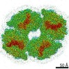 9918MC M: map data used to model this data C: citing same article ( |
|---|---|
| Similar structure data |


-Links
 PDBj
PDBj


- Assembly
| Deposited unit | 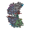
|
|---|---|
| 1 |
|
-Components
-Photosystem I ... , 12 types, 24 molecules AaXxBbCcDdEeFfJjKkIiLlMm
| #1: Protein | Mass: 83289.680 Da / Num. of mol.: 2 / Source method: isolated from a natural source / Source: (natural) Nostoc sp. PCC 7120 (bacteria) / Strain: PCC 7120 / References: UniProt: P58576, photosystem I#2: Protein/peptide | Mass: 4872.792 Da / Num. of mol.: 2 / Source method: isolated from a natural source / Source: (natural) Nostoc sp. PCC 7120 (bacteria) / Strain: PCC 7120 / References: UniProt: P58566#3: Protein | Mass: 83457.016 Da / Num. of mol.: 2 / Source method: isolated from a natural source / Source: (natural) Nostoc sp. PCC 7120 (bacteria) / Strain: PCC 7120 / References: UniProt: P58565, photosystem I#4: Protein | Mass: 8825.206 Da / Num. of mol.: 2 / Source method: isolated from a natural source / Source: (natural) Nostoc sp. PCC 7120 (bacteria) / Strain: PCC 7120 / References: UniProt: P0A410, photosystem I#5: Protein | Mass: 15176.240 Da / Num. of mol.: 2 / Source method: isolated from a natural source / Source: (natural) Nostoc sp. PCC 7120 (bacteria) / Strain: PCC 7120 / References: UniProt: P58573#6: Protein | Mass: 7897.051 Da / Num. of mol.: 2 / Source method: isolated from a natural source / Source: (natural) Nostoc sp. PCC 7120 (bacteria) / Strain: PCC 7120 / References: UniProt: P58575#7: Protein | Mass: 17850.568 Da / Num. of mol.: 2 / Source method: isolated from a natural source / Source: (natural) Nostoc sp. PCC 7120 (bacteria) / Strain: PCC 7120 / References: UniProt: P58564#8: Protein/peptide | Mass: 5499.422 Da / Num. of mol.: 2 / Source method: isolated from a natural source / Source: (natural) Nostoc sp. PCC 7120 (bacteria) / Strain: PCC 7120 / References: UniProt: P58568#9: Protein | Mass: 8852.457 Da / Num. of mol.: 2 / Source method: isolated from a natural source / Source: (natural) Nostoc sp. PCC 7120 (bacteria) / Strain: PCC 7120 / References: UniProt: P58583#10: Protein/peptide | Mass: 5066.925 Da / Num. of mol.: 2 / Source method: isolated from a natural source / Source: (natural) Nostoc sp. PCC 7120 (bacteria) / Strain: PCC 7120 / References: UniProt: P58560#11: Protein | Mass: 18130.725 Da / Num. of mol.: 2 / Source method: isolated from a natural source / Source: (natural) Nostoc sp. PCC 7120 (bacteria) / Strain: PCC 7120 / References: UniProt: P58577#12: Protein/peptide | Mass: 3538.204 Da / Num. of mol.: 2 / Source method: isolated from a natural source / Source: (natural) Nostoc sp. PCC 7120 (bacteria) / Strain: PCC 7120 / References: UniProt: Q8YNB0 |
|---|
-Non-polymers , 8 types, 259 molecules 


| #13: Chemical |  Mass: 893.489 Da / Num. of mol.: 2 / Source method: obtained synthetically / Formula: C55H72MgN4O5 / Feature type: SUBJECT OF INVESTIGATION Mass: 893.489 Da / Num. of mol.: 2 / Source method: obtained synthetically / Formula: C55H72MgN4O5 / Feature type: SUBJECT OF INVESTIGATION#14: Chemical | ChemComp-CLA /  Mass: 893.489 Da / Num. of mol.: 188 / Source method: obtained synthetically / Formula: C55H72MgN4O5 / Feature type: SUBJECT OF INVESTIGATION Mass: 893.489 Da / Num. of mol.: 188 / Source method: obtained synthetically / Formula: C55H72MgN4O5 / Feature type: SUBJECT OF INVESTIGATION#15: Chemical | ChemComp-PQN /  Mass: 450.696 Da / Num. of mol.: 4 / Source method: obtained synthetically / Formula: C31H46O2 / Feature type: SUBJECT OF INVESTIGATION Mass: 450.696 Da / Num. of mol.: 4 / Source method: obtained synthetically / Formula: C31H46O2 / Feature type: SUBJECT OF INVESTIGATION#16: Chemical | ChemComp-SF4 /  Mass: 351.640 Da / Num. of mol.: 6 / Source method: obtained synthetically / Formula: Fe4S4 / Feature type: SUBJECT OF INVESTIGATION Mass: 351.640 Da / Num. of mol.: 6 / Source method: obtained synthetically / Formula: Fe4S4 / Feature type: SUBJECT OF INVESTIGATION#17: Chemical | ChemComp-BCR /  Mass: 536.873 Da / Num. of mol.: 46 / Source method: obtained synthetically / Formula: C40H56 / Feature type: SUBJECT OF INVESTIGATION Mass: 536.873 Da / Num. of mol.: 46 / Source method: obtained synthetically / Formula: C40H56 / Feature type: SUBJECT OF INVESTIGATION#18: Chemical | ChemComp-LHG /  Mass: 722.970 Da / Num. of mol.: 6 / Source method: obtained synthetically / Formula: C38H75O10P / Feature type: SUBJECT OF INVESTIGATION / Comment: phospholipid*YM Mass: 722.970 Da / Num. of mol.: 6 / Source method: obtained synthetically / Formula: C38H75O10P / Feature type: SUBJECT OF INVESTIGATION / Comment: phospholipid*YM#19: Chemical | ChemComp-SQD /  Mass: 795.116 Da / Num. of mol.: 4 / Source method: obtained synthetically / Formula: C41H78O12S / Feature type: SUBJECT OF INVESTIGATION Mass: 795.116 Da / Num. of mol.: 4 / Source method: obtained synthetically / Formula: C41H78O12S / Feature type: SUBJECT OF INVESTIGATION#20: Chemical |  Mass: 787.158 Da / Num. of mol.: 3 / Source method: obtained synthetically / Formula: C45H86O10 / Feature type: SUBJECT OF INVESTIGATION Mass: 787.158 Da / Num. of mol.: 3 / Source method: obtained synthetically / Formula: C45H86O10 / Feature type: SUBJECT OF INVESTIGATION |
|---|
-Details
| Has ligand of interest | Y |
|---|
-Experimental details
-Experiment
| Experiment | Method: ELECTRON MICROSCOPY |
|---|---|
| EM experiment | Aggregation state: HELICAL ARRAY / 3D reconstruction method: single particle reconstruction |
- Sample preparation
| Component | Name: Photosystem I complex of cyanobacterium Anabaena sp. PCC 7120 Type: ORGANELLE OR CELLULAR COMPONENT / Entity ID: #1-#12 / Source: NATURAL |
|---|---|
| Molecular weight | Value: 1.3 MDa |
| Source (natural) | Organism: Nostoc sp. PCC 7120 (bacteria) |
| Buffer solution | pH: 6.5 |
| Specimen | Embedding applied: NO / Shadowing applied: NO / Staining applied: NO / Vitrification applied: YES |
| Vitrification | Instrument: FEI VITROBOT MARK IV / Cryogen name: ETHANE / Humidity: 100 % |
- Electron microscopy imaging
| Experimental equipment |  Model: Titan Krios / Image courtesy: FEI Company |
|---|---|
| Microscopy | Model: FEI TITAN KRIOS |
| Electron gun | Electron source:  FIELD EMISSION GUN / Accelerating voltage: 300 kV / Illumination mode: FLOOD BEAM FIELD EMISSION GUN / Accelerating voltage: 300 kV / Illumination mode: FLOOD BEAM |
| Electron lens | Mode: BRIGHT FIELD / Alignment procedure: COMA FREE |
| Specimen holder | Cryogen: NITROGEN / Specimen holder model: FEI TITAN KRIOS AUTOGRID HOLDER |
| Image recording | Electron dose: 58 e/Å2 / Film or detector model: GATAN K2 SUMMIT (4k x 4k) |
- Processing
| CTF correction | Type: PHASE FLIPPING AND AMPLITUDE CORRECTION |
|---|---|
| Symmetry | Point symmetry: C2 (2 fold cyclic) |
| 3D reconstruction | Resolution: 2.37 Å / Resolution method: FSC 0.143 CUT-OFF / Num. of particles: 71600 / Symmetry type: POINT |
