Movie
Movie Controller

[English] 日本語
 Yorodumi
- EMDB-9918: Cryo-EM structure of the tetrameric photosystem I from a heterocy... -
+ Open data

- Basic information
| Entry | Database: EMDB / ID: EMD-9918 | |||||||||
|---|---|---|---|---|---|---|---|---|---|---|
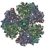
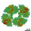
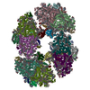
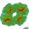
| Title | Cryo-EM structure of the tetrameric photosystem I from a heterocyst-forming cyanobacterium Anabaena sp. PCC7120 | |||||||||
 Map data Map data | ||||||||||
 Sample Sample |
| |||||||||
 Keywords Keywords | Photosystem I / PSI / PCC7120 / PHOTOSYNTHESIS | |||||||||
| Function / homology |  Function and homology information Function and homology informationphotosystem I reaction center / photosystem I / photosynthetic electron transport in photosystem I / photosystem I / plasma membrane-derived thylakoid membrane / chlorophyll binding / photosynthesis / 4 iron, 4 sulfur cluster binding / electron transfer activity / oxidoreductase activity ...photosystem I reaction center / photosystem I / photosynthetic electron transport in photosystem I / photosystem I / plasma membrane-derived thylakoid membrane / chlorophyll binding / photosynthesis / 4 iron, 4 sulfur cluster binding / electron transfer activity / oxidoreductase activity / magnesium ion binding / metal ion binding Similarity search - Function | |||||||||
| Biological species |  Nostoc sp. PCC 7120 (bacteria) Nostoc sp. PCC 7120 (bacteria) | |||||||||
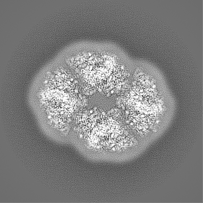
| Method | single particle reconstruction / cryo EM / Resolution: 2.37 Å | |||||||||
 Authors Authors | Zheng L / Li Y | |||||||||
| Funding support | 1 items
| |||||||||
 Citation Citation | Journal: Nat Plants / Year: 2019 Title: Structural and functional insights into the tetrameric photosystem I from heterocyst-forming cyanobacteria. Authors: Lvqin Zheng / Yanbing Li / Xiying Li / Qinglu Zhong / Ningning Li / Kun Zhang / Yuebin Zhang / Huiying Chu / Chengying Ma / Guohui Li / Jindong Zhao / Ning Gao /  Abstract: Two large protein-cofactor complexes, photosystem I and photosystem II, are the central components of photosynthesis in the thylakoid membranes. Here, we report the 2.37-Å structure of a tetrameric ...Two large protein-cofactor complexes, photosystem I and photosystem II, are the central components of photosynthesis in the thylakoid membranes. Here, we report the 2.37-Å structure of a tetrameric photosystem I complex from a heterocyst-forming cyanobacterium Anabaena sp. PCC 7120. Four photosystem I monomers, organized in a dimer of dimer, form two distinct interfaces that are largely mediated by specifically orientated polar lipids, such as sulfoquinovosyl diacylglycerol. The structure depicts a more closely connected network of chlorophylls across monomer interfaces than those seen in trimeric PSI from thermophilic cyanobacteria, possibly allowing a more efficient energy transfer between monomers. Our physiological data also revealed a functional link of photosystem I oligomerization to cyclic electron flow and thylakoid membrane organization. | |||||||||
| History |
|
- Structure visualization
| Movie |
Movie viewer |
|---|---|
| Structure viewer | EM map: SurfViewMolmilJmol/JSmol |
| Supplemental images |

- Downloads & links
-EMDB archive
| Map data | emd_9918.map.gz | 227.4 MB | EMDB map data format | |
|---|---|---|---|---|
| Header (meta data) | emd-9918-v30.xmlemd-9918.xml | 25 KB 25 KB | Display Display | EMDB header |
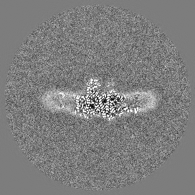
| Images | 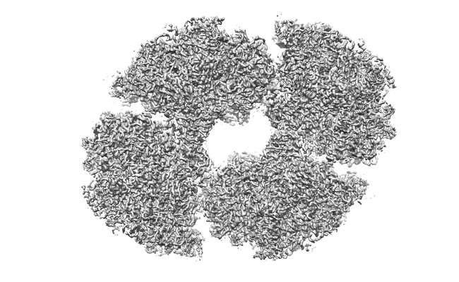 emd_9918.png emd_9918.png | 111.5 KB | ||
| Filedesc metadata | emd-9918.cif.gz | 7.5 KB | ||
| Archive directory |  http://ftp.pdbj.org/pub/emdb/structures/EMD-9918ftp://ftp.pdbj.org/pub/emdb/structures/EMD-9918 http://ftp.pdbj.org/pub/emdb/structures/EMD-9918ftp://ftp.pdbj.org/pub/emdb/structures/EMD-9918 | HTTPS FTP |
-Related structure data
| Related structure data | 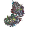 6k61MC M: atomic model generated by this map C: citing same article ( |
|---|---|
| Similar structure data |


-Links
| EMDB pages | EMDB (EBI/PDBe) / EMDataResource |
|---|---|
| Related items in Molecule of the Month |


-Map
| File | Download / File: emd_9918.map.gz / Format: CCP4 / Size: 244.1 MB / Type: IMAGE STORED AS FLOATING POINT NUMBER (4 BYTES) | ||||||||||||||||||||||||||||||||||||||||||||||||||||||||||||||||||||
|---|---|---|---|---|---|---|---|---|---|---|---|---|---|---|---|---|---|---|---|---|---|---|---|---|---|---|---|---|---|---|---|---|---|---|---|---|---|---|---|---|---|---|---|---|---|---|---|---|---|---|---|---|---|---|---|---|---|---|---|---|---|---|---|---|---|---|---|---|---|
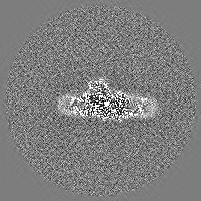
| Projections & slices | Image control
Images are generated by Spider. | ||||||||||||||||||||||||||||||||||||||||||||||||||||||||||||||||||||
| Voxel size | X=Y=Z: 1.052 Å | ||||||||||||||||||||||||||||||||||||||||||||||||||||||||||||||||||||
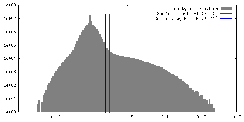
| Density |
| ||||||||||||||||||||||||||||||||||||||||||||||||||||||||||||||||||||
| Symmetry | Space group: 1 | ||||||||||||||||||||||||||||||||||||||||||||||||||||||||||||||||||||
| Details | EMDB XML:
CCP4 map header:
| ||||||||||||||||||||||||||||||||||||||||||||||||||||||||||||||||||||
 Z (Sec.)
 Y (Row.)
 X (Col.)


-Supplemental data
- Sample components
+Entire : Photosystem I complex of cyanobacterium Anabaena sp. PCC 7120
+Supramolecule #1: Photosystem I complex of cyanobacterium Anabaena sp. PCC 7120
+Macromolecule #1: Photosystem I P700 chlorophyll a apoprotein A1
+Macromolecule #2: Photosystem I 4.8 kDa protein
+Macromolecule #3: Photosystem I P700 chlorophyll a apoprotein A2 1
+Macromolecule #4: Photosystem I iron-sulfur center
+Macromolecule #5: Photosystem I reaction center subunit II
+Macromolecule #6: Photosystem I reaction center subunit IV
+Macromolecule #7: Photosystem I reaction center subunit III
+Macromolecule #8: Photosystem I reaction center subunit IX
+Macromolecule #9: Photosystem I reaction center subunit PsaK 1
+Macromolecule #10: Photosystem I reaction center subunit VIII
+Macromolecule #11: Photosystem I reaction center subunit XI
+Macromolecule #12: Photosystem I reaction center subunit XII
+Macromolecule #13: CHLOROPHYLL A ISOMER

+Macromolecule #14: CHLOROPHYLL A

+Macromolecule #15: PHYLLOQUINONE

+Macromolecule #16: IRON/SULFUR CLUSTER

+Macromolecule #17: BETA-CAROTENE

+Macromolecule #18: 1,2-DIPALMITOYL-PHOSPHATIDYL-GLYCEROLE

+Macromolecule #19: 1,2-DI-O-ACYL-3-O-[6-DEOXY-6-SULFO-ALPHA-D-GLUCOPYRANOSYL]-SN-GLYCEROL

+Macromolecule #20: 1,2-DISTEAROYL-MONOGALACTOSYL-DIGLYCERIDE

-Experimental details
-Structure determination
| Method | cryo EM |
|---|---|
 Processing Processing | single particle reconstruction |
| Aggregation state | helical array |
-Sample preparation
| Buffer | pH: 6.5 |
|---|---|
| Vitrification | Cryogen name: ETHANE / Chamber humidity: 100 % / Instrument: FEI VITROBOT MARK IV |
- Electron microscopy
| Microscope | FEI TITAN KRIOS |
|---|---|
| Image recording | Film or detector model: GATAN K2 SUMMIT (4k x 4k) / Average electron dose: 58.0 e/Å2 |
| Electron beam | Acceleration voltage: 300 kV / Electron source:  FIELD EMISSION GUN FIELD EMISSION GUN |
| Electron optics | Illumination mode: FLOOD BEAM / Imaging mode: BRIGHT FIELD |
| Sample stage | Specimen holder model: FEI TITAN KRIOS AUTOGRID HOLDER / Cooling holder cryogen: NITROGEN |
| Experimental equipment |  Model: Titan Krios / Image courtesy: FEI Company |
-Image processing
| Startup model | Type of model: PDB ENTRY PDB model - PDB ID: |
|---|---|
| Final reconstruction | Applied symmetry - Point group: C2 (2 fold cyclic) / Resolution.type: BY AUTHOR / Resolution: 2.37 Å / Resolution method: FSC 0.143 CUT-OFF / Number images used: 71600 |
| Initial angle assignment | Type: NOT APPLICABLE |
| Final angle assignment | Type: PROJECTION MATCHING |