Movie
Movie Controller


+ Open data

- Basic information
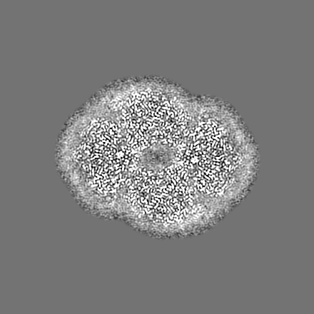
| Entry | Database: EMDB / ID: EMD-9807 | |||||||||
|---|---|---|---|---|---|---|---|---|---|---|
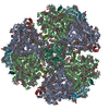
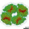
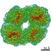
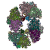
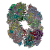
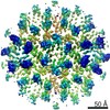
| Title | Structure of PSI tetramer from Anabaena | |||||||||
 Map data Map data | ||||||||||
 Sample Sample |
| |||||||||
 Keywords Keywords | Photosystem / PHOTOSYNTHESIS | |||||||||
| Function / homology |  Function and homology information Function and homology informationphotosystem I reaction center / photosystem I / photosynthetic electron transport in photosystem I / photosystem I / plasma membrane-derived thylakoid membrane / chlorophyll binding / photosynthesis / 4 iron, 4 sulfur cluster binding / electron transfer activity / oxidoreductase activity ...photosystem I reaction center / photosystem I / photosynthetic electron transport in photosystem I / photosystem I / plasma membrane-derived thylakoid membrane / chlorophyll binding / photosynthesis / 4 iron, 4 sulfur cluster binding / electron transfer activity / oxidoreductase activity / magnesium ion binding / metal ion binding Similarity search - Function | |||||||||
| Biological species |  Nostoc sp. PCC 7120 (bacteria) / Nostoc sp. (strain PCC 7120 / SAG 25.82 / UTEX 2576) (bacteria) Nostoc sp. PCC 7120 (bacteria) / Nostoc sp. (strain PCC 7120 / SAG 25.82 / UTEX 2576) (bacteria) | |||||||||
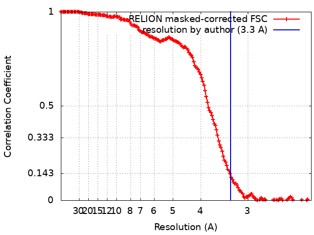
| Method | single particle reconstruction / cryo EM / Resolution: 3.3 Å | |||||||||
 Authors Authors | Kato K / Nagao R | |||||||||
 Citation Citation | Journal: Nat Commun / Year: 2019 Title: Structure of a cyanobacterial photosystem I tetramer revealed by cryo-electron microscopy. Authors: Koji Kato / Ryo Nagao / Tian-Yi Jiang / Yoshifumi Ueno / Makio Yokono / Siu Kit Chan / Mai Watanabe / Masahiko Ikeuchi / Jian-Ren Shen / Seiji Akimoto / Naoyuki Miyazaki / Fusamichi Akita /  Abstract: Photosystem I (PSI) functions to harvest light energy for conversion into chemical energy. The organisation of PSI is variable depending on the species of organism. Here we report the structure of a ...Photosystem I (PSI) functions to harvest light energy for conversion into chemical energy. The organisation of PSI is variable depending on the species of organism. Here we report the structure of a tetrameric PSI core isolated from a cyanobacterium, Anabaena sp. PCC 7120, analysed by single-particle cryo-electron microscopy (cryo-EM) at 3.3 Å resolution. The PSI tetramer has a C2 symmetry and is organised in a dimer of dimers form. The structure reveals interactions at the dimer-dimer interface and the existence of characteristic pigment orientations and inter-pigment distances within the dimer units that are important for unique excitation energy transfer. In particular, characteristic residues of PsaL are identified to be responsible for the formation of the tetramer. Time-resolved fluorescence analyses showed that the PSI tetramer has an enhanced excitation-energy quenching. These structural and spectroscopic findings provide insights into the physiological significance of the PSI tetramer and evolutionary changes of the PSI organisations. | |||||||||
| History |
|
- Structure visualization
| Movie |
Movie viewer |
|---|---|
| Structure viewer | EM map: SurfViewMolmilJmol/JSmol |
| Supplemental images |

- Downloads & links
-EMDB archive
| Map data | emd_9807.map.gz | 28.2 MB | EMDB map data format | |
|---|---|---|---|---|
| Header (meta data) | emd-9807-v30.xmlemd-9807.xml | 36.5 KB 36.5 KB | Display Display | EMDB header |
| FSC (resolution estimation) | emd_9807_fsc.xml | 15.6 KB | Display | FSC data file |
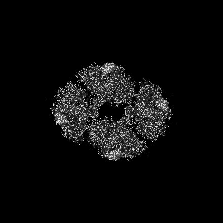
| Images | 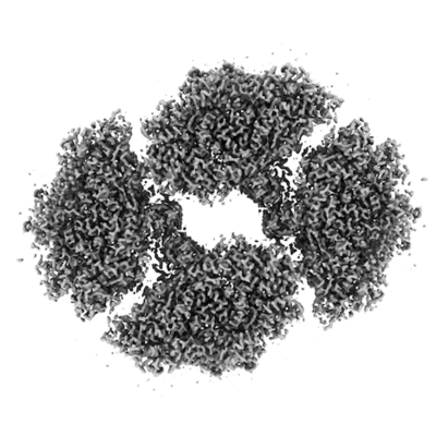 emd_9807.png emd_9807.png | 72 KB | ||
| Filedesc metadata | emd-9807.cif.gz | 8.9 KB | ||
| Archive directory |  http://ftp.pdbj.org/pub/emdb/structures/EMD-9807ftp://ftp.pdbj.org/pub/emdb/structures/EMD-9807 http://ftp.pdbj.org/pub/emdb/structures/EMD-9807ftp://ftp.pdbj.org/pub/emdb/structures/EMD-9807 | HTTPS FTP |
-Related structure data
| Related structure data | 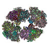 6jeoMC 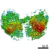 9877C M: atomic model generated by this map C: citing same article ( |
|---|---|
| Similar structure data |

-Links
| EMDB pages | EMDB (EBI/PDBe) / EMDataResource |
|---|---|
| Related items in Molecule of the Month |


-Map
| File | Download / File: emd_9807.map.gz / Format: CCP4 / Size: 325 MB / Type: IMAGE STORED AS FLOATING POINT NUMBER (4 BYTES) | ||||||||||||||||||||||||||||||||||||||||||||||||||||||||||||||||||||
|---|---|---|---|---|---|---|---|---|---|---|---|---|---|---|---|---|---|---|---|---|---|---|---|---|---|---|---|---|---|---|---|---|---|---|---|---|---|---|---|---|---|---|---|---|---|---|---|---|---|---|---|---|---|---|---|---|---|---|---|---|---|---|---|---|---|---|---|---|---|
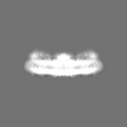
| Projections & slices | Image control
Images are generated by Spider. | ||||||||||||||||||||||||||||||||||||||||||||||||||||||||||||||||||||
| Voxel size | X=Y=Z: 1.12 Å | ||||||||||||||||||||||||||||||||||||||||||||||||||||||||||||||||||||
| Density |
| ||||||||||||||||||||||||||||||||||||||||||||||||||||||||||||||||||||
| Symmetry | Space group: 1 | ||||||||||||||||||||||||||||||||||||||||||||||||||||||||||||||||||||
| Details | EMDB XML:
CCP4 map header:
| ||||||||||||||||||||||||||||||||||||||||||||||||||||||||||||||||||||
 Z (Sec.)
 Y (Row.)
 X (Col.)


-Supplemental data
- Sample components
+Entire : PSI tetramer
+Supramolecule #1: PSI tetramer
+Macromolecule #1: Photosystem I P700 chlorophyll a apoprotein A1
+Macromolecule #2: Photosystem I P700 chlorophyll a apoprotein A2 1
+Macromolecule #3: Photosystem I iron-sulfur center
+Macromolecule #4: Photosystem I reaction center subunit II
+Macromolecule #5: Photosystem I reaction center subunit IV
+Macromolecule #6: Photosystem I reaction center subunit III
+Macromolecule #7: Photosystem I reaction center subunit VIII
+Macromolecule #8: Photosystem I reaction center subunit IX
+Macromolecule #9: Photosystem I reaction center subunit PsaK 1
+Macromolecule #10: Photosystem I reaction center subunit XI
+Macromolecule #11: Photosystem I reaction center subunit XII
+Macromolecule #12: Photosystem I 4.8 kDa protein
+Macromolecule #13: CHLOROPHYLL A ISOMER

+Macromolecule #14: CHLOROPHYLL A

+Macromolecule #15: PHYLLOQUINONE

+Macromolecule #16: IRON/SULFUR CLUSTER

+Macromolecule #17: BETA-CAROTENE

+Macromolecule #18: 1,2-DIPALMITOYL-PHOSPHATIDYL-GLYCEROLE

+Macromolecule #19: 1,2-DISTEAROYL-MONOGALACTOSYL-DIGLYCERIDE

-Experimental details
-Structure determination
| Method | cryo EM |
|---|---|
 Processing Processing | single particle reconstruction |
| Aggregation state | particle |
-Sample preparation
| Concentration | 0.014 mg/mL | ||||||
|---|---|---|---|---|---|---|---|
| Buffer | pH: 6.5 / Component:
| ||||||
| Grid | Model: Quantifoil R2/1 / Material: COPPER / Mesh: 300 / Support film - Material: CARBON / Pretreatment - Type: PLASMA CLEANING / Pretreatment - Time: 30 sec. | ||||||
| Vitrification | Cryogen name: ETHANE / Chamber humidity: 100 % / Chamber temperature: 277 K / Instrument: FEI VITROBOT MARK IV |
- Electron microscopy
| Microscope | FEI TITAN KRIOS |
|---|---|
| Image recording | Film or detector model: FEI FALCON III (4k x 4k) / Average electron dose: 40.0 e/Å2 |
| Electron beam | Acceleration voltage: 300 kV / Electron source:  FIELD EMISSION GUN FIELD EMISSION GUN |
| Electron optics | Illumination mode: FLOOD BEAM / Imaging mode: BRIGHT FIELD |
| Experimental equipment |  Model: Titan Krios / Image courtesy: FEI Company |