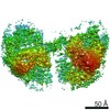
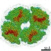
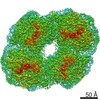
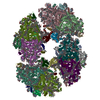
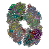
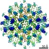
- EMDB-9807: Structure of PSI tetramer from Anabaena -
+
データを開く
IDまたはキーワード:
読み込み中...
-
基本情報
登録情報
データベース: EMDB / ID: EMD-9807
タイトル
Structure of PSI tetramer from Anabaena
マップデータ
試料
複合体: PSI tetramer
タンパク質・ペプチド: x 12種
リガンド: x 7種
機能・相同性
機能・相同性情報
photosystem I reaction center / 光化学系I / photosynthetic electron transport in photosystem I / 光化学系I / chlorophyll binding / plasma membrane-derived thylakoid membrane / 光合成 / 4 iron, 4 sulfur cluster binding / electron transfer activity / magnesium ion binding / metal ion binding 類似検索 - 分子機能
Photosystem I PsaX / Photosystem I PsaX superfamily / PsaX family / Photosystem I reaction center subunit PsaK / Photosystem I reaction centre subunit PsaK / Photosystem I PsaM, reaction centre superfamily / Photosystem I PsaM, reaction centre / Photosystem I protein M (PsaM) / Photosystem I reaction centre subunit PsaK superfamily / Photosystem I reaction center subunit V/PsaK ...Photosystem I PsaX / Photosystem I PsaX superfamily / PsaX family / Photosystem I reaction center subunit PsaK / Photosystem I reaction centre subunit PsaK / Photosystem I PsaM, reaction centre superfamily / Photosystem I PsaM, reaction centre / Photosystem I protein M (PsaM) / Photosystem I reaction centre subunit PsaK superfamily / Photosystem I reaction center subunit V/PsaK / Photosystem I psaG / psaK / Photosystem I PsaL, reaction centre subunit XI / Photosystem I, reaction centre subunit XI / Photosystem I PsaL, reaction centre subunit XI superfamily / Photosystem I reaction centre subunit XI / Photosystem I reaction centre subunit VIII / Photosystem I reaction centre subunit VIII / Photosystem I reaction centre subunit VIII superfamily / Photosystem I PsaF, reaction centre subunit III / Photosystem I PsaF, reaction centre subunit III superfamily / Photosystem I reaction centre subunit III / Photosystem I PsaJ, reaction centre subunit IX / Photosystem I PsaD / Photosystem I PsaJ, reaction centre subunit IX superfamily / Photosystem I, reaction centre subunit PsaD superfamily / Photosystem I reaction centre subunit IX / PsaJ / PsaD / Photosystem I PsaE, reaction centre subunit IV / Photosystem I reaction centre subunit IV / PsaE / Photosystem I protein PsaC / Photosystem I PsaA / Photosystem I PsaB / Photosystem I PsaA/PsaB, conserved site / Photosystem I psaA and psaB proteins signature. / Photosystem I PsaA/PsaB / Photosystem I PsaA/PsaB superfamily / Photosystem I psaA/psaB protein / Electron transport accessory-like domain superfamily / 4Fe-4S dicluster domain / 4Fe-4S ferredoxin, iron-sulphur binding, conserved site / 4Fe-4S ferredoxin-type iron-sulfur binding region signature. / 4Fe-4S ferredoxin-type iron-sulfur binding domain profile. / 4Fe-4S ferredoxin-type, iron-sulphur binding domain 類似検索 - ドメイン・相同性
Photosystem I iron-sulfur center / Photosystem I reaction center subunit VIII / Photosystem I reaction center subunit III / Photosystem I P700 chlorophyll a apoprotein A2 1 / Photosystem I 4.8 kDa protein / Photosystem I reaction center subunit IX / Photosystem I reaction center subunit II / Photosystem I reaction center subunit IV / Photosystem I P700 chlorophyll a apoprotein A1 / Photosystem I reaction center subunit XI ...Photosystem I iron-sulfur center / Photosystem I reaction center subunit VIII / Photosystem I reaction center subunit III / Photosystem I P700 chlorophyll a apoprotein A2 1 / Photosystem I 4.8 kDa protein / Photosystem I reaction center subunit IX / Photosystem I reaction center subunit II / Photosystem I reaction center subunit IV / Photosystem I P700 chlorophyll a apoprotein A1 / Photosystem I reaction center subunit XI / Photosystem I reaction center subunit PsaK 1 / Photosystem I reaction center subunit XII 類似検索 - 構成要素
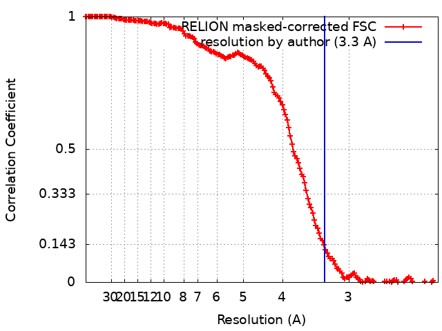
ジャーナル: Nat Commun / 年: 2019 タイトル: Structure of a cyanobacterial photosystem I tetramer revealed by cryo-electron microscopy. 著者: Koji Kato / Ryo Nagao / Tian-Yi Jiang / Yoshifumi Ueno / Makio Yokono / Siu Kit Chan / Mai Watanabe / Masahiko Ikeuchi / Jian-Ren Shen / Seiji Akimoto / Naoyuki Miyazaki / Fusamichi Akita / 要旨: Photosystem I (PSI) functions to harvest light energy for conversion into chemical energy. The organisation of PSI is variable depending on the species of organism. Here we report the structure of a ...Photosystem I (PSI) functions to harvest light energy for conversion into chemical energy. The organisation of PSI is variable depending on the species of organism. Here we report the structure of a tetrameric PSI core isolated from a cyanobacterium, Anabaena sp. PCC 7120, analysed by single-particle cryo-electron microscopy (cryo-EM) at 3.3 Å resolution. The PSI tetramer has a C2 symmetry and is organised in a dimer of dimers form. The structure reveals interactions at the dimer-dimer interface and the existence of characteristic pigment orientations and inter-pigment distances within the dimer units that are important for unique excitation energy transfer. In particular, characteristic residues of PsaL are identified to be responsible for the formation of the tetramer. Time-resolved fluorescence analyses showed that the PSI tetramer has an enhanced excitation-energy quenching. These structural and spectroscopic findings provide insights into the physiological significance of the PSI tetramer and evolutionary changes of the PSI organisations.
 ムービー
ムービー コントローラー


 データを開く
 基本情報
基本情報 マップデータ
 試料
試料 機能・相同性情報
 photosystem I reaction center /
  Nostoc sp. PCC 7120 (バクテリア) /
  データ登録者
 引用
引用
 構造の表示
構造の表示
 ダウンロードとリンク
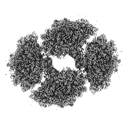 emd_9807.png
 http://ftp.pdbj.org/pub/emdb/structures/EMD-9807


 試料の構成要素


 解析
解析 電子顕微鏡法