 Movie
Movie Controller

[English] 日本語
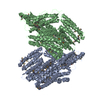
 Yorodumi
- PDB-7pil: Cryo-EM structure of the Rhodobacter sphaeroides RC-LH1-PufXY mon... -
+ Open data

- Basic information
| Entry | Database: PDB / ID: 7pil | ||||||||||||
|---|---|---|---|---|---|---|---|---|---|---|---|---|---|
| Title | Cryo-EM structure of the Rhodobacter sphaeroides RC-LH1-PufXY monomer complex at 2.5 A | ||||||||||||
 Components Components |
| ||||||||||||
 Keywords Keywords | PHOTOSYNTHESIS / Photosynthetic bacteria / light harvesting complex / Cryo-EM / RC-LH1 / RC-LH1-PufX / RC-LH1-PufX-Y | ||||||||||||
| Function / homology |  Function and homology information Function and homology informationorganelle inner membrane / plasma membrane-derived chromatophore membrane / plasma membrane light-harvesting complex / bacteriochlorophyll binding / photosynthetic electron transport in photosystem II / photosynthesis, light reaction / photosynthesis / metal ion binding / plasma membrane Similarity search - Function | ||||||||||||
| Biological species |  Rhodobacter sphaeroides (bacteria) Rhodobacter sphaeroides (bacteria) | ||||||||||||
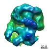
| Method | ELECTRON MICROSCOPY / single particle reconstruction / cryo EM / Resolution: 2.5 Å | ||||||||||||
 Authors Authors | Qian, P. / Hunter, C.N. | ||||||||||||
| Funding support |  United Kingdom, 3items United Kingdom, 3items
| ||||||||||||
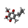
 Citation Citation | Journal: Biochem J / Year: 2021 Title: Cryo-EM structure of the monomeric Rhodobacter sphaeroides RC-LH1 core complex at 2.5 Å. Authors: Pu Qian / David J K Swainsbury / Tristan I Croll / Jack H Salisbury / Elizabeth C Martin / Philip J Jackson / Andrew Hitchcock / Pablo Castro-Hartmann / Kasim Sader / C Neil Hunter /  Abstract: Reaction centre light-harvesting 1 (RC-LH1) complexes are the essential components of bacterial photosynthesis. The membrane-intrinsic LH1 complex absorbs light and the energy migrates to an enclosed ...Reaction centre light-harvesting 1 (RC-LH1) complexes are the essential components of bacterial photosynthesis. The membrane-intrinsic LH1 complex absorbs light and the energy migrates to an enclosed RC where a succession of electron and proton transfers conserves the energy as a quinol, which is exported to the cytochrome bc1 complex. In some RC-LH1 variants quinols can diffuse through small pores in a fully circular, 16-subunit LH1 ring, while in others missing LH1 subunits create a gap for quinol export. We used cryogenic electron microscopy to obtain a 2.5 Å resolution structure of one such RC-LH1, a monomeric complex from Rhodobacter sphaeroides. The structure shows that the RC is partly enclosed by a 14-subunit LH1 ring in which each αβ heterodimer binds two bacteriochlorophylls and, unusually for currently reported complexes, two carotenoids rather than one. Although the extra carotenoids confer an advantage in terms of photoprotection and light harvesting, they could impede passage of quinones through small, transient pores in the LH1 ring, necessitating a mechanism to create a dedicated quinone channel. The structure shows that two transmembrane proteins play a part in stabilising an open ring structure; one of these components, the PufX polypeptide, is augmented by a hitherto undescribed protein subunit we designate as protein-Y, which lies against the transmembrane regions of the thirteenth and fourteenth LH1α polypeptides. Protein-Y prevents LH1 subunits 11-14 adjacent to the RC QB site from bending inwards towards the RC and, with PufX preventing complete encirclement of the RC, this pair of polypeptides ensures unhindered quinone diffusion. #1: Journal: Acta Crystallogr., Sect. D: Biol. Crystallogr. / Year: 2018Title: Real-space refinement in PHENIX for cryo-EM and crystallography Authors: qian, P. / Hunter, C.N. #2: Journal: To Be PublishedTitle: Cryo-EM structure of the Rhodobacter sphaeroides RC-LH1-PufXY monomer complex at 2.5 A Authors: Qian, P. / Hunter, C.N. | ||||||||||||
| History |
|
- Structure visualization
| Movie |
Movie viewer |
|---|---|
| Structure viewer | Molecule: MolmilJmol/JSmol |
- Downloads & links
-Download
| PDBx/mmCIF format | 7pil.cif.gz | 552 KB | Display | PDBx/mmCIF format |
|---|---|---|---|---|
| PDB format | pdb7pil.ent.gz | Display | PDB format | |
| PDBx/mmJSON format | 7pil.json.gz | Tree view | PDBx/mmJSON format | |
| Others |  Other downloads Other downloads |
-Validation report
| Arichive directory | https://data.pdbj.org/pub/pdb/validation_reports/pi/7pilftp://data.pdbj.org/pub/pdb/validation_reports/pi/7pil | HTTPS FTP |
|---|
-Related structure data
| Related structure data |  13441MC M: map data used to model this data C: citing same article ( |
|---|---|
| Similar structure data |


-Links
 PDBj
PDBj


- Assembly
| Deposited unit | 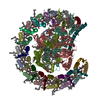
|
|---|---|
| 1 |
|
-Components
-Light-harvesting protein B-875 ... , 2 types, 28 molecules AAABACADAEAFAGAHAIAJAKALAMANBABBBCBDBEBFBGBHBIBJBKBLBMBN
| #1: Protein | Mass: 6516.847 Da / Num. of mol.: 14 / Source method: isolated from a natural source Source: (natural) Rhodobacter sphaeroides (strain ATCC 17023 / DSM 158 / JCM 6121 / NBRC 12203 / NCIMB 8253 / ATH 2.4.1.) (bacteria)Strain: ATCC 17023 / DSM 158 / JCM 6121 / NBRC 12203 / NCIMB 8253 / ATH 2.4.1. References: UniProt: Q3J1A4 #2: Protein/peptide | Mass: 4943.656 Da / Num. of mol.: 14 / Source method: isolated from a natural source Source: (natural) Rhodobacter sphaeroides (strain ATCC 17023 / DSM 158 / JCM 6121 / NBRC 12203 / NCIMB 8253 / ATH 2.4.1.) (bacteria)Strain: ATCC 17023 / DSM 158 / JCM 6121 / NBRC 12203 / NCIMB 8253 / ATH 2.4.1. References: UniProt: Q3J1A3 |
|---|
-Reaction center protein ... , 3 types, 3 molecules HLM
| #3: Protein | Mass: 26544.471 Da / Num. of mol.: 1 / Source method: isolated from a natural source Source: (natural) Rhodobacter sphaeroides (strain ATCC 17023 / DSM 158 / JCM 6121 / NBRC 12203 / NCIMB 8253 / ATH 2.4.1.) (bacteria)Strain: ATCC 17023 / DSM 158 / JCM 6121 / NBRC 12203 / NCIMB 8253 / ATH 2.4.1. References: UniProt: Q3J170 |
|---|---|
| #4: Protein | Mass: 31346.389 Da / Num. of mol.: 1 / Source method: isolated from a natural source Source: (natural) Rhodobacter sphaeroides (strain ATCC 17023 / DSM 158 / JCM 6121 / NBRC 12203 / NCIMB 8253 / ATH 2.4.1.) (bacteria)Strain: ATCC 17023 / DSM 158 / JCM 6121 / NBRC 12203 / NCIMB 8253 / ATH 2.4.1. References: UniProt: Q3J1A5 |
| #5: Protein | Mass: 34398.543 Da / Num. of mol.: 1 / Source method: isolated from a natural source Source: (natural) Rhodobacter sphaeroides (strain ATCC 17023 / 2.4.1 / NCIB 8253 / DSM 158) (bacteria)Strain: ATCC 17023 / 2.4.1 / NCIB 8253 / DSM 158 / References: UniProt: Q3J1A6 |
-Protein/peptide / Protein / Sugars , 3 types, 6 molecules UUX
| #10: Sugar | ChemComp-LMT /  Type: D-saccharide / Mass: 510.615 Da / Num. of mol.: 4 / Source method: obtained synthetically / Formula: C24H46O11 / Comment: detergent*YM Type: D-saccharide / Mass: 510.615 Da / Num. of mol.: 4 / Source method: obtained synthetically / Formula: C24H46O11 / Comment: detergent*YM#6: Protein/peptide | | Mass: 5126.067 Da / Num. of mol.: 1 / Source method: isolated from a natural source Source: (natural) Rhodobacter sphaeroides (strain ATCC 17023 / DSM 158 / JCM 6121 / NBRC 12203 / NCIMB 8253 / ATH 2.4.1.) (bacteria)Strain: ATCC 17023 / DSM 158 / JCM 6121 / NBRC 12203 / NCIMB 8253 / ATH 2.4.1. References: UniProt: U5NME9 #7: Protein | | Mass: 5980.121 Da / Num. of mol.: 1 / Source method: isolated from a natural source Source: (natural) Rhodobacter sphaeroides (strain ATCC 17023 / DSM 158 / JCM 6121 / NBRC 12203 / NCIMB 8253 / ATH 2.4.1.) (bacteria)Strain: ATCC 17023 / DSM 158 / JCM 6121 / NBRC 12203 / NCIMB 8253 / ATH 2.4.1. References: UniProt: P13402 |
|---|
-Non-polymers , 9 types, 73 molecules 


| #8: Chemical | ChemComp-BCL /  Mass: 911.504 Da / Num. of mol.: 32 / Source method: obtained synthetically / Formula: C55H74MgN4O6 / Feature type: SUBJECT OF INVESTIGATION Mass: 911.504 Da / Num. of mol.: 32 / Source method: obtained synthetically / Formula: C55H74MgN4O6 / Feature type: SUBJECT OF INVESTIGATION#9: Chemical | ChemComp-SPO /  Mass: 568.914 Da / Num. of mol.: 26 / Source method: obtained synthetically / Formula: C41H60O Mass: 568.914 Da / Num. of mol.: 26 / Source method: obtained synthetically / Formula: C41H60O#11: Chemical | ChemComp-3PE /  Mass: 748.065 Da / Num. of mol.: 4 / Source method: obtained synthetically / Formula: C41H82NO8P / Comment: phospholipid*YM Mass: 748.065 Da / Num. of mol.: 4 / Source method: obtained synthetically / Formula: C41H82NO8P / Comment: phospholipid*YM#12: Chemical |  Mass: 889.215 Da / Num. of mol.: 2 / Source method: obtained synthetically / Formula: C55H76N4O6 / Feature type: SUBJECT OF INVESTIGATION Mass: 889.215 Da / Num. of mol.: 2 / Source method: obtained synthetically / Formula: C55H76N4O6 / Feature type: SUBJECT OF INVESTIGATION#13: Chemical |  Mass: 863.343 Da / Num. of mol.: 2 / Source method: obtained synthetically / Formula: C59H90O4 / Feature type: SUBJECT OF INVESTIGATION Mass: 863.343 Da / Num. of mol.: 2 / Source method: obtained synthetically / Formula: C59H90O4 / Feature type: SUBJECT OF INVESTIGATION#14: Chemical | ChemComp-UQ1 / |  Mass: 250.290 Da / Num. of mol.: 1 / Source method: obtained synthetically / Formula: C14H18O4 Mass: 250.290 Da / Num. of mol.: 1 / Source method: obtained synthetically / Formula: C14H18O4#15: Chemical | ChemComp-CD4 / ( |  Mass: 1241.633 Da / Num. of mol.: 1 / Source method: obtained synthetically / Formula: C65H126O17P2 Mass: 1241.633 Da / Num. of mol.: 1 / Source method: obtained synthetically / Formula: C65H126O17P2#16: Chemical | ChemComp-FE / |  Mass: 55.845 Da / Num. of mol.: 1 / Source method: obtained synthetically / Formula: Fe Mass: 55.845 Da / Num. of mol.: 1 / Source method: obtained synthetically / Formula: Fe#17: Water | ChemComp-HOH / | Mass: 18.015 Da / Num. of mol.: 4 / Source method: isolated from a natural source / Formula: H2O |
|---|
-Details
| Has ligand of interest | Y |
|---|
-Experimental details
-Experiment
| Experiment | Method: ELECTRON MICROSCOPY |
|---|---|
| EM experiment | Aggregation state: PARTICLE / 3D reconstruction method: single particle reconstruction |
- Sample preparation
| Component | Name: Light harvesting complex / Type: COMPLEX / Entity ID: #1-#7 / Source: NATURAL |
|---|---|
| Source (natural) | Organism: Cereibacter sphaeroides 2.4.1 (bacteria) |
| Buffer solution | pH: 7.8 / Details: 20 mM HEPES, pH 7.8 , 0.03% beta-DDM |
| Buffer component | Conc.: 20 mMol / Formula: HEPES |
| Specimen | Conc.: 4 mg/ml / Embedding applied: NO / Shadowing applied: NO / Staining applied: NO / Vitrification applied: YES / Details: in 0.03% beta-DDM detergent |
| Specimen support | Grid material: COPPER / Grid mesh size: 300 divisions/in. / Grid type: Quantifoil R1.2/1.3 |
| Vitrification | Instrument: FEI VITROBOT MARK III / Cryogen name: ETHANE / Humidity: 100 % / Chamber temperature: 277 K / Details: QF R1.2/1.3 grid coated graphene oxide |
- Electron microscopy imaging
| Experimental equipment |  Model: Titan Krios / Image courtesy: FEI Company |
|---|---|
| Microscopy | Model: FEI TITAN KRIOS |
| Electron gun | Electron source:  FIELD EMISSION GUN / Accelerating voltage: 300 kV / Illumination mode: FLOOD BEAM FIELD EMISSION GUN / Accelerating voltage: 300 kV / Illumination mode: FLOOD BEAM |
| Electron lens | Mode: BRIGHT FIELD / Nominal magnification: 120000 X / Nominal defocus max: 2200 nm / Nominal defocus min: 800 nm / Cs: 2.7 mm / C2 aperture diameter: 50 µm / Alignment procedure: COMA FREE |
| Specimen holder | Cryogen: NITROGEN / Specimen holder model: FEI TITAN KRIOS AUTOGRID HOLDER |
| Image recording | Average exposure time: 12.21 sec. / Electron dose: 44.94 e/Å2 / Film or detector model: FEI FALCON IV (4k x 4k) / Num. of grids imaged: 1 / Num. of real images: 3180 |
- Processing
| Software |
| ||||||||||||||||||||||||
|---|---|---|---|---|---|---|---|---|---|---|---|---|---|---|---|---|---|---|---|---|---|---|---|---|---|
| EM software |
| ||||||||||||||||||||||||
| CTF correction | Type: PHASE FLIPPING AND AMPLITUDE CORRECTION | ||||||||||||||||||||||||
| Particle selection | Num. of particles selected: 1057624 | ||||||||||||||||||||||||
| Symmetry | Point symmetry: C1 (asymmetric) | ||||||||||||||||||||||||
| 3D reconstruction | Resolution: 2.5 Å / Resolution method: FSC 0.143 CUT-OFF / Num. of particles: 250613 / Algorithm: BACK PROJECTION / Num. of class averages: 1 / Symmetry type: POINT |
