 Movie
Movie Controller


+ Open data

- Basic information
| Entry | Database: EMDB / ID: EMD-1112 | |||||||||
|---|---|---|---|---|---|---|---|---|---|---|
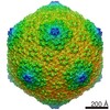
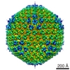
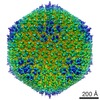
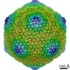
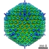
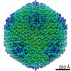
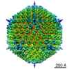
| Title | A quasi-atomic model of human adenovirus type 5 capsid. | |||||||||
 Map data Map data | Structure of Adenovirus pIX deletion mutant (single frozen and thawed sample) | |||||||||
 Sample Sample |
| |||||||||
| Function / homology |  Function and homology information Function and homology informationT=25 icosahedral viral capsid / microtubule-dependent intracellular transport of viral material towards nucleus / viral capsid / host cell / clathrin-dependent endocytosis of virus by host cell / symbiont entry into host cell / virion attachment to host cell / host cell nucleus / structural molecule activity Similarity search - Function | |||||||||
| Biological species |  unidentified adenovirus unidentified adenovirus | |||||||||
| Method | single particle reconstruction / cryo EM / negative staining / Resolution: 15.5 Å | |||||||||
 Authors Authors | Fabry CM / Rosa-Calatrava M / Conway JF / Zubieta C / Cusack S / Ruigrok RW / Schoehn G | |||||||||
 Citation Citation | Journal: EMBO J / Year: 2005 Title: A quasi-atomic model of human adenovirus type 5 capsid. Authors: Céline M S Fabry / Manuel Rosa-Calatrava / James F Conway / Chloé Zubieta / Stephen Cusack / Rob W H Ruigrok / Guy Schoehn /  Abstract: Adenoviruses infect a wide range of vertebrates including humans. Their icosahedral capsids are composed of three major proteins: the trimeric hexon forms the facets and the penton, a noncovalent ...Adenoviruses infect a wide range of vertebrates including humans. Their icosahedral capsids are composed of three major proteins: the trimeric hexon forms the facets and the penton, a noncovalent complex of the pentameric penton base and trimeric fibre proteins, is located at the 12 capsid vertices. Several proteins (IIIa, VI, VIII and IX) stabilise the capsid. We have obtained a 10 A resolution map of the human adenovirus 5 by image analysis from cryo-electron micrographs (cryoEMs). This map, in combination with the X-ray structures of the penton base and hexon, was used to build a quasi-atomic model of the arrangement of the two major capsid components and to analyse the hexon-hexon and hexon-penton interactions. The secondary proteins, notably VIII, were located by comparing cryoEM maps of native and pIX deletion mutant virions. Minor proteins IX and IIIa are located on the outside of the capsid, whereas protein VIII is organised with a T=2 lattice on the inner face of the capsid. The capsid organisation is compared with the known X-ray structure of bacteriophage PRD1. | |||||||||
| History |
|
- Structure visualization
| Movie |
Movie viewer |
|---|---|
| Structure viewer | EM map: SurfViewMolmilJmol/JSmol |
| Supplemental images |
 UCSF Chimera
- Downloads & links
-EMDB archive
| Map data | emd_1112.map.gz | 84.1 MB | EMDB map data format | |
|---|---|---|---|---|
| Header (meta data) | emd-1112-v30.xmlemd-1112.xml | 9.8 KB 9.8 KB | Display Display | EMDB header |
| Images |  1112.gif 1112.gif | 35.6 KB | ||
| Archive directory |  http://ftp.pdbj.org/pub/emdb/structures/EMD-1112ftp://ftp.pdbj.org/pub/emdb/structures/EMD-1112 http://ftp.pdbj.org/pub/emdb/structures/EMD-1112ftp://ftp.pdbj.org/pub/emdb/structures/EMD-1112 | HTTPS FTP |
-Related structure data
| Related structure data |  4v4uMC  1111C  1113C M: atomic model generated by this map C: citing same article ( |
|---|---|
| Similar structure data |


-Links
| EMDB pages | EMDB (EBI/PDBe) / EMDataResource |
|---|---|
| Related items in Molecule of the Month |


-Map
| File | Download / File: emd_1112.map.gz / Format: CCP4 / Size: 335 MB / Type: IMAGE STORED AS FLOATING POINT NUMBER (4 BYTES) | ||||||||||||||||||||||||||||||||||||||||||||||||||||||||||||||||||||
|---|---|---|---|---|---|---|---|---|---|---|---|---|---|---|---|---|---|---|---|---|---|---|---|---|---|---|---|---|---|---|---|---|---|---|---|---|---|---|---|---|---|---|---|---|---|---|---|---|---|---|---|---|---|---|---|---|---|---|---|---|---|---|---|---|---|---|---|---|---|
| Annotation | Structure of Adenovirus pIX deletion mutant (single frozen and thawed sample) | ||||||||||||||||||||||||||||||||||||||||||||||||||||||||||||||||||||
| Projections & slices | Image control
Images are generated by Spider. | ||||||||||||||||||||||||||||||||||||||||||||||||||||||||||||||||||||
| Voxel size | X=Y=Z: 2.45 Å | ||||||||||||||||||||||||||||||||||||||||||||||||||||||||||||||||||||
| Density |
| ||||||||||||||||||||||||||||||||||||||||||||||||||||||||||||||||||||
| Symmetry | Space group: 1 | ||||||||||||||||||||||||||||||||||||||||||||||||||||||||||||||||||||
| Details | EMDB XML:
CCP4 map header:
| ||||||||||||||||||||||||||||||||||||||||||||||||||||||||||||||||||||
 Y (Sec.)
 X (Row.)
 Z (Col.)


-Supplemental data
- Sample components
-Entire : Human adenovirus type 5 pIX deletion mutant
| Entire | Name: Human adenovirus type 5 pIX deletion mutant |
|---|---|
| Components |
|
-Supramolecule #1000: Human adenovirus type 5 pIX deletion mutant
| Supramolecule | Name: Human adenovirus type 5 pIX deletion mutant / type: sample / ID: 1000 / Number unique components: 1 |
|---|---|
| Molecular weight | Theoretical: 150 MDa |
-Supramolecule #1: unidentified adenovirus
| Supramolecule | Name: unidentified adenovirus / type: virus / ID: 1 / NCBI-ID: 10535 / Sci species name: unidentified adenovirus / Virus type: VIRION / Virus isolate: SEROTYPE / Virus enveloped: No / Virus empty: No |
|---|---|
| Host (natural) | Organism:  Homo sapiens (human) / synonym: VERTEBRATES Homo sapiens (human) / synonym: VERTEBRATES |
| Molecular weight | Experimental: 150 MDa / Theoretical: 150 MDa |
| Virus shell | Shell ID: 1 / Name: adenovirus / Diameter: 900 Å / T number (triangulation number): 25 |
-Experimental details
-Structure determination
| Method | negative staining, cryo EM |
|---|---|
 Processing Processing | single particle reconstruction |
| Aggregation state | particle |
-Sample preparation
| Concentration | 1.0 mg/mL |
|---|---|
| Buffer | pH: 7.5 / Details: 150 mM NaCl 20 mM Tris-HCl pH 7.5 |
| Staining | Type: NEGATIVE / Details: Cryo EM |
| Grid | Details: Quantifoil grid |
| Vitrification | Cryogen name: ETHANE / Chamber temperature: 90 K / Instrument: OTHER / Details: Vitrification instrument: Zeiss plunger / Method: Manual blot for 1-2 seconds before plunging |
- Electron microscopy
| Microscope | FEI/PHILIPS CM200T |
|---|---|
| Temperature | Min: 90 K / Max: 90 K / Average: 90 K |
| Alignment procedure | Legacy - Astigmatism: objective lens astigmatism was corrected at 100,000 times magnification |
| Date | Dec 1, 2003 |
| Image recording | Category: FILM / Film or detector model: KODAK SO-163 FILM / Digitization - Scanner: ZEISS SCAI / Digitization - Sampling interval: 7 µm / Number real images: 22 / Average electron dose: 10 e/Å2 / Od range: 1.2 / Bits/pixel: 8 |
| Electron beam | Acceleration voltage: 200 kV / Electron source: LAB6 |
| Electron optics | Calibrated magnification: 28600 / Illumination mode: OTHER / Imaging mode: BRIGHT FIELD / Cs: 2 mm / Nominal defocus max: 0.25 µm / Nominal defocus min: 0.1 µm / Nominal magnification: 27500 |
| Sample stage | Specimen holder: Eucentric / Specimen holder model: GATAN LIQUID NITROGEN |
-Image processing
| CTF correction | Details: CTFMIX |
|---|---|
| Final reconstruction | Applied symmetry - Point group: I (icosahedral) / Algorithm: OTHER / Resolution.type: BY AUTHOR / Resolution: 15.5 Å / Resolution method: FSC 0.33 CUT-OFF / Software - Name: PFT EM3DR / Details: Final maps were calculated from 1060 single images / Number images used: 1060 |
