 Movie
Movie Controller


+ Open data

- Basic information
| Entry | Database: PDB / ID: 4v4u | |||||||||
|---|---|---|---|---|---|---|---|---|---|---|
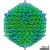
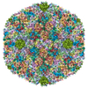
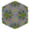
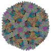
| Title | The quasi-atomic model of Human Adenovirus type 5 capsid | |||||||||
 Components Components |
| |||||||||
 Keywords Keywords | VIRUS / ADENOVIRUS / MINOR CAPSID PROTEIN / QUASI ATOMIC | |||||||||
| Function / homology |  Function and homology information Function and homology informationT=25 icosahedral viral capsid / microtubule-dependent intracellular transport of viral material towards nucleus / viral capsid / host cell / clathrin-dependent endocytosis of virus by host cell / symbiont entry into host cell / virion attachment to host cell / host cell nucleus / structural molecule activity Similarity search - Function | |||||||||
| Biological species |   HUMAN ADENOVIRUS 2 HUMAN ADENOVIRUS 2 HUMAN ADENOVIRUS TYPE 5HUMAN ADENOVIRUS TYPE 2 HUMAN ADENOVIRUS TYPE 5HUMAN ADENOVIRUS TYPE 2 | |||||||||
| Method | ELECTRON MICROSCOPY / single particle reconstruction / cryo EM / Resolution: 10 Å | |||||||||
 Authors Authors | Fabry, C.M.S. / Rosa-Calatrava, M. / Conway, J.F. / Zubieta, C. / Cusack, S. / Ruigrok, R.W.H. / Schoehn, G. | |||||||||
 Citation Citation | Journal: EMBO J / Year: 2005 Title: A quasi-atomic model of human adenovirus type 5 capsid. Authors: Céline M S Fabry / Manuel Rosa-Calatrava / James F Conway / Chloé Zubieta / Stephen Cusack / Rob W H Ruigrok / Guy Schoehn /  Abstract: Adenoviruses infect a wide range of vertebrates including humans. Their icosahedral capsids are composed of three major proteins: the trimeric hexon forms the facets and the penton, a noncovalent ...Adenoviruses infect a wide range of vertebrates including humans. Their icosahedral capsids are composed of three major proteins: the trimeric hexon forms the facets and the penton, a noncovalent complex of the pentameric penton base and trimeric fibre proteins, is located at the 12 capsid vertices. Several proteins (IIIa, VI, VIII and IX) stabilise the capsid. We have obtained a 10 A resolution map of the human adenovirus 5 by image analysis from cryo-electron micrographs (cryoEMs). This map, in combination with the X-ray structures of the penton base and hexon, was used to build a quasi-atomic model of the arrangement of the two major capsid components and to analyse the hexon-hexon and hexon-penton interactions. The secondary proteins, notably VIII, were located by comparing cryoEM maps of native and pIX deletion mutant virions. Minor proteins IX and IIIa are located on the outside of the capsid, whereas protein VIII is organised with a T=2 lattice on the inner face of the capsid. The capsid organisation is compared with the known X-ray structure of bacteriophage PRD1. | |||||||||
| History |
|
- Structure visualization
| Movie |
Movie viewer |
|---|---|
| Structure viewer | Molecule: MolmilJmol/JSmol |
 UCSF Chimera
- Downloads & links
-Download
| PDBx/mmCIF format | 4v4u.cif.gz | 2.4 MB | Display | PDBx/mmCIF format |
|---|---|---|---|---|
| PDB format | pdb4v4u.ent.gz | Display | PDB format | |
| PDBx/mmJSON format | 4v4u.json.gz | Tree view | PDBx/mmJSON format | |
| Others |  Other downloads Other downloads |
-Validation report
| Arichive directory | https://data.pdbj.org/pub/pdb/validation_reports/v4/4v4uftp://data.pdbj.org/pub/pdb/validation_reports/v4/4v4u | HTTPS FTP |
|---|
-Related structure data
| Related structure data |  1111MC 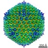 1112MC  1113MC M: map data used to model this data C: citing same article ( |
|---|---|
| Similar structure data |


-Links
 PDBj
PDBj


- Assembly
| Deposited unit | 
|
|---|---|
| 1 | x 12 x 60 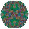
|
| 2 |
|
| 3 | 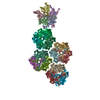
|
| Symmetry | Point symmetry: (Schoenflies symbol: I (icosahedral)) |
-Components
| #1: Protein | Mass: 58218.684 Da / Num. of mol.: 5 / Fragment: RESIDUES 49-571 Source method: isolated from a genetically manipulated source Details: VIRION COMPONENT III, PENTON BASE PROTEIN / Source: (gene. exp.) HUMAN ADENOVIRUS 2 / Production host:  TRICHOPLUSIA NI (cabbage looper) / References: UniProt: P03276 TRICHOPLUSIA NI (cabbage looper) / References: UniProt: P03276#2: Protein/peptide | Mass: 1216.295 Da / Num. of mol.: 5 / Source method: obtained synthetically / Details: VIRION COMPONENT IV, FIBER PROTEIN / Source: (synth.) HUMAN ADENOVIRUS TYPE 2#3: Protein | Mass: 107976.422 Da / Num. of mol.: 12 Source method: isolated from a genetically manipulated source Details: VIRION COMPONENT II, HEXON PROTEIN / Source: (gene. exp.) HUMAN ADENOVIRUS TYPE 5 / Cell line (production host): HELA S3 / Production host:  HOMO SAPIENS (human) / References: UniProt: P04133 HOMO SAPIENS (human) / References: UniProt: P04133 |
|---|
-Experimental details
-Experiment
| Experiment | Method: ELECTRON MICROSCOPY |
|---|---|
| EM experiment | Aggregation state: PARTICLE / 3D reconstruction method: single particle reconstruction |
- Sample preparation
| Component | Name: HUMAN ADENOVIRUS TYPE 5 / Type: VIRUS |
|---|---|
| Buffer solution | Name: TRIS / pH: 7.5 / Details: TRIS |
| Specimen | Conc.: 3 mg/ml / Embedding applied: NO / Shadowing applied: NO / Staining applied: NO / Vitrification applied: YES |
| Vitrification | Instrument: ZEISS PLUNGE FREEZER CRYOBOX / Cryogen name: ETHANE / Details: LIQUID ETHANE |
- Electron microscopy imaging
| Microscopy | Model: FEI/PHILIPS CM200T / Date: Oct 1, 2003 |
|---|---|
| Electron gun | Electron source: LAB6 / Accelerating voltage: 200 kV / Illumination mode: FLOOD BEAM |
| Electron lens | Mode: BRIGHT FIELD / Nominal magnification: 27500 X / Calibrated magnification: 28600 X / Nominal defocus max: 250 nm / Nominal defocus min: 100 nm / Cs: 2 mm |
| Specimen holder | Temperature: 95 K / Tilt angle max: 0 ° / Tilt angle min: 0 ° |
| Image recording | Electron dose: 10 e/Å2 / Film or detector model: KODAK SO-163 FILM |
| Image scans | Num. digital images: 21 |
| Radiation wavelength | Relative weight: 1 |
- Processing
| EM software |
| ||||||||||||||||||||||||||||||||
|---|---|---|---|---|---|---|---|---|---|---|---|---|---|---|---|---|---|---|---|---|---|---|---|---|---|---|---|---|---|---|---|---|---|
| CTF correction | Details: AMPLITUDE, PHASE TMV AND COMPARISON WITH X-RAY DATA | ||||||||||||||||||||||||||||||||
| Symmetry | Point symmetry: I (icosahedral) | ||||||||||||||||||||||||||||||||
| 3D reconstruction | Method: POLAR FOURIER TRANSFORM, FOURIER BESSELS RECONSTRUCTION Resolution: 10 Å / Num. of particles: 4100 / Nominal pixel size: 2.55 Å / Actual pixel size: 2.45 Å Details: THE NUMBER OF ATOMS IN THIS STRUCTURE EXCEEDS THE MAXIMUM ALLOWED BY THE PDB FORMAT. IT HAS THEREFORE BEEN SPLIT ACROSS TWO PDB ENTRIES: 2BLD CONTAINS THE PENTON BASE, 2BVI CONTAINS THE FOUR ...Details: THE NUMBER OF ATOMS IN THIS STRUCTURE EXCEEDS THE MAXIMUM ALLOWED BY THE PDB FORMAT. IT HAS THEREFORE BEEN SPLIT ACROSS TWO PDB ENTRIES: 2BLD CONTAINS THE PENTON BASE, 2BVI CONTAINS THE FOUR UNIQUE COPIES OF THE HEXON. THE COMPLETE CAPSID CAN BE GENERATED BY APPLYING ICOSAHEDRAL SYMMETRY OPERATORS TO THE COORDINATES IN THE TWO ENTRIES AND THEN COMBINING THE RESULTING CAPSID FRAGMENTS. THE PENTON BASE COMPRISES CHAINS A,B,C,D,E,S,T,U,V AND W AND MUST BE TRANSFORMED BY 12 SPECIFIC MATRICES DEFINED IN REMARK 350. THE HEXONS COMPRISE CHAINS F,G,H,I,J,K,L M,N,O,P AND Q AND MUST BE TRANSFORMED BY ALL ICOSAHEDRAL OPERATORS. Symmetry type: POINT | ||||||||||||||||||||||||||||||||
| Atomic model building | Protocol: RIGID BODY FIT / Space: RECIPROCAL / Target criteria: R-FACTOR, CORRELATION COEFFICIENT / Details: METHOD--RIGID BODY REFINEMENT PROTOCOL--X-RAY | ||||||||||||||||||||||||||||||||
| Refinement | Highest resolution: 10 Å | ||||||||||||||||||||||||||||||||
| Refinement step | Cycle: LAST / Highest resolution: 10 Å
|
