 Movie
Movie Controller


+ Open data

- Basic information
| Entry | Database: EMDB / ID: EMD-20123 | |||||||||
|---|---|---|---|---|---|---|---|---|---|---|
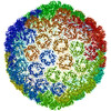
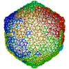
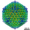
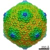
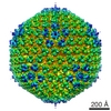
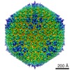
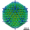
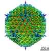
| Title | decorated head of the phage T5 | |||||||||
 Map data Map data | empty expanded head with decoration protein | |||||||||
 Sample Sample |
| |||||||||
| Biological species |  Escherichia phage T5 (virus) Escherichia phage T5 (virus) | |||||||||
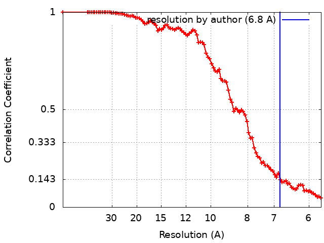
| Method | single particle reconstruction / cryo EM / Resolution: 6.8 Å | |||||||||
 Authors Authors | Huet A / Duda RL / Boulanger P / Conway JF | |||||||||
| Funding support |  United States, 2 items United States, 2 items
| |||||||||
 Citation Citation | Journal: Proc Natl Acad Sci U S A / Year: 2019 Title: Capsid expansion of bacteriophage T5 revealed by high resolution cryoelectron microscopy. Authors: Alexis Huet / Robert L Duda / Pascale Boulanger / James F Conway /  Abstract: The large (90-nm) icosahedral capsid of bacteriophage T5 is composed of 775 copies of the major capsid protein (mcp) together with portal, protease, and decoration proteins. Its assembly is a ...The large (90-nm) icosahedral capsid of bacteriophage T5 is composed of 775 copies of the major capsid protein (mcp) together with portal, protease, and decoration proteins. Its assembly is a regulated process that involves several intermediates, including a thick-walled round precursor prohead that expands as the viral DNA is packaged to yield a thin-walled and angular mature capsid. We investigated capsid maturation by comparing cryoelectron microscopy (cryo-EM) structures of the prohead, the empty expanded capsid both with and without decoration protein, and the virion capsid at a resolution of 3.8 Å for the latter. We detail the molecular structure of the mcp, its complex pattern of interactions, and their evolution during maturation. The bacteriophage T5 mcp is a variant of the canonical HK97-fold with a high level of plasticity that allows for the precise assembly of a giant macromolecule and the adaptability needed to interact with other proteins and the packaged DNA. | |||||||||
| History |
|
- Structure visualization
| Movie |
 Movie viewer Movie viewer |
|---|---|
| Structure viewer | EM map: SurfViewMolmilJmol/JSmol |
| Supplemental images |

- Downloads & links
-EMDB archive
| Map data | emd_20123.map.gz | 1.3 GB | EMDB map data format | |
|---|---|---|---|---|
| Header (meta data) | emd-20123-v30.xmlemd-20123.xml | 11.9 KB 11.9 KB | Display Display | EMDB header |
| FSC (resolution estimation) | emd_20123_fsc.xml | 9 KB | Display | FSC data file |
| Images | 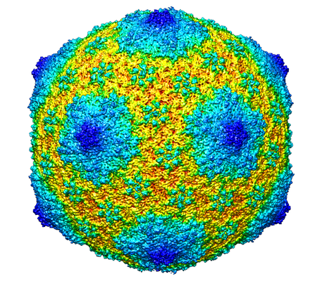 emd_20123.png emd_20123.png | 312.5 KB | ||
| Archive directory |  http://ftp.pdbj.org/pub/emdb/structures/EMD-20123ftp://ftp.pdbj.org/pub/emdb/structures/EMD-20123 http://ftp.pdbj.org/pub/emdb/structures/EMD-20123ftp://ftp.pdbj.org/pub/emdb/structures/EMD-20123 | HTTPS FTP |
-Related structure data


-Links
| EMDB pages | EMDB (EBI/PDBe) / EMDataResource |
|---|
-Map
| File | Download / File: emd_20123.map.gz / Format: CCP4 / Size: 3.7 GB / Type: IMAGE STORED AS FLOATING POINT NUMBER (4 BYTES) | ||||||||||||||||||||||||||||||||||||||||||||||||||||||||||||
|---|---|---|---|---|---|---|---|---|---|---|---|---|---|---|---|---|---|---|---|---|---|---|---|---|---|---|---|---|---|---|---|---|---|---|---|---|---|---|---|---|---|---|---|---|---|---|---|---|---|---|---|---|---|---|---|---|---|---|---|---|---|
| Annotation | empty expanded head with decoration protein | ||||||||||||||||||||||||||||||||||||||||||||||||||||||||||||
| Projections & slices | Image control
Images are generated by Spider. | ||||||||||||||||||||||||||||||||||||||||||||||||||||||||||||
| Voxel size | X=Y=Z: 1.35 Å | ||||||||||||||||||||||||||||||||||||||||||||||||||||||||||||
| Density |
| ||||||||||||||||||||||||||||||||||||||||||||||||||||||||||||
| Symmetry | Space group: 1 | ||||||||||||||||||||||||||||||||||||||||||||||||||||||||||||
| Details | EMDB XML:
CCP4 map header:
| ||||||||||||||||||||||||||||||||||||||||||||||||||||||||||||
 Z (Sec.)
 Y (Row.)
 X (Col.)


-Supplemental data
- Sample components
-Entire : Escherichia phage T5
| Entire | Name: Escherichia phage T5 (virus) |
|---|---|
| Components |
|
-Supramolecule #1: Escherichia phage T5
| Supramolecule | Name: Escherichia phage T5 / type: virus / ID: 1 / Parent: 0 / Macromolecule list: #1 / NCBI-ID: 10726 / Sci species name: Escherichia phage T5 / Virus type: VIRUS-LIKE PARTICLE / Virus isolate: OTHER / Virus enveloped: No / Virus empty: Yes |
|---|---|
| Host (natural) | Organism:  |
| Molecular weight | Theoretical: 28 MDa |
| Virus shell | Shell ID: 1 / Name: D-head / Diameter: 700.0 Å / T number (triangulation number): 13 |
-Experimental details
-Structure determination
| Method | cryo EM |
|---|---|
 Processing Processing | single particle reconstruction |
| Aggregation state | particle |
-Sample preparation
| Concentration | 2 mg/mL | |||||||||||||||
|---|---|---|---|---|---|---|---|---|---|---|---|---|---|---|---|---|
| Buffer | pH: 7.6 Component:
| |||||||||||||||
| Grid | Details: unspecified | |||||||||||||||
| Vitrification | Cryogen name: ETHANE-PROPANE / Chamber humidity: 100 % / Chamber temperature: 298 K / Instrument: FEI VITROBOT MARK III |
- Electron microscopy
| Microscope | FEI POLARA 300 |
|---|---|
| Image recording | Film or detector model: FEI FALCON II (4k x 4k) / Detector mode: INTEGRATING / Number grids imaged: 1 / Number real images: 1939 / Average electron dose: 30.0 e/Å2 |
| Electron beam | Acceleration voltage: 300 kV / Electron source:  FIELD EMISSION GUN FIELD EMISSION GUN |
| Electron optics | Illumination mode: SPOT SCAN / Imaging mode: BRIGHT FIELD |
| Sample stage | Specimen holder model: GATAN LIQUID NITROGEN / Cooling holder cryogen: NITROGEN |
| Experimental equipment |  Model: Tecnai Polara / Image courtesy: FEI Company |
+Image processing
-Atomic model buiding 1
| Initial model | PDB ID: |
|---|---|
| Refinement | Space: REAL / Protocol: FLEXIBLE FIT |
