 Movie
Movie Controller

[English] 日本語
 Yorodumi
- EMDB-11072: Reconstruction of bovine Ryanodine receptor 2 dimer of tetramers ... -
+ Open data

- Basic information
| Entry | Database: EMDB / ID: EMD-11072 | |||||||||
|---|---|---|---|---|---|---|---|---|---|---|
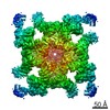
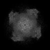
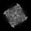
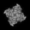
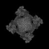
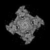
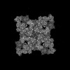
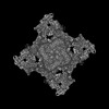
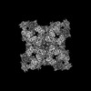
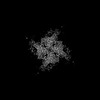
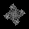
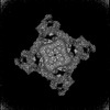
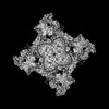
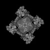
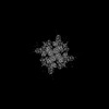
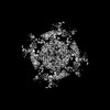
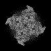
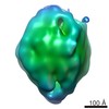
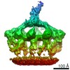
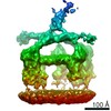
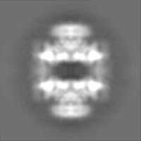
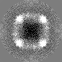
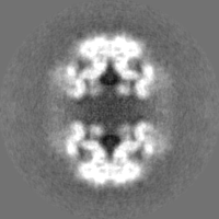
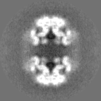
| Title | Reconstruction of bovine Ryanodine receptor 2 dimer of tetramers in complex with FKBP12.6 and nanobodies | |||||||||
 Map data Map data | ||||||||||
 Sample Sample |
| |||||||||
 Keywords Keywords | nanodisc / nanobody / FKBP12.6 / RyR2 / MEMBRANE PROTEIN | |||||||||
| Function / homology |  Function and homology information Function and homology informationcyclic nucleotide binding / Stimuli-sensing channels / negative regulation of insulin secretion involved in cellular response to glucose stimulus / : / type B pancreatic cell apoptotic process / Purkinje myocyte to ventricular cardiac muscle cell signaling / regulation of atrial cardiac muscle cell action potential / neuronal action potential propagation / Ion homeostasis / suramin binding ...cyclic nucleotide binding / Stimuli-sensing channels / negative regulation of insulin secretion involved in cellular response to glucose stimulus / : / type B pancreatic cell apoptotic process / Purkinje myocyte to ventricular cardiac muscle cell signaling / regulation of atrial cardiac muscle cell action potential / neuronal action potential propagation / Ion homeostasis / suramin binding / regulation of AV node cell action potential / regulation of SA node cell action potential / positive regulation of axon regeneration / ventricular cardiac muscle cell action potential / embryonic heart tube morphogenesis / regulation of ventricular cardiac muscle cell action potential / calcium ion transport into cytosol / ryanodine-sensitive calcium-release channel activity / T cell proliferation / release of sequestered calcium ion into cytosol by sarcoplasmic reticulum / insulin secretion / negative regulation of release of sequestered calcium ion into cytosol / response to redox state / cellular response to caffeine / negative regulation of heart rate / protein peptidyl-prolyl isomerization / smooth muscle contraction / response to vitamin E / response to muscle activity / FK506 binding / negative regulation of ryanodine-sensitive calcium-release channel activity / protein kinase A regulatory subunit binding / protein kinase A catalytic subunit binding / positive regulation of the force of heart contraction / smooth endoplasmic reticulum / intracellular membrane-bounded organelle / regulation of ryanodine-sensitive calcium-release channel activity / detection of calcium ion / response to glucose / regulation of cytosolic calcium ion concentration / calcium channel inhibitor activity / regulation of release of sequestered calcium ion into cytosol by sarcoplasmic reticulum / release of sequestered calcium ion into cytosol / response to muscle stretch / regulation of cardiac muscle contraction by regulation of the release of sequestered calcium ion / cellular response to epinephrine stimulus / sarcoplasmic reticulum membrane / calcium channel complex / regulation of heart rate / striated muscle contraction / peptidylprolyl isomerase / sarcoplasmic reticulum / peptidyl-prolyl cis-trans isomerase activity / response to hydrogen peroxide / sarcolemma / intracellular calcium ion homeostasis / Z disc / positive regulation of cytosolic calcium ion concentration / protein folding / transmembrane transporter binding / response to hypoxia / calmodulin binding / signaling receptor binding / calcium ion binding / membrane / identical protein binding / cytoplasm Similarity search - Function | |||||||||
| Biological species |  | |||||||||
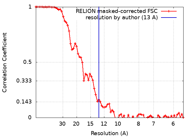
| Method | single particle reconstruction / cryo EM / Resolution: 13.0 Å | |||||||||
 Authors Authors | Willegems K / Efremov R | |||||||||
| Funding support |  Belgium, 2 items Belgium, 2 items
| |||||||||
 Citation Citation | Journal: J Biol Chem / Year: 2024 Title: Rapid small-scale nanobody-assisted purification of ryanodine receptors for cryo-EM. Authors: Chenyao Li / Katrien Willegems / Tomasz Uchański / Els Pardon / Jan Steyaert / Rouslan G Efremov / Abstract: Ryanodine receptors (RyRs) are large Ca release channels residing in the endoplasmic or sarcoplasmic reticulum membrane. Three isoforms of RyRs have been identified in mammals, the disfunction of ...Ryanodine receptors (RyRs) are large Ca release channels residing in the endoplasmic or sarcoplasmic reticulum membrane. Three isoforms of RyRs have been identified in mammals, the disfunction of which has been associated with a series of life-threatening diseases. The need for large amounts of native tissue or eukaryotic cell cultures limits advances in structural studies of RyRs. Here, we report a method that utilizes nanobodies to purify RyRs from only 5 mg of total protein. The purification process, from isolated membranes to cryo-EM grade protein, is achieved within 4 h on the bench, yielding protein usable for cryo-EM analysis. This is demonstrated by solving the structures of rabbit RyR1, solubilized in detergent, reconstituted into lipid nanodiscs or liposomes, and bovine RyR2 reconstituted in nanodisc, and mouse RyR2 in detergent. The reported method facilitates structural studies of RyRs directed toward drug development and is useful in cases where the amount of starting material is limited. | |||||||||
| History |
|
- Structure visualization
| Movie |
Movie viewer |
|---|---|
| Structure viewer | EM map: SurfViewMolmilJmol/JSmol |
| Supplemental images |

- Downloads & links
-EMDB archive
| Map data | emd_11072.map.gz | 6 MB | EMDB map data format | |
|---|---|---|---|---|
| Header (meta data) | emd-11072-v30.xmlemd-11072.xml | 17.8 KB 17.8 KB | Display Display | EMDB header |
| FSC (resolution estimation) | emd_11072_fsc.xml | 7.2 KB | Display | FSC data file |
| Images | 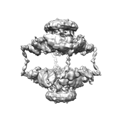 emd_11072.png emd_11072.png | 84.9 KB | ||
| Masks | emd_11072_msk_1.map | 30.5 MB | Mask map | |
| Filedesc metadata | emd-11072.cif.gz | 4.8 KB | ||
| Others | emd_11072_half_map_1.map.gzemd_11072_half_map_2.map.gz | 20.3 MB 20.3 MB | ||
| Archive directory |  http://ftp.pdbj.org/pub/emdb/structures/EMD-11072ftp://ftp.pdbj.org/pub/emdb/structures/EMD-11072 http://ftp.pdbj.org/pub/emdb/structures/EMD-11072ftp://ftp.pdbj.org/pub/emdb/structures/EMD-11072 | HTTPS FTP |
-Related structure data
| Related structure data |  8rrsC  8rrtC  8rruC 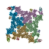 8rrvC 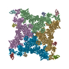 8rrwC 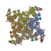 8rrxC  8rs0C C: citing same article ( |
|---|---|
| Similar structure data |


-Links
| EMDB pages | EMDB (EBI/PDBe) / EMDataResource |
|---|---|
| Related items in Molecule of the Month |


-Map
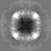
| File | Download / File: emd_11072.map.gz / Format: CCP4 / Size: 30.5 MB / Type: IMAGE STORED AS FLOATING POINT NUMBER (4 BYTES) | ||||||||||||||||||||||||||||||||||||||||||||||||||||||||||||
|---|---|---|---|---|---|---|---|---|---|---|---|---|---|---|---|---|---|---|---|---|---|---|---|---|---|---|---|---|---|---|---|---|---|---|---|---|---|---|---|---|---|---|---|---|---|---|---|---|---|---|---|---|---|---|---|---|---|---|---|---|---|
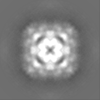
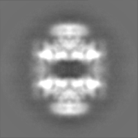
| Projections & slices | Image control
Images are generated by Spider. | ||||||||||||||||||||||||||||||||||||||||||||||||||||||||||||
| Voxel size | X=Y=Z: 2.8 Å | ||||||||||||||||||||||||||||||||||||||||||||||||||||||||||||
| Density |
| ||||||||||||||||||||||||||||||||||||||||||||||||||||||||||||
| Symmetry | Space group: 1 | ||||||||||||||||||||||||||||||||||||||||||||||||||||||||||||
| Details | EMDB XML:
CCP4 map header:
| ||||||||||||||||||||||||||||||||||||||||||||||||||||||||||||
 Z (Sec.)
 Y (Row.)
 X (Col.)


-Supplemental data
-Mask #1
| File | emd_11072_msk_1.map | ||||||||||||
|---|---|---|---|---|---|---|---|---|---|---|---|---|---|
| Projections & Slices |
| ||||||||||||
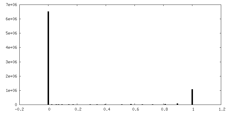
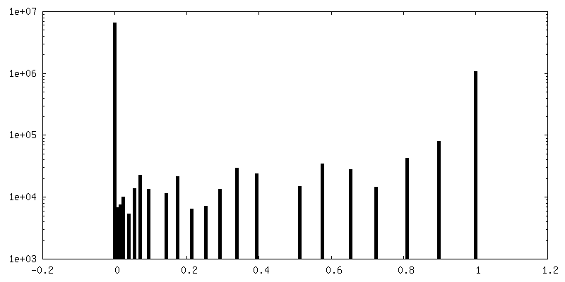
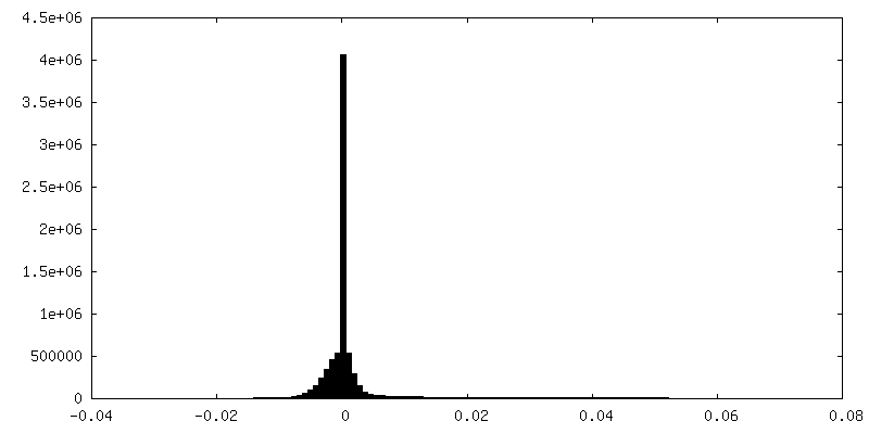
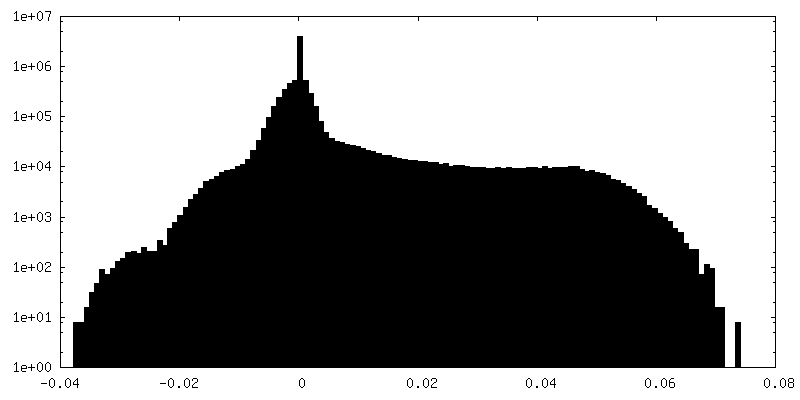
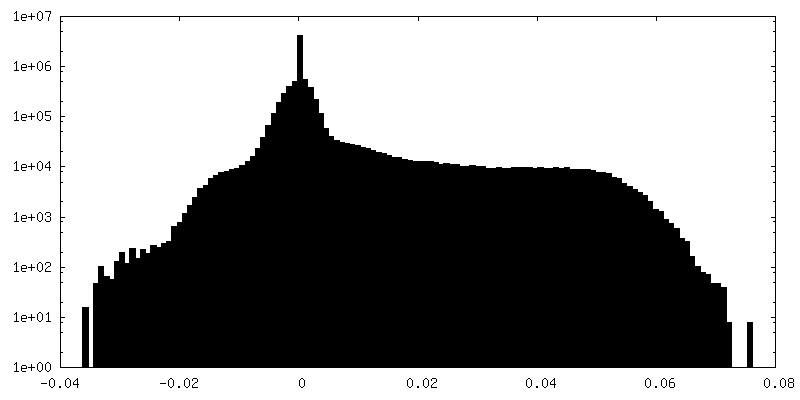
| Density Histograms |


-Half map: half map 1
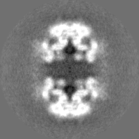
| File | emd_11072_half_map_1.map | ||||||||||||
|---|---|---|---|---|---|---|---|---|---|---|---|---|---|
| Annotation | half map 1 | ||||||||||||
| Projections & Slices |
| ||||||||||||
| Density Histograms |
-Half map: half map 2
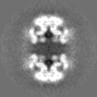
| File | emd_11072_half_map_2.map | ||||||||||||
|---|---|---|---|---|---|---|---|---|---|---|---|---|---|
| Annotation | half map 2 | ||||||||||||
| Projections & Slices |
| ||||||||||||
| Density Histograms |


- Sample components
-Entire : Dimer of bovine Ryanodine receptor 2
| Entire | Name: Dimer of bovine Ryanodine receptor 2 |
|---|---|
| Components |
|
-Supramolecule #1: Dimer of bovine Ryanodine receptor 2
| Supramolecule | Name: Dimer of bovine Ryanodine receptor 2 / type: complex / ID: 1 / Parent: 0 / Macromolecule list: #1 Details: Complex of Ryanodine receptor 2 with auxiliary protein FKBP12.6 and high affinity nanobody |
|---|---|
| Source (natural) | Organism: |
| Molecular weight | Theoretical: 2.2 MDa |
-Experimental details
-Structure determination
| Method | cryo EM |
|---|---|
 Processing Processing | single particle reconstruction |
| Aggregation state | particle |
-Sample preparation
| Concentration | 1 mg/mL | ||||||||||||||||||
|---|---|---|---|---|---|---|---|---|---|---|---|---|---|---|---|---|---|---|---|
| Buffer | pH: 7.4 Component:
| ||||||||||||||||||
| Grid | Model: Quantifoil R2/1 / Material: COPPER / Mesh: 400 / Support film - #0 - Film type ID: 1 / Support film - #0 - Material: CARBON / Support film - #0 - topology: HOLEY / Support film - #1 - Film type ID: 2 / Support film - #1 - Material: GRAPHENE OXIDE / Support film - #1 - topology: CONTINUOUS | ||||||||||||||||||
| Vitrification | Cryogen name: ETHANE / Chamber humidity: 95 % / Chamber temperature: 293.15 K / Instrument: GATAN CRYOPLUNGE 3 Details: .Grid was blotted manually from the back in CP3 cryoplunge (Gatan) for 2s. | ||||||||||||||||||
| Details | The RyR2 tetrameric channels formed channel-dimers with the nanobody at the dimer-interface bound to the repeat12 domain of the individual channels |
- Electron microscopy
| Microscope | FEI TITAN KRIOS |
|---|---|
| Specialist optics | Spherical aberration corrector: Microscope was modified with a Cs corrector |
| Image recording | Film or detector model: FEI FALCON III (4k x 4k) / Detector mode: INTEGRATING / Number grids imaged: 1 / Number real images: 4088 / Average electron dose: 47.0 e/Å2 |
| Electron beam | Acceleration voltage: 300 kV / Electron source:  FIELD EMISSION GUN FIELD EMISSION GUN |
| Electron optics | Illumination mode: FLOOD BEAM / Imaging mode: BRIGHT FIELD / Cs: 0.0 mm / Nominal defocus max: 4.0 µm / Nominal defocus min: 2.0 µm / Nominal magnification: 59000 |
| Sample stage | Specimen holder model: FEI TITAN KRIOS AUTOGRID HOLDER / Cooling holder cryogen: NITROGEN |
| Experimental equipment |  Model: Titan Krios / Image courtesy: FEI Company |
+Image processing
-Atomic model buiding 1
| Initial model | PDB ID: Chain - Source name: PDB / Chain - Initial model type: experimental model |
|---|---|
| Refinement | Space: REAL / Protocol: RIGID BODY FIT |

