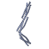
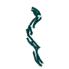
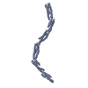
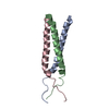
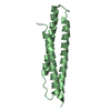
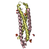
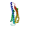
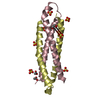
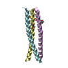
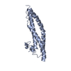
Journal: J Biol Chem / Year: 2018 Title: Dystrophin's central domain forms a complex filament that becomes disorganized by in-frame deletions. Authors: Olivier Delalande / Anne-Elisabeth Molza / Raphael Dos Santos Morais / Angélique Chéron / Émeline Pollet / Céline Raguenes-Nicol / Christophe Tascon / Emmanuel Giudice / Marine Guilbaud ...Authors: Olivier Delalande / Anne-Elisabeth Molza / Raphael Dos Santos Morais / Angélique Chéron / Émeline Pollet / Céline Raguenes-Nicol / Christophe Tascon / Emmanuel Giudice / Marine Guilbaud / Aurélie Nicolas / Arnaud Bondon / France Leturcq / Nicolas Férey / Marc Baaden / Javier Perez / Pierre Roblin / France Piétri-Rouxel / Jean-François Hubert / Mirjam Czjzek / Elisabeth Le Rumeur / Abstract: Dystrophin, encoded by the gene, is critical for maintaining plasma membrane integrity during muscle contraction events. Mutations in the gene disrupting the reading frame prevent dystrophin ...Dystrophin, encoded by the gene, is critical for maintaining plasma membrane integrity during muscle contraction events. Mutations in the gene disrupting the reading frame prevent dystrophin production and result in severe Duchenne muscular dystrophy (DMD); in-frame internal deletions allow production of partly functional internally deleted dystrophin and result in less severe Becker muscular dystrophy (BMD). Many known BMD deletions occur in dystrophin's central domain, generally considered to be a monotonous rod-shaped domain based on the knowledge of spectrin family proteins. However, the effects caused by these deletions, ranging from asymptomatic to severe BMD, argue against the central domain serving only as a featureless scaffold. We undertook structural studies combining small-angle X-ray scattering and molecular modeling in an effort to uncover the structure of the central domain, as dystrophin has been refractory to characterization. We show that this domain appears to be a tortuous and complex filament that is profoundly disorganized by the most severe BMD deletion (loss of exons 45-47). Despite the preservation of large parts of the binding site for neuronal nitric oxide synthase (nNOS) in this deletion, computational approaches failed to recreate the association of dystrophin with nNOS. This observation is in agreement with a strong decrease of nNOS immunolocalization in muscle biopsies, a parameter related to the severity of BMD phenotypes. The structural description of the whole dystrophin central domain we present here is a first necessary step to improve the design of microdystrophin constructs toward the goal of a successful gene therapy for DMD.
Contact author
Elisabeth Le Rumeur (Université de Rennes 1, Institut Génétique et Développement de Rennes, France)
Instrument name: SOLEIL SWING / City: Saint-Aubin / 国: France / Type of source: X-ray synchrotron / Wavelength: 0.1 Å / Dist. spec. to detc.: 1.82 mm
Detector
Name: AVIEX PCCD170170 / Type: CCD
Scan
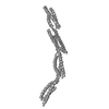
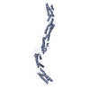
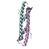
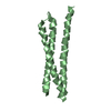
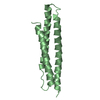
Title: Human dystrophin central domain single repeat 23 / Measurement date: Oct 7, 2011 / Storage temperature: 15 °C / Exposure time: 1.5 sec. / Number of frames: 30 / Unit: 1/nm /
Min
Max
Q
0.0504
5.392
Distance distribution function P(R)
Sofotware P(R): GNOM 4.6 / Number of points: 476 /
Min
Max
Q
0.009576
0.2488
P(R) point
10
485
R
0
74
Result
D max: 7.4 / Type of curve: single_conc /
Experimental
Porod
MW
12.5 kDa
12.5 kDa
Volume
-
20 nm3
P(R)
Guinier
Forward scattering, I0
0.0351
0.0353
Radius of gyration, Rg
2.2 nm
2.2 nm
+
About Yorodumi
-
News
-
Feb 9, 2022. New format data for meta-information of EMDB entries
New format data for meta-information of EMDB entries
Version 3 of the EMDB header file is now the official format.
The previous official version 1.9 will be removed from the archive.
In the structure databanks used in Yorodumi, some data are registered as the other names, "COVID-19 virus" and "2019-nCoV". Here are the details of the virus and the list of structure data.
Jan 31, 2019. EMDB accession codes are about to change! (news from PDBe EMDB page)
EMDB accession codes are about to change! (news from PDBe EMDB page)
The allocation of 4 digits for EMDB accession codes will soon come to an end. Whilst these codes will remain in use, new EMDB accession codes will include an additional digit and will expand incrementally as the available range of codes is exhausted. The current 4-digit format prefixed with “EMD-” (i.e. EMD-XXXX) will advance to a 5-digit format (i.e. EMD-XXXXX), and so on. It is currently estimated that the 4-digit codes will be depleted around Spring 2019, at which point the 5-digit format will come into force.
The EM Navigator/Yorodumi systems omit the EMD- prefix.
Related info.:Q: What is EMD? / ID/Accession-code notation in Yorodumi/EM Navigator
Yorodumi is a browser for structure data from EMDB, PDB, SASBDB, etc.
This page is also the successor to EM Navigator detail page, and also detail information page/front-end page for Omokage search.
The word "yorodu" (or yorozu) is an old Japanese word meaning "ten thousand". "mi" (miru) is to see.
Related info.:EMDB / PDB / SASBDB / Comparison of 3 databanks / Yorodumi Search / Aug 31, 2016. New EM Navigator & Yorodumi / Yorodumi Papers / Jmol/JSmol / Function and homology information / Changes in new EM Navigator and Yorodumi
 Movie
Movie Controller
 Yorodumi
 Open data
 Basic information
 Sample
 Function and homology information
 Homo sapiens (human)
 Citation
 Contact author
 Structure visualization
 Downloads & links
 SASDBB3
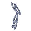


 Search similar-shape structures of this assembly by Omokage search (details)


