 Movie
Movie Controller

[English] 日本語
 Yorodumi
- PDB-3fzh: Crystal Structures of Hsc70/Bag1 in Complex with Small Molecule I... -
+ Open data

- Basic information
| Entry | Database: PDB / ID: 3fzh | ||||||
|---|---|---|---|---|---|---|---|
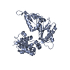
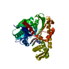
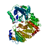
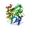
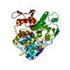
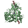
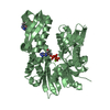
| Title | Crystal Structures of Hsc70/Bag1 in Complex with Small Molecule Inhibitors | ||||||
 Components Components |
| ||||||
 Keywords Keywords | CHAPERONE / HSP70 / HSC70 / BAG1 / heat shock / protein folding / adenosine / nucleotide / nucleotide exchange factor / small molecule inhibitor / ATP-binding / nucleotide-binding / stress response / apoptosis | ||||||
| Function / homology |  Function and homology information Function and homology informationlumenal side of lysosomal membrane / regulation of protein import / negative regulation of supramolecular fiber organization / chaperone-mediated autophagy translocation complex disassembly / clathrin-sculpted gamma-aminobutyric acid transport vesicle membrane / positive regulation of ferroptosis / Lipophagy / GABA synthesis, release, reuptake and degradation / protein targeting to lysosome involved in chaperone-mediated autophagy / Respiratory syncytial virus genome transcription ...lumenal side of lysosomal membrane / regulation of protein import / negative regulation of supramolecular fiber organization / chaperone-mediated autophagy translocation complex disassembly / clathrin-sculpted gamma-aminobutyric acid transport vesicle membrane / positive regulation of ferroptosis / Lipophagy / GABA synthesis, release, reuptake and degradation / protein targeting to lysosome involved in chaperone-mediated autophagy / Respiratory syncytial virus genome transcription / adenyl-nucleotide exchange factor activity / clathrin coat disassembly / C3HC4-type RING finger domain binding / positive regulation of smooth muscle cell apoptotic process / CHL1 interactions / regulation of protein complex stability / negative regulation of NLRP3 inflammasome complex assembly / ATP-dependent protein disaggregase activity / membrane organization / cellular response to steroid hormone stimulus / Lysosome Vesicle Biogenesis / chaperone-mediated autophagy / Golgi Associated Vesicle Biogenesis / non-chaperonin molecular chaperone ATPase / Prp19 complex / HSF1-dependent transactivation / Regulation of HSF1-mediated heat shock response / protein folding chaperone complex / response to unfolded protein / regulation of protein-containing complex assembly / Attenuation phase / ATP metabolic process / Protein methylation / heat shock protein binding / mRNA Splicing - Major Pathway / protein folding chaperone / lysosomal lumen / HSP90 chaperone cycle for steroid hormone receptors (SHR) in the presence of ligand / cellular response to starvation / spliceosomal complex / ATP-dependent protein folding chaperone / AUF1 (hnRNP D0) binds and destabilizes mRNA / regulation of protein stability / mRNA splicing, via spliceosome / Late endosomal microautophagy / PKR-mediated signaling / G protein-coupled receptor binding / Chaperone Mediated Autophagy / : / melanosome / MHC class II protein complex binding / Clathrin-mediated endocytosis / protein refolding / protein folding / protein-folding chaperone binding / Interleukin-4 and Interleukin-13 signaling / secretory granule lumen / blood microparticle / ficolin-1-rich granule lumen / protein-macromolecule adaptor activity / cell surface receptor signaling pathway / protein stabilization / positive regulation of cell migration / cadherin binding / ribonucleoprotein complex / receptor ligand activity / lysosomal membrane / focal adhesion / negative regulation of DNA-templated transcription / apoptotic process / Neutrophil degranulation / ubiquitin protein ligase binding / negative regulation of apoptotic process / nucleolus / enzyme binding / ATP hydrolysis activity / : / RNA binding / extracellular exosome / extracellular region / nucleoplasm / ATP binding / membrane / nucleus / plasma membrane / cytoplasm / cytosol Similarity search - Function | ||||||
| Biological species |  Homo sapiens (human) Homo sapiens (human) | ||||||
| Method |  X-RAY DIFFRACTION / MOLECULAR REPLACEMENT / Resolution: 2 Å X-RAY DIFFRACTION / MOLECULAR REPLACEMENT / Resolution: 2 Å | ||||||
 Authors Authors | Dokurno, P. / Williamson, D.S. / Murray, J.B. / Surgenor, A.E. | ||||||
 Citation Citation | Journal: J.Med.Chem. / Year: 2009 Title: Novel adenosine-derived inhibitors of 70 kDa heat shock protein, discovered through structure-based design Authors: Williamson, D.S. / Borgognoni, J. / Clay, A. / Daniels, Z. / Dokurno, P. / Drysdale, M.J. / Foloppe, N. / Francis, G.L. / Graham, C.J. / Howes, R. / Macias, A.T. / Murray, J.B. / Parsons, R. ...Authors: Williamson, D.S. / Borgognoni, J. / Clay, A. / Daniels, Z. / Dokurno, P. / Drysdale, M.J. / Foloppe, N. / Francis, G.L. / Graham, C.J. / Howes, R. / Macias, A.T. / Murray, J.B. / Parsons, R. / Shaw, T. / Surgenor, A.E. / Terry, L. / Wang, Y. / Wood, M. / Massey, A.J. | ||||||
| History |
|
- Structure visualization
| Structure viewer | Molecule: MolmilJmol/JSmol |
|---|
- Downloads & links
-Download
| PDBx/mmCIF format | 3fzh.cif.gz | 114.6 KB | Display | PDBx/mmCIF format |
|---|---|---|---|---|
| PDB format | pdb3fzh.ent.gz | 87.1 KB | Display | PDB format |
| PDBx/mmJSON format | 3fzh.json.gz | Tree view | PDBx/mmJSON format | |
| Others |  Other downloads Other downloads |
-Validation report
| Arichive directory | https://data.pdbj.org/pub/pdb/validation_reports/fz/3fzhftp://data.pdbj.org/pub/pdb/validation_reports/fz/3fzh | HTTPS FTP |
|---|
-Related structure data
| Related structure data | 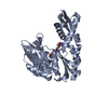 3fzfC 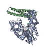 3fzkC  3fzlC 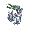 3fzmC 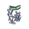 1hx1S C: citing same article ( S: Starting model for refinement |
|---|---|
| Similar structure data |


-Links
 PDBj
PDBj


- Assembly
| Deposited unit | 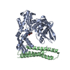
| ||||||||
|---|---|---|---|---|---|---|---|---|---|
| 1 | 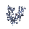
| ||||||||
| 2 | 
| ||||||||
| Unit cell |
|
-Components
| #1: Protein | Mass: 42086.547 Da / Num. of mol.: 1 / Fragment: UNP residues 4-381 Source method: isolated from a genetically manipulated source Source: (gene. exp.) Homo sapiens (human) / Gene: HSPA8, HSC70, HSP73, HSPA10 / Plasmid: pET101 / Production host:  |
|---|---|
| #2: Protein | Mass: 13157.168 Da / Num. of mol.: 1 / Fragment: UNP residues 222-334 Source method: isolated from a genetically manipulated source Source: (gene. exp.) Homo sapiens (human) / Gene: BAG1, HAP / Plasmid: pGEX-2T / Production host: |
| #3: Chemical | ChemComp-3BH / (  Mass: 282.256 Da / Num. of mol.: 1 / Source method: obtained synthetically / Formula: C10H14N6O4 Mass: 282.256 Da / Num. of mol.: 1 / Source method: obtained synthetically / Formula: C10H14N6O4 |
| #4: Water | ChemComp-HOH /  Mass: 18.015 Da / Num. of mol.: 309 / Source method: isolated from a natural source / Formula: H2O Mass: 18.015 Da / Num. of mol.: 309 / Source method: isolated from a natural source / Formula: H2O |
| Has protein modification | Y |
-Experimental details
-Experiment
| Experiment | Method: X-RAY DIFFRACTION / Number of used crystals: 1 |
|---|
- Sample preparation
| Crystal | Density Matthews: 2.28 Å3/Da / Density % sol: 45.95 % |
|---|---|
| Crystal grow | Temperature: 293 K / Method: vapor diffusion, hanging drop / pH: 8.5 Details: 15% PEG3350, 0.1M Tris buffer, 25mM sodium-potassium tartrate, pH8.5, VAPOR DIFFUSION, HANGING DROP, temperature 293K |
-Data collection
| Diffraction | Mean temperature: 100 K |
|---|---|
| Diffraction source | Source: ROTATING ANODE / Type: RIGAKU RUH3R / Wavelength: 1.5418 Å |
| Detector | Type: RIGAKU RAXIS IV++ / Detector: IMAGE PLATE / Date: Nov 14, 2008 / Details: mirrors |
| Radiation | Monochromator: mirrors / Protocol: SINGLE WAVELENGTH / Monochromatic (M) / Laue (L): M / Scattering type: x-ray |
| Radiation wavelength | Wavelength: 1.5418 Å / Relative weight: 1 |
| Reflection | Resolution: 1.9→25 Å / Num. all: 37186 / Num. obs: 37186 / % possible obs: 95.8 % / Observed criterion σ(F): 1 / Observed criterion σ(I): 1 / Redundancy: 4.17 % / Rmerge(I) obs: 0.127 / Net I/σ(I): 4.8 |
| Reflection shell | Resolution: 1.9→1.97 Å / Redundancy: 4.01 % / Rmerge(I) obs: 0.54 / Mean I/σ(I) obs: 1.1 / % possible all: 87.2 |
- Processing
| Software |
| ||||||||||||||||||||||||||||||||||||||||||||||||||||||||||||||||||||||||||||||||||||||||||
|---|---|---|---|---|---|---|---|---|---|---|---|---|---|---|---|---|---|---|---|---|---|---|---|---|---|---|---|---|---|---|---|---|---|---|---|---|---|---|---|---|---|---|---|---|---|---|---|---|---|---|---|---|---|---|---|---|---|---|---|---|---|---|---|---|---|---|---|---|---|---|---|---|---|---|---|---|---|---|---|---|---|---|---|---|---|---|---|---|---|---|---|
| Refinement | Method to determine structure: MOLECULAR REPLACEMENT Starting model: PDB ENRTY 1HX1 Resolution: 2→25 Å / Cor.coef. Fo:Fc: 0.945 / Cor.coef. Fo:Fc free: 0.913 / SU B: 5.19 / SU ML: 0.146 / Cross valid method: THROUGHOUT / σ(F): 1 / σ(I): 1 / ESU R: 0.248 / ESU R Free: 0.21 / Stereochemistry target values: MAXIMUM LIKELIHOOD / Details: HYDROGENS HAVE BEEN ADDED IN THE RIDING POSITIONS
| ||||||||||||||||||||||||||||||||||||||||||||||||||||||||||||||||||||||||||||||||||||||||||
| Solvent computation | Ion probe radii: 0.8 Å / Shrinkage radii: 0.8 Å / VDW probe radii: 1.2 Å / Solvent model: MASK | ||||||||||||||||||||||||||||||||||||||||||||||||||||||||||||||||||||||||||||||||||||||||||
| Displacement parameters | Biso mean: 31.728 Å2
| ||||||||||||||||||||||||||||||||||||||||||||||||||||||||||||||||||||||||||||||||||||||||||
| Refinement step | Cycle: LAST / Resolution: 2→25 Å
| ||||||||||||||||||||||||||||||||||||||||||||||||||||||||||||||||||||||||||||||||||||||||||
| Refine LS restraints |
| ||||||||||||||||||||||||||||||||||||||||||||||||||||||||||||||||||||||||||||||||||||||||||
| LS refinement shell | Resolution: 2→2.052 Å / Total num. of bins used: 20
|
