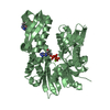
- PDB-4io8: Crystal structure of human HSP70 complexed with 4-{(2R,3S,4R)-5-[... -
+
Open data
ID or keywords:
Loading...
-
Basic information
Entry
Database: PDB / ID: 4io8
Title
Crystal structure of human HSP70 complexed with 4-{(2R,3S,4R)-5-[(R)-6-Amino-8-(3,4-dichloro-benzylamino)-purin-9-yl]-3,4-dihydroxy-tetrahydro-furan-2-ylmethoxymethyl}-benzonitrile
Components
Heat shock 70kDa protein 1A variant
Keywords
CHAPERONE / Hsp70 / ATPase / molecular chaperone
Function / homology
Function and homology information
: / cellular heat acclimation / negative regulation of inclusion body assembly / Viral RNP Complexes in the Host Cell Nucleus / C3HC4-type RING finger domain binding / positive regulation of nucleotide-binding oligomerization domain containing 2 signaling pathway / positive regulation of microtubule nucleation / ATP-dependent protein disaggregase activity / misfolded protein binding / negative regulation of mitochondrial outer membrane permeabilization involved in apoptotic signaling pathway ...: / cellular heat acclimation / negative regulation of inclusion body assembly / Viral RNP Complexes in the Host Cell Nucleus / C3HC4-type RING finger domain binding / positive regulation of nucleotide-binding oligomerization domain containing 2 signaling pathway / positive regulation of microtubule nucleation / ATP-dependent protein disaggregase activity / misfolded protein binding / negative regulation of mitochondrial outer membrane permeabilization involved in apoptotic signaling pathway / regulation of mitotic spindle assembly / positive regulation of tumor necrosis factor-mediated signaling pathway / aggresome / lysosomal transport / cellular response to steroid hormone stimulus / mRNA catabolic process / regulation of protein ubiquitination / cellular response to unfolded protein / HSF1-dependent transactivation / Regulation of HSF1-mediated heat shock response / response to unfolded protein / negative regulation of extrinsic apoptotic signaling pathway in absence of ligand / Mitochondrial unfolded protein response (UPRmt) / Attenuation phase / negative regulation of endoplasmic reticulum stress-induced intrinsic apoptotic signaling pathway / chaperone-mediated protein complex assembly / transcription regulator inhibitor activity / ATP metabolic process / endoplasmic reticulum unfolded protein response / heat shock protein binding / protein folding chaperone / inclusion body / negative regulation of protein ubiquitination / positive regulation of erythrocyte differentiation / HSP90 chaperone cycle for steroid hormone receptors (SHR) in the presence of ligand / positive regulation of RNA splicing / : / positive regulation of interleukin-8 production / ATP-dependent protein folding chaperone / centriole / negative regulation of transforming growth factor beta receptor signaling pathway / AUF1 (hnRNP D0) binds and destabilizes mRNA / negative regulation of cell growth / PKR-mediated signaling / G protein-coupled receptor binding / histone deacetylase binding / disordered domain specific binding / : / transcription corepressor activity / positive regulation of proteasomal ubiquitin-dependent protein catabolic process / cellular response to heat / protein refolding / virus receptor activity / cellular response to oxidative stress / blood microparticle / vesicle / ficolin-1-rich granule lumen / nuclear speck / protein stabilization / cadherin binding / ribonucleoprotein complex / receptor ligand activity / signaling receptor binding / negative regulation of cell population proliferation / focal adhesion / Neutrophil degranulation / ubiquitin protein ligase binding / positive regulation of gene expression / centrosome / negative regulation of apoptotic process / perinuclear region of cytoplasm / enzyme binding / negative regulation of transcription by RNA polymerase II / endoplasmic reticulum / ATP hydrolysis activity / protein-containing complex / mitochondrion / : / RNA binding / extracellular exosome / extracellular region / nucleoplasm / ATP binding / nucleus / plasma membrane / cytoplasm / cytosol Similarity search - Function
Defensin A-like - #30 / Defensin A-like / Heat shock hsp70 proteins family signature 2. / Heat shock hsp70 proteins family signature 1. / Heat shock hsp70 proteins family signature 3. / Heat shock protein 70, conserved site / Heat shock protein 70kD, peptide-binding domain superfamily / Heat shock protein 70kD, C-terminal domain superfamily / Heat shock protein 70 family / Hsp70 protein ...Defensin A-like - #30 / Defensin A-like / Heat shock hsp70 proteins family signature 2. / Heat shock hsp70 proteins family signature 1. / Heat shock hsp70 proteins family signature 3. / Heat shock protein 70, conserved site / Heat shock protein 70kD, peptide-binding domain superfamily / Heat shock protein 70kD, C-terminal domain superfamily / Heat shock protein 70 family / Hsp70 protein / ATPase, substrate binding domain, subdomain 4 / Actin; Chain A, domain 4 / ATPase, nucleotide binding domain / ATPase, nucleotide binding domain / Nucleotidyltransferase; domain 5 / Alpha-Beta Complex / 2-Layer Sandwich / Alpha Beta Similarity search - Domain/homology
In the structure databanks used in Yorodumi, some data are registered as the other names, "COVID-19 virus" and "2019-nCoV". Here are the details of the virus and the list of structure data.
Jan 31, 2019. EMDB accession codes are about to change! (news from PDBe EMDB page)
EMDB accession codes are about to change! (news from PDBe EMDB page)
The allocation of 4 digits for EMDB accession codes will soon come to an end. Whilst these codes will remain in use, new EMDB accession codes will include an additional digit and will expand incrementally as the available range of codes is exhausted. The current 4-digit format prefixed with “EMD-” (i.e. EMD-XXXX) will advance to a 5-digit format (i.e. EMD-XXXXX), and so on. It is currently estimated that the 4-digit codes will be depleted around Spring 2019, at which point the 5-digit format will come into force.
The EM Navigator/Yorodumi systems omit the EMD- prefix.
Related info.:Q: What is EMD? / ID/Accession-code notation in Yorodumi/EM Navigator
Yorodumi is a browser for structure data from EMDB, PDB, SASBDB, etc.
This page is also the successor to EM Navigator detail page, and also detail information page/front-end page for Omokage search.
The word "yorodu" (or yorozu) is an old Japanese word meaning "ten thousand". "mi" (miru) is to see.
Related info.:EMDB / PDB / SASBDB / Comparison of 3 databanks / Yorodumi Search / Aug 31, 2016. New EM Navigator & Yorodumi / Yorodumi Papers / Jmol/JSmol / Function and homology information / Changes in new EM Navigator and Yorodumi
 Movie
Movie Controller

 Yorodumi
 Open data
 Basic information
 Components
 Keywords
 Function and homology information
 Homo sapiens (human)
 X-RAY DIFFRACTION /
  Authors
 Citation
 Structure visualization
 Downloads & links
 Other downloads
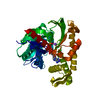


 PDBj
PDBj


 Assembly
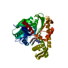

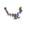
 Mass: 556.401 Da / Num. of mol.: 1 / Source method: obtained synthetically / Formula: C25H23Cl2N7O4
 Mass: 18.015 Da / Num. of mol.: 121 / Source method: isolated from a natural source / Formula: H2O
 Sample preparation
 / Beamline: X06SA / Wavelength: 1 Å
 Processing