 Movie
Movie Controller

[English] 日本語
 Yorodumi
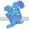
- PDB-9e8h: Human proteasome in resting state conformation bound to TXNL1 in ... -
+ Open data

- Basic information
| Entry | Database: PDB / ID: 9e8h | |||||||||||||||||||||||||||||||||
|---|---|---|---|---|---|---|---|---|---|---|---|---|---|---|---|---|---|---|---|---|---|---|---|---|---|---|---|---|---|---|---|---|---|---|
| Title | Human proteasome in resting state conformation bound to TXNL1 in backward conformation | |||||||||||||||||||||||||||||||||
 Components Components |
| |||||||||||||||||||||||||||||||||
 Keywords Keywords | MOTOR PROTEIN / HYDROLASE/PROTEIN BINDING / 26S Proteasome / Nub1 / Fat10 / HYDROLASE-PROTEIN BINDING complex | |||||||||||||||||||||||||||||||||
| Function / homology |  Function and homology information Function and homology informationregulation of ubiquitin-dependent protein catabolic process / disulfide oxidoreductase activity / positive regulation of inclusion body assembly / thyrotropin-releasing hormone receptor binding / nuclear proteasome complex / host-mediated perturbation of viral transcription / Impaired BRCA2 translocation to the nucleus / Impaired BRCA2 binding to SEM1 (DSS1) / proteasome accessory complex / purine ribonucleoside triphosphate binding ...regulation of ubiquitin-dependent protein catabolic process / disulfide oxidoreductase activity / positive regulation of inclusion body assembly / thyrotropin-releasing hormone receptor binding / nuclear proteasome complex / host-mediated perturbation of viral transcription / Impaired BRCA2 translocation to the nucleus / Impaired BRCA2 binding to SEM1 (DSS1) / proteasome accessory complex / purine ribonucleoside triphosphate binding / integrator complex / proteasome regulatory particle / CD8-positive, alpha-beta T cell differentiation / thymic T cell selection / cytosolic proteasome complex / CD8-positive, alpha-beta T cell homeostasis / positive regulation of proteasomal protein catabolic process / proteasome-activating activity / Antigen processing: Ub, ATP-independent proteasomal degradation / RND1 GTPase cycle / RND2 GTPase cycle / RND3 GTPase cycle / proteasome regulatory particle, lid subcomplex / proteasome regulatory particle, base subcomplex / negative regulation of programmed cell death / RHOBTB1 GTPase cycle / negative regulation of regulatory T cell differentiation / T-helper 1 cell differentiation / protein K63-linked deubiquitination / RHOV GTPase cycle / cellular response to type I interferon / metal-dependent deubiquitinase activity / Regulation of ornithine decarboxylase (ODC) / proteasome core complex / Proteasome assembly / T-helper 17 cell differentiation / retrograde vesicle-mediated transport, Golgi to endoplasmic reticulum / Cross-presentation of soluble exogenous antigens (endosomes) / transcription factor binding / Somitogenesis / flagellated sperm motility / K63-linked deubiquitinase activity / Homologous DNA Pairing and Strand Exchange / Defective homologous recombination repair (HRR) due to BRCA1 loss of function / Defective HDR through Homologous Recombination Repair (HRR) due to PALB2 loss of BRCA1 binding function / Defective HDR through Homologous Recombination Repair (HRR) due to PALB2 loss of BRCA2/RAD51/RAD51C binding function / Resolution of D-loop Structures through Synthesis-Dependent Strand Annealing (SDSA) / Resolution of D-loop Structures through Holliday Junction Intermediates / Lewy body / proteasome binding / RHOU GTPase cycle / Impaired BRCA2 binding to RAD51 / myofibril / protein-disulfide reductase activity / response to tumor necrosis factor / positive regulation of RNA polymerase II transcription preinitiation complex assembly / proteasomal ubiquitin-independent protein catabolic process / general transcription initiation factor binding / proteasome storage granule / Presynaptic phase of homologous DNA pairing and strand exchange / AMPK-induced ERAD and lysosome mediated degradation of PD-L1(CD274) / protein deubiquitination / polyubiquitin modification-dependent protein binding / NF-kappaB binding / RHOBTB2 GTPase cycle / endopeptidase activator activity / proteasome core complex, alpha-subunit complex / proteasome assembly / mRNA export from nucleus / GSK3B-mediated proteasomal degradation of PD-L1(CD274) / SARS-CoV-1 targets host intracellular signalling and regulatory pathways / SPOP-mediated proteasomal degradation of PD-L1(CD274) / immune system process / regulation of G1/S transition of mitotic cell cycle / enzyme regulator activity / regulation of macroautophagy / positive regulation of interleukin-2 production / ERAD pathway / ciliary tip / Ribosome Quality Control (RQC) complex extracts and degrades nascent peptide / response to type II interferon / inclusion body / TBP-class protein binding / stem cell differentiation / proteasome complex / : / regulation of proteasomal protein catabolic process / sarcomere / sperm end piece / Regulation of activated PAK-2p34 by proteasome mediated degradation / ubiquitin binding / proteasomal protein catabolic process / Autodegradation of Cdh1 by Cdh1:APC/C / negative regulation of inflammatory response to antigenic stimulus / APC/C:Cdc20 mediated degradation of Securin / N-glycan trimming in the ER and Calnexin/Calreticulin cycle / Asymmetric localization of PCP proteins / lipopolysaccharide binding / Ubiquitin-dependent degradation of Cyclin D / SCF-beta-TrCP mediated degradation of Emi1 Similarity search - Function | |||||||||||||||||||||||||||||||||
| Biological species |  Homo sapiens (human) Homo sapiens (human) | |||||||||||||||||||||||||||||||||
| Method | ELECTRON MICROSCOPY / single particle reconstruction / cryo EM / Resolution: 2.9 Å | |||||||||||||||||||||||||||||||||
 Authors Authors | Arkinson, C. / Gee, C.L. / Martin, A. | |||||||||||||||||||||||||||||||||
| Funding support |  United States, 1items United States, 1items
| |||||||||||||||||||||||||||||||||
 Citation Citation | Journal: Nat Struct Mol Biol / Year: 2025 Title: Structural landscape of the degrading 26S proteasome reveals conformation-specific binding of TXNL1. Authors: Connor Arkinson / Christine L Gee / Zeyuan Zhang / Ken C Dong / Andreas Martin / Abstract: The 26S proteasome targets many cellular proteins for degradation during homeostasis and quality control. Proteasome-interacting cofactors modulate these functions and aid in substrate degradation. ...The 26S proteasome targets many cellular proteins for degradation during homeostasis and quality control. Proteasome-interacting cofactors modulate these functions and aid in substrate degradation. Here we solve high-resolution structures of the redox active cofactor TXNL1 bound to the human 26S proteasome at saturating and substoichiometric concentrations by time-resolved cryo-electron microscopy (cryo-EM). We identify distinct binding modes of TXNL1 that depend on the proteasome conformation and ATPase motor states. Together with biophysical and biochemical experiments, we show that the resting-state proteasome binds TXNL1 with low affinity and in variable positions on top of the Rpn11 deubiquitinase. In contrast, in the actively degrading proteasome, TXNL1 uses additional interactions for high-affinity binding, whereby its C-terminal tail covers the catalytic groove of Rpn11 and coordinates the active-site Zn. Furthermore, these cryo-EM structures of the degrading proteasome capture the ATPase hexamer in several spiral-staircase arrangements that indicate temporally asymmetric hydrolysis and conformational changes in bursts during mechanical substrate unfolding and translocation. Remarkably, we catch the proteasome in the act of unfolding the β-barrel mEos3.2 substrate while the ATPase hexamer is in a particular staircase register. Our findings advance current models for protein translocation through hexameric AAA+ motors and reveal how the proteasome uses its distinct conformational states to coordinate cofactor binding and substrate processing. #1: Journal: bioRxiv / Year: 2024 Title: Structural landscape of AAA+ ATPase motor states in the substrate-degrading human 26S proteasome reveals conformation-specific binding of TXNL1. Authors: Connor Arkinson / Christine L Gee / Zeyuan Zhang / Ken C Dong / Andreas Martin / Abstract: The 26S proteasome targets many cellular proteins for degradation during general homeostasis, protein quality control, and the regulation of vital processes. A broad range of proteasome-interacting ...The 26S proteasome targets many cellular proteins for degradation during general homeostasis, protein quality control, and the regulation of vital processes. A broad range of proteasome-interacting cofactors thereby modulates these functions and aids in substrate degradation. Here, we solved several high-resolution structures of the redox active cofactor TXNL1 bound to the human 26S proteasome at saturating and sub-stoichiometric concentrations by time resolved cryo-EM. We identified distinct binding modes of TXNL1 that depend on the proteasome conformational and ATPase motor states. Together with biophysical and biochemical experiments, our structural studies reveal that the resting-state proteasome prior to substrate engagement with the ATPase motor binds TXNL1 with low affinity and in variable positions on top of the Rpn11 deubiquitinase. In contrast, the actively degrading proteasome shows additional interactions leading to high-affinity TXNL1 binding, whereby TXNL1's C-terminal tail covers the catalytic groove of the Rpn11 deubiquitinase and coordinates the active-site Zn. Furthermore, these cryo-EM structures of the degrading proteasome capture the ATPase hexamer in all registers of spiral-staircase arrangements and thus visualize the complete ATP-hydrolysis cycle of the AAA+ motor, indicating temporally asymmetric hydrolysis and conformational changes in bursts during mechanical substrate unfolding and translocation. Remarkably, we catch the proteasome in the act of unfolding the beta-barrel mEos3.2 substrate while the ATPase hexamer is in a particular spiral staircase register. Our findings challenge current models for protein translocation through hexameric AAA+ motors and reveal how the proteasome uses its distinct but broad range of conformational states to coordinate cofactor binding and substrate processing. | |||||||||||||||||||||||||||||||||
| History |
|
- Structure visualization
| Structure viewer | Molecule: MolmilJmol/JSmol |
|---|
- Downloads & links
-Download
| PDBx/mmCIF format | 9e8h.cif.gz | 1.7 MB | Display | PDBx/mmCIF format |
|---|---|---|---|---|
| PDB format | pdb9e8h.ent.gz | Display | PDB format | |
| PDBx/mmJSON format | 9e8h.json.gz | Tree view | PDBx/mmJSON format | |
| Others |  Other downloads Other downloads |
-Validation report
| Arichive directory | https://data.pdbj.org/pub/pdb/validation_reports/e8/9e8hftp://data.pdbj.org/pub/pdb/validation_reports/e8/9e8h | HTTPS FTP |
|---|
-Related structure data
| Related structure data |  47720MC  9e8gC  9e8iC  9e8jC  9e8kC  9e8lC  9e8nC  9e8oC 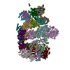 9e8qC  9pdiC  9pdlC  9pdnC  9pf1C M: map data used to model this data C: citing same article ( |
|---|---|
| Similar structure data |


-Links
 PDBj
PDBj









- Assembly
| Deposited unit | 
|
|---|---|
| 1 |
|
-Components
-26S proteasome regulatory subunit ... , 4 types, 4 molecules ABDF
| #1: Protein | Mass: 48700.805 Da / Num. of mol.: 1 / Source method: isolated from a natural source / Source: (natural) Homo sapiens (human) / Cell line: HEK293 / References: UniProt: P35998 |
|---|---|
| #2: Protein | Mass: 49260.504 Da / Num. of mol.: 1 / Source method: isolated from a natural source / Source: (natural) Homo sapiens (human) / Cell line: HEK293 / References: UniProt: P62191 |
| #4: Protein | Mass: 47426.141 Da / Num. of mol.: 1 / Source method: isolated from a natural source / Source: (natural) Homo sapiens (human) / Cell line: HEK293 / References: UniProt: P43686 |
| #6: Protein | Mass: 49266.457 Da / Num. of mol.: 1 / Source method: isolated from a natural source / Source: (natural) Homo sapiens (human) / Cell line: HEK293 / References: UniProt: P17980 |
-26S protease regulatory subunit ... , 2 types, 2 molecules CE
| #3: Protein | Mass: 45694.047 Da / Num. of mol.: 1 / Source method: isolated from a natural source / Source: (natural) Homo sapiens (human) / References: UniProt: P62195 |
|---|---|
| #5: Protein | Mass: 44241.008 Da / Num. of mol.: 1 / Source method: isolated from a natural source / Source: (natural) Homo sapiens (human) / Cell line: HEK293 / References: UniProt: P62333 |
-Proteasome subunit alpha type- ... , 7 types, 7 molecules GHIJKLM
| #7: Protein | Mass: 27432.459 Da / Num. of mol.: 1 / Source method: isolated from a natural source / Source: (natural) Homo sapiens (human) / Cell line: HEK293References: UniProt: P60900, proteasome endopeptidase complex |
|---|---|
| #8: Protein | Mass: 25927.535 Da / Num. of mol.: 1 / Source method: isolated from a natural source / Source: (natural) Homo sapiens (human)References: UniProt: P25787, proteasome endopeptidase complex |
| #9: Protein | Mass: 29525.842 Da / Num. of mol.: 1 / Source method: isolated from a natural source / Source: (natural) Homo sapiens (human) / Cell line: HEK293References: UniProt: P25789, proteasome endopeptidase complex |
| #10: Protein | Mass: 27929.891 Da / Num. of mol.: 1 / Source method: isolated from a natural source / Source: (natural) Homo sapiens (human)References: UniProt: O14818, proteasome endopeptidase complex |
| #11: Protein | Mass: 26435.977 Da / Num. of mol.: 1 / Source method: isolated from a natural source / Source: (natural) Homo sapiens (human) / Cell line: HEK293References: UniProt: P28066, proteasome endopeptidase complex |
| #12: Protein | Mass: 29595.627 Da / Num. of mol.: 1 / Source method: isolated from a natural source / Source: (natural) Homo sapiens (human) / Cell line: HEK293References: UniProt: P25786, proteasome endopeptidase complex |
| #13: Protein | Mass: 28469.252 Da / Num. of mol.: 1 / Source method: isolated from a natural source / Source: (natural) Homo sapiens (human) / Cell line: HEK293References: UniProt: P25788, proteasome endopeptidase complex |
-26S proteasome non-ATPase regulatory subunit ... , 11 types, 11 molecules UVWXYZabcdf
| #14: Protein | Mass: 105958.234 Da / Num. of mol.: 1 / Source method: isolated from a natural source / Source: (natural) Homo sapiens (human) / References: UniProt: Q99460 |
|---|---|
| #15: Protein | Mass: 61066.500 Da / Num. of mol.: 1 / Source method: isolated from a natural source / Source: (natural) Homo sapiens (human) / Cell line: HEK293 / References: UniProt: O43242 |
| #16: Protein | Mass: 52979.359 Da / Num. of mol.: 1 / Source method: isolated from a natural source / Source: (natural) Homo sapiens (human) / Cell line: HEK293 / References: UniProt: O00232 |
| #17: Protein | Mass: 47526.688 Da / Num. of mol.: 1 / Source method: isolated from a natural source / Source: (natural) Homo sapiens (human) / Cell line: HEK293 / References: UniProt: O00231 |
| #18: Protein | Mass: 45592.285 Da / Num. of mol.: 1 / Source method: isolated from a natural source / Source: (natural) Homo sapiens (human) / Cell line: HEK293 / References: UniProt: Q15008 |
| #19: Protein | Mass: 37086.441 Da / Num. of mol.: 1 / Source method: isolated from a natural source / Source: (natural) Homo sapiens (human) / Cell line: HEK293 / References: UniProt: P51665 |
| #20: Protein | Mass: 42995.359 Da / Num. of mol.: 1 / Source method: isolated from a natural source / Source: (natural) Homo sapiens (human) / Cell line: HEK293 / References: UniProt: Q9UNM6 |
| #21: Protein | Mass: 40781.590 Da / Num. of mol.: 1 / Source method: isolated from a natural source / Source: (natural) Homo sapiens (human) / Cell line: HEK293 / References: UniProt: P55036 |
| #22: Protein | Mass: 46940.898 Da / Num. of mol.: 1 Source method: isolated from a genetically manipulated source Source: (gene. exp.) Homo sapiens (human) / Cell line: HEK293 / Gene: PSMD14 / Cell (production host): HEK293 / Production host: Homo sapiens (human) / Strain (production host): PSMD14-httb / References: UniProt: O00487 |
| #23: Protein | Mass: 39667.871 Da / Num. of mol.: 1 / Source method: isolated from a natural source / Source: (natural) Homo sapiens (human) / Cell line: HEK293 / References: UniProt: P48556 |
| #25: Protein | Mass: 100313.625 Da / Num. of mol.: 1 / Source method: isolated from a natural source / Source: (natural) Homo sapiens (human) / Cell line: HEK293 / References: UniProt: Q13200 |
-Protein , 3 types, 3 molecules egu
| #24: Protein | Mass: 7706.940 Da / Num. of mol.: 1 / Source method: isolated from a natural source / Source: (natural) Homo sapiens (human) / References: UniProt: P60896 |
|---|---|
| #26: Protein | Mass: 69218.562 Da / Num. of mol.: 1 Source method: isolated from a genetically manipulated source Source: (gene. exp.) Homo sapiens (human) / Gene: NUB1, NYREN18 / Production host:   Escherichia coli BL21(DE3) (bacteria) / References: UniProt: Q9Y5A7 Escherichia coli BL21(DE3) (bacteria) / References: UniProt: Q9Y5A7 |
| #27: Protein | Mass: 32284.229 Da / Num. of mol.: 1 Source method: isolated from a genetically manipulated source Source: (gene. exp.) Homo sapiens (human) / Gene: TXNL1, TRP32, TXL, TXNL / Production host: Escherichia coli BL21(DE3) (bacteria) / References: UniProt: O43396 |
-Non-polymers , 4 types, 13 molecules 



| #28: Chemical | ChemComp-ATP / 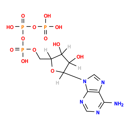 Mass: 507.181 Da / Num. of mol.: 5 / Source method: obtained synthetically / Formula: C10H16N5O13P3 / Feature type: SUBJECT OF INVESTIGATION / Comment: ATP, energy-carrying molecule*YM Mass: 507.181 Da / Num. of mol.: 5 / Source method: obtained synthetically / Formula: C10H16N5O13P3 / Feature type: SUBJECT OF INVESTIGATION / Comment: ATP, energy-carrying molecule*YM#29: Chemical | ChemComp-MG /  Mass: 24.305 Da / Num. of mol.: 6 / Source method: obtained synthetically / Formula: Mg / Feature type: SUBJECT OF INVESTIGATION Mass: 24.305 Da / Num. of mol.: 6 / Source method: obtained synthetically / Formula: Mg / Feature type: SUBJECT OF INVESTIGATION#30: Chemical | ChemComp-ADP / | 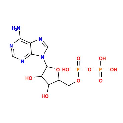 Mass: 427.201 Da / Num. of mol.: 1 / Source method: obtained synthetically / Formula: C10H15N5O10P2 / Feature type: SUBJECT OF INVESTIGATION / Comment: ADP, energy-carrying molecule*YM Mass: 427.201 Da / Num. of mol.: 1 / Source method: obtained synthetically / Formula: C10H15N5O10P2 / Feature type: SUBJECT OF INVESTIGATION / Comment: ADP, energy-carrying molecule*YM#31: Chemical | ChemComp-ZN / |  Mass: 65.409 Da / Num. of mol.: 1 / Source method: obtained synthetically / Formula: Zn / Feature type: SUBJECT OF INVESTIGATION Mass: 65.409 Da / Num. of mol.: 1 / Source method: obtained synthetically / Formula: Zn / Feature type: SUBJECT OF INVESTIGATION |
|---|
-Details
| Has ligand of interest | Y |
|---|---|
| Has protein modification | Y |
-Experimental details
-Experiment
| Experiment | Method: ELECTRON MICROSCOPY |
|---|---|
| EM experiment | Aggregation state: PARTICLE / 3D reconstruction method: single particle reconstruction |
- Sample preparation
| Component | Name: Human 26S proteasome complexed with Nub1, Fat10 and TXNL1 Type: COMPLEX / Entity ID: #1-#27 / Source: MULTIPLE SOURCES |
|---|---|
| Molecular weight | Value: 2.6 MDa / Experimental value: NO |
| Source (natural) | Organism: Homo sapiens (human) |
| Buffer solution | pH: 7.4 Details: 30 mM HEPES pH7.4, 25 mM NaCl, 25 mM KCl, 3% (v/v) glycerol, 5 mM MgCl2 2 mM ATP and 0.5 mM TCEP. |
| Specimen | Embedding applied: NO / Shadowing applied: NO / Staining applied: NO / Vitrification applied: YES |
| Vitrification | Instrument: FEI VITROBOT MARK IV / Cryogen name: ETHANE / Humidity: 100 % / Chamber temperature: 285 K |
- Electron microscopy imaging
| Experimental equipment |  Model: Titan Krios / Image courtesy: FEI Company |
|---|---|
| Microscopy | Model: TFS KRIOS |
| Electron gun | Electron source:  FIELD EMISSION GUN / Accelerating voltage: 300 kV / Illumination mode: FLOOD BEAM FIELD EMISSION GUN / Accelerating voltage: 300 kV / Illumination mode: FLOOD BEAM |
| Electron lens | Mode: BRIGHT FIELD / Nominal defocus max: 1700 nm / Nominal defocus min: 500 nm / Alignment procedure: BASIC |
| Specimen holder | Cryogen: NITROGEN / Specimen holder model: FEI TITAN KRIOS AUTOGRID HOLDER |
| Image recording | Electron dose: 50 e/Å2 / Film or detector model: GATAN K3 (6k x 4k) |
- Processing
| CTF correction | Type: PHASE FLIPPING AND AMPLITUDE CORRECTION |
|---|---|
| 3D reconstruction | Resolution: 2.9 Å / Resolution method: FSC 0.143 CUT-OFF / Num. of particles: 53010 / Symmetry type: POINT |
