[English] 日本語
 Yorodumi
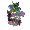
- PDB-9pf1: Nub1/Fat10-processing human 26S proteasome with Rpt4 at top of sp... -
+ Open data
- Basic information
| Entry | Database: PDB / ID: 9pf1 | |||||||||||||||||||||||||||
|---|---|---|---|---|---|---|---|---|---|---|---|---|---|---|---|---|---|---|---|---|---|---|---|---|---|---|---|---|
| Title | Nub1/Fat10-processing human 26S proteasome with Rpt4 at top of spiral staircase (AAA+ motor locally refined) | |||||||||||||||||||||||||||
 Components Components |
| |||||||||||||||||||||||||||
 Keywords Keywords | MOTOR PROTEIN / 26S Proteasome / HYDROLASE-PROTEIN BINDING complex | |||||||||||||||||||||||||||
| Function / homology |  Function and homology information Function and homology informationthyrotropin-releasing hormone receptor binding / nuclear proteasome complex / host-mediated perturbation of viral transcription / positive regulation of inclusion body assembly / Hydrolases; Acting on peptide bonds (peptidases); Omega peptidases / proteasome accessory complex / purine ribonucleoside triphosphate binding / cytosolic proteasome complex / positive regulation of proteasomal protein catabolic process / proteasome-activating activity ...thyrotropin-releasing hormone receptor binding / nuclear proteasome complex / host-mediated perturbation of viral transcription / positive regulation of inclusion body assembly / Hydrolases; Acting on peptide bonds (peptidases); Omega peptidases / proteasome accessory complex / purine ribonucleoside triphosphate binding / cytosolic proteasome complex / positive regulation of proteasomal protein catabolic process / proteasome-activating activity / proteasome regulatory particle, lid subcomplex / proteasome regulatory particle, base subcomplex / protein K63-linked deubiquitination / metal-dependent deubiquitinase activity / negative regulation of programmed cell death / Regulation of ornithine decarboxylase (ODC) / Proteasome assembly / Cross-presentation of soluble exogenous antigens (endosomes) / proteasome core complex / Somitogenesis / K63-linked deubiquitinase activity / proteasome binding / transcription factor binding / myofibril / general transcription initiation factor binding / positive regulation of RNA polymerase II transcription preinitiation complex assembly / protein deubiquitination / blastocyst development / immune system process / NF-kappaB binding / endopeptidase activator activity / proteasome core complex, alpha-subunit complex / SARS-CoV-1 targets host intracellular signalling and regulatory pathways / ERAD pathway / regulation of proteasomal protein catabolic process / inclusion body / proteasome complex / : / TBP-class protein binding / sarcomere / Regulation of activated PAK-2p34 by proteasome mediated degradation / Autodegradation of Cdh1 by Cdh1:APC/C / APC/C:Cdc20 mediated degradation of Securin / N-glycan trimming in the ER and Calnexin/Calreticulin cycle / Asymmetric localization of PCP proteins / SCF-beta-TrCP mediated degradation of Emi1 / NIK-->noncanonical NF-kB signaling / Ubiquitin-dependent degradation of Cyclin D / TNFR2 non-canonical NF-kB pathway / AUF1 (hnRNP D0) binds and destabilizes mRNA / Vpu mediated degradation of CD4 / Assembly of the pre-replicative complex / Ubiquitin-Mediated Degradation of Phosphorylated Cdc25A / negative regulation of inflammatory response to antigenic stimulus / Degradation of DVL / Dectin-1 mediated noncanonical NF-kB signaling / P-body / Cdc20:Phospho-APC/C mediated degradation of Cyclin A / Degradation of AXIN / lipopolysaccharide binding / Hh mutants are degraded by ERAD / Activation of NF-kappaB in B cells / Degradation of GLI1 by the proteasome / G2/M Checkpoints / Hedgehog ligand biogenesis / Defective CFTR causes cystic fibrosis / Autodegradation of the E3 ubiquitin ligase COP1 / Regulation of RUNX3 expression and activity / GSK3B and BTRC:CUL1-mediated-degradation of NFE2L2 / Negative regulation of NOTCH4 signaling / Hedgehog 'on' state / Vif-mediated degradation of APOBEC3G / APC/C:Cdh1 mediated degradation of Cdc20 and other APC/C:Cdh1 targeted proteins in late mitosis/early G1 / FBXL7 down-regulates AURKA during mitotic entry and in early mitosis / double-strand break repair via homologous recombination / Degradation of GLI2 by the proteasome / GLI3 is processed to GLI3R by the proteasome / MAPK6/MAPK4 signaling / Degradation of beta-catenin by the destruction complex / Oxygen-dependent proline hydroxylation of Hypoxia-inducible Factor Alpha / double-strand break repair via nonhomologous end joining / ABC-family proteins mediated transport / CDK-mediated phosphorylation and removal of Cdc6 / CLEC7A (Dectin-1) signaling / SCF(Skp2)-mediated degradation of p27/p21 / response to virus / FCERI mediated NF-kB activation / nuclear matrix / Regulation of expression of SLITs and ROBOs / Metalloprotease DUBs / Regulation of PTEN stability and activity / Interleukin-1 signaling / cytoplasmic ribonucleoprotein granule / Orc1 removal from chromatin / metallopeptidase activity / Regulation of RAS by GAPs / Regulation of RUNX2 expression and activity / osteoblast differentiation / The role of GTSE1 in G2/M progression after G2 checkpoint / Separation of Sister Chromatids Similarity search - Function | |||||||||||||||||||||||||||
| Biological species |  Homo sapiens (human) Homo sapiens (human) | |||||||||||||||||||||||||||
| Method | ELECTRON MICROSCOPY / single particle reconstruction / cryo EM / Resolution: 3.57 Å | |||||||||||||||||||||||||||
 Authors Authors | Arkinson, C. / Gee, C.L. / Martin, A. | |||||||||||||||||||||||||||
| Funding support |  United States, 1items United States, 1items
| |||||||||||||||||||||||||||
 Citation Citation |  Journal: Nat Struct Mol Biol / Year: 2025 Journal: Nat Struct Mol Biol / Year: 2025Title: Structural landscape of the degrading 26S proteasome reveals conformation-specific binding of TXNL1. Authors: Connor Arkinson / Christine L Gee / Zeyuan Zhang / Ken C Dong / Andreas Martin /  Abstract: The 26S proteasome targets many cellular proteins for degradation during homeostasis and quality control. Proteasome-interacting cofactors modulate these functions and aid in substrate degradation. ...The 26S proteasome targets many cellular proteins for degradation during homeostasis and quality control. Proteasome-interacting cofactors modulate these functions and aid in substrate degradation. Here we solve high-resolution structures of the redox active cofactor TXNL1 bound to the human 26S proteasome at saturating and substoichiometric concentrations by time-resolved cryo-electron microscopy (cryo-EM). We identify distinct binding modes of TXNL1 that depend on the proteasome conformation and ATPase motor states. Together with biophysical and biochemical experiments, we show that the resting-state proteasome binds TXNL1 with low affinity and in variable positions on top of the Rpn11 deubiquitinase. In contrast, in the actively degrading proteasome, TXNL1 uses additional interactions for high-affinity binding, whereby its C-terminal tail covers the catalytic groove of Rpn11 and coordinates the active-site Zn. Furthermore, these cryo-EM structures of the degrading proteasome capture the ATPase hexamer in several spiral-staircase arrangements that indicate temporally asymmetric hydrolysis and conformational changes in bursts during mechanical substrate unfolding and translocation. Remarkably, we catch the proteasome in the act of unfolding the β-barrel mEos3.2 substrate while the ATPase hexamer is in a particular staircase register. Our findings advance current models for protein translocation through hexameric AAA+ motors and reveal how the proteasome uses its distinct conformational states to coordinate cofactor binding and substrate processing. | |||||||||||||||||||||||||||
| History |
|
- Structure visualization
| Structure viewer | Molecule:  Molmil Molmil Jmol/JSmol Jmol/JSmol |
|---|
- Downloads & links
- Download
| PDBx/mmCIF format |  9pf1.cif.gz 9pf1.cif.gz | 749.2 KB | Display |  PDBx/mmCIF format PDBx/mmCIF format |
|---|---|---|---|---|
| PDB format |  pdb9pf1.ent.gz pdb9pf1.ent.gz | Display |  PDB format PDB format | |
| PDBx/mmJSON format |  9pf1.json.gz 9pf1.json.gz | Tree view |  PDBx/mmJSON format PDBx/mmJSON format | |
| Others |  Other downloads Other downloads |
-Validation report
| Summary document |  9pf1_validation.pdf.gz 9pf1_validation.pdf.gz | 2.3 MB | Display |  wwPDB validaton report wwPDB validaton report |
|---|---|---|---|---|
| Full document |  9pf1_full_validation.pdf.gz 9pf1_full_validation.pdf.gz | 2.4 MB | Display | |
| Data in XML |  9pf1_validation.xml.gz 9pf1_validation.xml.gz | 130.4 KB | Display | |
| Data in CIF |  9pf1_validation.cif.gz 9pf1_validation.cif.gz | 199.7 KB | Display | |
| Arichive directory |  https://data.pdbj.org/pub/pdb/validation_reports/pf/9pf1 https://data.pdbj.org/pub/pdb/validation_reports/pf/9pf1 ftp://data.pdbj.org/pub/pdb/validation_reports/pf/9pf1 ftp://data.pdbj.org/pub/pdb/validation_reports/pf/9pf1 | HTTPS FTP |
-Related structure data
| Related structure data |  71584MC  9e8gC  9e8hC  9e8iC  9e8jC  9e8kC  9e8lC  9e8nC  9e8oC  9e8qC  9pdiC  9pdlC  9pdnC M: map data used to model this data C: citing same article ( |
|---|---|
| Similar structure data | Similarity search - Function & homology  F&H Search F&H Search |
- Links
Links
- Assembly
| Deposited unit | 
|
|---|---|
| 1 |
|
- Components
-26S proteasome regulatory subunit ... , 4 types, 4 molecules ABDF
| #1: Protein | Mass: 48700.805 Da / Num. of mol.: 1 Source method: isolated from a genetically manipulated source Source: (gene. exp.)  Homo sapiens (human) / Gene: PSMC2, MSS1 / Cell line (production host): HEK293 / Production host: Homo sapiens (human) / Gene: PSMC2, MSS1 / Cell line (production host): HEK293 / Production host:  Homo sapiens (human) / References: UniProt: P35998 Homo sapiens (human) / References: UniProt: P35998 |
|---|---|
| #11: Protein | Mass: 49260.504 Da / Num. of mol.: 1 Source method: isolated from a genetically manipulated source Source: (gene. exp.)  Homo sapiens (human) / Gene: PSMC1 / Cell line (production host): HEK293 / Production host: Homo sapiens (human) / Gene: PSMC1 / Cell line (production host): HEK293 / Production host:  Homo sapiens (human) / References: UniProt: P62191 Homo sapiens (human) / References: UniProt: P62191 |
| #12: Protein | Mass: 47368.105 Da / Num. of mol.: 1 Source method: isolated from a genetically manipulated source Source: (gene. exp.)  Homo sapiens (human) / Gene: PSMC4, MIP224, TBP7 / Cell line (production host): HEK293 / Production host: Homo sapiens (human) / Gene: PSMC4, MIP224, TBP7 / Cell line (production host): HEK293 / Production host:  Homo sapiens (human) / References: UniProt: P43686 Homo sapiens (human) / References: UniProt: P43686 |
| #14: Protein | Mass: 49266.457 Da / Num. of mol.: 1 Source method: isolated from a genetically manipulated source Source: (gene. exp.)  Homo sapiens (human) / Gene: PSMC3, TBP1 / Cell line (production host): HEK293 / Production host: Homo sapiens (human) / Gene: PSMC3, TBP1 / Cell line (production host): HEK293 / Production host:  Homo sapiens (human) / References: UniProt: P17980 Homo sapiens (human) / References: UniProt: P17980 |
-26S protease regulatory subunit ... , 2 types, 2 molecules CE
| #2: Protein | Mass: 45694.047 Da / Num. of mol.: 1 Source method: isolated from a genetically manipulated source Source: (gene. exp.)  Homo sapiens (human) / Gene: PSMC5, SUG1 / Cell line (production host): HEK293 / Production host: Homo sapiens (human) / Gene: PSMC5, SUG1 / Cell line (production host): HEK293 / Production host:  Homo sapiens (human) / References: UniProt: P62195 Homo sapiens (human) / References: UniProt: P62195 |
|---|---|
| #13: Protein | Mass: 44241.008 Da / Num. of mol.: 1 Source method: isolated from a genetically manipulated source Source: (gene. exp.)  Homo sapiens (human) / Gene: PSMC6, SUG2 / Cell line (production host): HEK293 / Production host: Homo sapiens (human) / Gene: PSMC6, SUG2 / Cell line (production host): HEK293 / Production host:  Homo sapiens (human) / References: UniProt: P62333 Homo sapiens (human) / References: UniProt: P62333 |
-Proteasome subunit alpha type- ... , 7 types, 7 molecules GHIJLMK
| #3: Protein | Mass: 27432.459 Da / Num. of mol.: 1 Source method: isolated from a genetically manipulated source Source: (gene. exp.)  Homo sapiens (human) / Gene: PSMA6, PROS27 / Cell line (production host): HEK293 / Production host: Homo sapiens (human) / Gene: PSMA6, PROS27 / Cell line (production host): HEK293 / Production host:  Homo sapiens (human) Homo sapiens (human)References: UniProt: P60900, proteasome endopeptidase complex |
|---|---|
| #4: Protein | Mass: 25934.486 Da / Num. of mol.: 1 Source method: isolated from a genetically manipulated source Source: (gene. exp.)  Homo sapiens (human) / Gene: PSMA2, HC3, PSC3 / Cell line (production host): HEK293 / Production host: Homo sapiens (human) / Gene: PSMA2, HC3, PSC3 / Cell line (production host): HEK293 / Production host:  Homo sapiens (human) / References: UniProt: P25787 Homo sapiens (human) / References: UniProt: P25787 |
| #5: Protein | Mass: 29525.842 Da / Num. of mol.: 1 Source method: isolated from a genetically manipulated source Source: (gene. exp.)  Homo sapiens (human) / Gene: PSMA4, HC9, PSC9 / Cell line (production host): HEK293 / Production host: Homo sapiens (human) / Gene: PSMA4, HC9, PSC9 / Cell line (production host): HEK293 / Production host:  Homo sapiens (human) Homo sapiens (human)References: UniProt: P25789, proteasome endopeptidase complex |
| #6: Protein | Mass: 27929.891 Da / Num. of mol.: 1 Source method: isolated from a genetically manipulated source Source: (gene. exp.)  Homo sapiens (human) / Gene: PSMA7, HSPC / Cell line (production host): HEK293 / Production host: Homo sapiens (human) / Gene: PSMA7, HSPC / Cell line (production host): HEK293 / Production host:  Homo sapiens (human) / References: UniProt: O14818 Homo sapiens (human) / References: UniProt: O14818 |
| #7: Protein | Mass: 29595.627 Da / Num. of mol.: 1 Source method: isolated from a genetically manipulated source Source: (gene. exp.)  Homo sapiens (human) / Gene: PSMA1, HC2, NU, PROS30, PSC2 / Cell line (production host): HEK293 / Production host: Homo sapiens (human) / Gene: PSMA1, HC2, NU, PROS30, PSC2 / Cell line (production host): HEK293 / Production host:  Homo sapiens (human) Homo sapiens (human)References: UniProt: P25786, proteasome endopeptidase complex |
| #8: Protein | Mass: 28469.252 Da / Num. of mol.: 1 Source method: isolated from a genetically manipulated source Source: (gene. exp.)  Homo sapiens (human) / Gene: PSMA3, HC8, PSC8 / Cell line (production host): HEK293 / Production host: Homo sapiens (human) / Gene: PSMA3, HC8, PSC8 / Cell line (production host): HEK293 / Production host:  Homo sapiens (human) Homo sapiens (human)References: UniProt: P25788, proteasome endopeptidase complex |
| #15: Protein | Mass: 26435.977 Da / Num. of mol.: 1 Source method: isolated from a genetically manipulated source Source: (gene. exp.)  Homo sapiens (human) / Gene: PSMA5 / Cell line (production host): HEK293 / Production host: Homo sapiens (human) / Gene: PSMA5 / Cell line (production host): HEK293 / Production host:  Homo sapiens (human) / References: UniProt: P28066 Homo sapiens (human) / References: UniProt: P28066 |
-Protein / Protein/peptide , 2 types, 2 molecules cv
| #10: Protein/peptide | Mass: 1039.273 Da / Num. of mol.: 1 Source method: isolated from a genetically manipulated source Source: (gene. exp.)  Homo sapiens (human) / Production host: Homo sapiens (human) / Production host:  |
|---|---|
| #9: Protein | Mass: 46940.898 Da / Num. of mol.: 1 Source method: isolated from a genetically manipulated source Source: (gene. exp.)  Homo sapiens (human) / Gene: PSMD14 / Cell line (production host): HEK293 / Production host: Homo sapiens (human) / Gene: PSMD14 / Cell line (production host): HEK293 / Production host:  Homo sapiens (human) / References: UniProt: O00487 Homo sapiens (human) / References: UniProt: O00487 |
-Non-polymers , 4 types, 9 molecules 


| #16: Chemical | ChemComp-ADP / #17: Chemical | ChemComp-ZN / | #18: Chemical | #19: Chemical | |
|---|
-Details
| Has ligand of interest | Y |
|---|---|
| Has protein modification | Y |
-Experimental details
-Experiment
| Experiment | Method: ELECTRON MICROSCOPY |
|---|---|
| EM experiment | Aggregation state: PARTICLE / 3D reconstruction method: single particle reconstruction |
- Sample preparation
| Component | Name: Human 26S proteasome complexed with Nub1 and Fat 10 with RPT4 at the top Type: COMPLEX / Entity ID: #1-#15 / Source: MULTIPLE SOURCES |
|---|---|
| Molecular weight | Value: 2.6 MDa / Experimental value: NO |
| Source (natural) | Organism:  Homo sapiens (human) Homo sapiens (human) |
| Source (recombinant) | Organism:  Homo sapiens (human) / Cell: HEK293 Homo sapiens (human) / Cell: HEK293 |
| Buffer solution | pH: 7.4 Details: 30 mM HEPES pH7.4, 25 mM NaCl, 25 mM KCl, 3% (v/v) glycerol, 5 mM MgCl2 2 mM ATP and 0.5 mM TCEP |
| Specimen | Embedding applied: NO / Shadowing applied: NO / Staining applied: NO / Vitrification applied: YES |
| Specimen support | Grid material: GOLD / Grid mesh size: 200 divisions/in. / Grid type: UltrAuFoil R2/2 |
| Vitrification | Instrument: FEI VITROBOT MARK IV / Cryogen name: ETHANE / Humidity: 100 % / Chamber temperature: 298 K |
- Electron microscopy imaging
| Experimental equipment |  Model: Titan Krios / Image courtesy: FEI Company |
|---|---|
| Microscopy | Model: TFS KRIOS |
| Electron gun | Electron source:  FIELD EMISSION GUN / Accelerating voltage: 300 kV / Illumination mode: FLOOD BEAM FIELD EMISSION GUN / Accelerating voltage: 300 kV / Illumination mode: FLOOD BEAM |
| Electron lens | Mode: BRIGHT FIELD / Nominal defocus max: 1700 nm / Nominal defocus min: 500 nm / Alignment procedure: BASIC |
| Specimen holder | Cryogen: NITROGEN / Specimen holder model: FEI TITAN KRIOS AUTOGRID HOLDER |
| Image recording | Electron dose: 50 e/Å2 / Film or detector model: GATAN K3 (6k x 4k) |
- Processing
| EM software |
| ||||||||||||||||
|---|---|---|---|---|---|---|---|---|---|---|---|---|---|---|---|---|---|
| CTF correction | Type: PHASE FLIPPING AND AMPLITUDE CORRECTION | ||||||||||||||||
| 3D reconstruction | Resolution: 3.57 Å / Resolution method: FSC 0.143 CUT-OFF / Num. of particles: 16398 / Symmetry type: POINT |
 Movie
Movie Controller


 PDBj
PDBj


