 Movie
Movie Controller

[English] 日本語
 Yorodumi
- EMDB-71581: Nub1/Fat10-processing human 26S proteasome bound to TXNL1 with Rp... -
+ Open data

- Basic information
| Entry |  | |||||||||
|---|---|---|---|---|---|---|---|---|---|---|
| Title | Nub1/Fat10-processing human 26S proteasome bound to TXNL1 with Rpt3 at top of spiral staircase | |||||||||
 Map data Map data | ||||||||||
 Sample Sample |
| |||||||||
 Keywords Keywords | 26S Proteasome / MOTOR PROTEIN / HYDROLASE-PROTEIN BINDING complex | |||||||||
| Biological species |  Homo sapiens (human) Homo sapiens (human) | |||||||||
| Method | single particle reconstruction / cryo EM / Resolution: 3.49 Å | |||||||||
 Authors Authors | Arkinson C / Gee CL / Martin A | |||||||||
| Funding support |  United States, 1 items United States, 1 items
| |||||||||
 Citation Citation | Journal: Nat Struct Mol Biol / Year: 2025 Title: Structural landscape of the degrading 26S proteasome reveals conformation-specific binding of TXNL1. Authors: Connor Arkinson / Christine L Gee / Zeyuan Zhang / Ken C Dong / Andreas Martin / Abstract: The 26S proteasome targets many cellular proteins for degradation during homeostasis and quality control. Proteasome-interacting cofactors modulate these functions and aid in substrate degradation. ...The 26S proteasome targets many cellular proteins for degradation during homeostasis and quality control. Proteasome-interacting cofactors modulate these functions and aid in substrate degradation. Here we solve high-resolution structures of the redox active cofactor TXNL1 bound to the human 26S proteasome at saturating and substoichiometric concentrations by time-resolved cryo-electron microscopy (cryo-EM). We identify distinct binding modes of TXNL1 that depend on the proteasome conformation and ATPase motor states. Together with biophysical and biochemical experiments, we show that the resting-state proteasome binds TXNL1 with low affinity and in variable positions on top of the Rpn11 deubiquitinase. In contrast, in the actively degrading proteasome, TXNL1 uses additional interactions for high-affinity binding, whereby its C-terminal tail covers the catalytic groove of Rpn11 and coordinates the active-site Zn. Furthermore, these cryo-EM structures of the degrading proteasome capture the ATPase hexamer in several spiral-staircase arrangements that indicate temporally asymmetric hydrolysis and conformational changes in bursts during mechanical substrate unfolding and translocation. Remarkably, we catch the proteasome in the act of unfolding the β-barrel mEos3.2 substrate while the ATPase hexamer is in a particular staircase register. Our findings advance current models for protein translocation through hexameric AAA+ motors and reveal how the proteasome uses its distinct conformational states to coordinate cofactor binding and substrate processing. | |||||||||
| History |
|
- Structure visualization
| Supplemental images |
|---|

- Downloads & links
-EMDB archive
| Map data | emd_71581.map.gz | 75.3 MB |  EMDB map data format EMDB map data format | |
|---|---|---|---|---|
| Header (meta data) | emd-71581-v30.xmlemd-71581.xml | 17.3 KB 17.3 KB | Display Display | EMDB header |
| FSC (resolution estimation) | emd_71581_fsc.xml | 11.2 KB | Display | FSC data file |
| Images |  emd_71581.png emd_71581.png | 60.3 KB | ||
| Filedesc metadata | emd-71581.cif.gz | 4.3 KB | ||
| Others | emd_71581_additional_1.map.gzemd_71581_additional_2.map.gzemd_71581_half_map_1.map.gzemd_71581_half_map_2.map.gz | 134.5 MB 141.8 MB 138.9 MB 138.9 MB | ||
| Archive directory |  http://ftp.pdbj.org/pub/emdb/structures/EMD-71581ftp://ftp.pdbj.org/pub/emdb/structures/EMD-71581 http://ftp.pdbj.org/pub/emdb/structures/EMD-71581ftp://ftp.pdbj.org/pub/emdb/structures/EMD-71581 | HTTPS FTP |
-Related structure data
| Related structure data |  9e8gC  9e8hC  9e8iC  9e8jC  9e8kC  9e8lC  9e8nC  9e8oC 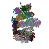 9e8qC  9pdiC  9pdlC  9pdnC  9pf1C C: citing same article ( |
|---|


-Links
| EMDB pages | EMDB (EBI/PDBe) / EMDataResource |
|---|
-Map
| File | Download / File: emd_71581.map.gz / Format: CCP4 / Size: 149.9 MB / Type: IMAGE STORED AS FLOATING POINT NUMBER (4 BYTES) | ||||||||||||||||||||||||||||||||||||
|---|---|---|---|---|---|---|---|---|---|---|---|---|---|---|---|---|---|---|---|---|---|---|---|---|---|---|---|---|---|---|---|---|---|---|---|---|---|
| Projections & slices | Image control
Images are generated by Spider. | ||||||||||||||||||||||||||||||||||||
| Voxel size | X=Y=Z: 1.048 Å | ||||||||||||||||||||||||||||||||||||
| Density |
| ||||||||||||||||||||||||||||||||||||
| Symmetry | Space group: 1 | ||||||||||||||||||||||||||||||||||||
| Details | EMDB XML:
|
 Z (Sec.)
 Y (Row.)
 X (Col.)


-Supplemental data
-Additional map: Enhanced Map.
| File | emd_71581_additional_1.map | ||||||||||||
|---|---|---|---|---|---|---|---|---|---|---|---|---|---|
| Annotation | Enhanced Map. | ||||||||||||
| Projections & Slices |
| ||||||||||||
| Density Histograms |


-Additional map: globally Sharpened
| File | emd_71581_additional_2.map | ||||||||||||
|---|---|---|---|---|---|---|---|---|---|---|---|---|---|
| Annotation | globally Sharpened | ||||||||||||
| Projections & Slices |
| ||||||||||||
| Density Histograms |


-Half map: #1
| File | emd_71581_half_map_1.map | ||||||||||||
|---|---|---|---|---|---|---|---|---|---|---|---|---|---|
| Projections & Slices |
| ||||||||||||
| Density Histograms |


-Half map: #2
| File | emd_71581_half_map_2.map | ||||||||||||
|---|---|---|---|---|---|---|---|---|---|---|---|---|---|
| Projections & Slices |
| ||||||||||||
| Density Histograms |


- Sample components
-Entire : Human 26S proteasome complexed with Nub1 and Fat 10 with RPT3 at ...
| Entire | Name: Human 26S proteasome complexed with Nub1 and Fat 10 with RPT3 at the top and TXNL1 bound |
|---|---|
| Components |
|
-Supramolecule #1: Human 26S proteasome complexed with Nub1 and Fat 10 with RPT3 at ...
| Supramolecule | Name: Human 26S proteasome complexed with Nub1 and Fat 10 with RPT3 at the top and TXNL1 bound type: complex / ID: 1 / Parent: 0 |
|---|---|
| Source (natural) | Organism: Homo sapiens (human) / Strain: HEK293 |
| Molecular weight | Theoretical: 2.6 MDa |
-Experimental details
-Structure determination
| Method | cryo EM |
|---|---|
 Processing Processing | single particle reconstruction |
| Aggregation state | particle |
-Sample preparation
| Buffer | pH: 7.4 Details: 30 mM HEPES pH7.4, 25 mM NaCl, 25 mM KCl, 3% (v/v) glycerol, 5 mM MgCl2 2 mM ATP and 0.5 mM TCEP |
|---|---|
| Grid | Model: UltrAuFoil R2/2 / Material: GOLD / Mesh: 200 / Support film - Material: GOLD |
| Vitrification | Cryogen name: ETHANE / Chamber humidity: 100 % / Chamber temperature: 298 K / Instrument: FEI VITROBOT MARK IV |
- Electron microscopy
| Microscope | TFS KRIOS |
|---|---|
| Image recording | Film or detector model: GATAN K3 (6k x 4k) / Average electron dose: 50.0 e/Å2 |
| Electron beam | Acceleration voltage: 300 kV / Electron source:  FIELD EMISSION GUN FIELD EMISSION GUN |
| Electron optics | Illumination mode: FLOOD BEAM / Imaging mode: BRIGHT FIELD / Nominal defocus max: 1.7 µm / Nominal defocus min: 0.5 µm |
| Sample stage | Specimen holder model: FEI TITAN KRIOS AUTOGRID HOLDER / Cooling holder cryogen: NITROGEN |
| Experimental equipment |  Model: Titan Krios / Image courtesy: FEI Company |

