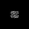
 ムービー
ムービー コントローラー


+ データを開く

- 基本情報
基本情報
| 登録情報 | データベース: PDB / ID: 8ems | ||||||
|---|---|---|---|---|---|---|---|
| タイトル | Cryo-EM structure of human liver glycogen phosphorylase | ||||||
 要素 要素 | Glycogen phosphorylase, liver form | ||||||
 キーワード キーワード | HYDROLASE / Glycogen Phosphorylase / PLP | ||||||
| 機能・相同性 |  機能・相同性情報 機能・相同性情報purine nucleobase binding / vitamin binding / D-glucose binding / bile acid binding / glycogen phosphorylase / glycogen phosphorylase activity / glycogen catabolic process / glycogen metabolic process / Glycogen breakdown (glycogenolysis) / AMP binding ...purine nucleobase binding / vitamin binding / D-glucose binding / bile acid binding / glycogen phosphorylase / glycogen phosphorylase activity / glycogen catabolic process / glycogen metabolic process / Glycogen breakdown (glycogenolysis) / AMP binding / necroptotic process / response to bacterium / pyridoxal phosphate binding / glucose homeostasis / secretory granule lumen / ficolin-1-rich granule lumen / Neutrophil degranulation / extracellular exosome / extracellular region / ATP binding / identical protein binding / cytoplasm / cytosol 類似検索 - 分子機能 | ||||||
| 生物種 |  Homo sapiens (ヒト) Homo sapiens (ヒト) | ||||||
| 手法 | 電子顕微鏡法 / 単粒子再構成法 / クライオ電子顕微鏡法 / 解像度: 2.65 Å | ||||||
 データ登録者 データ登録者 | Su, C. / Lyu, M. / Zhang, Z. / Yu, E.W. | ||||||
| 資金援助 |  米国, 1件 米国, 1件
| ||||||
 引用 引用 | ジャーナル: Cell Rep / 年: 2023 タイトル: High-resolution structural-omics of human liver enzymes. 著者: Chih-Chia Su / Meinan Lyu / Zhemin Zhang / Masaru Miyagi / Wei Huang / Derek J Taylor / Edward W Yu / 要旨: We applied raw human liver microsome lysate to a holey carbon grid and used cryo-electron microscopy (cryo-EM) to define its composition. From this sample we identified and simultaneously determined ...We applied raw human liver microsome lysate to a holey carbon grid and used cryo-electron microscopy (cryo-EM) to define its composition. From this sample we identified and simultaneously determined high-resolution structural information for ten unique human liver enzymes involved in diverse cellular processes. Notably, we determined the structure of the endoplasmic bifunctional protein H6PD, where the N- and C-terminal domains independently possess glucose-6-phosphate dehydrogenase and 6-phosphogluconolactonase enzymatic activity, respectively. We also obtained the structure of heterodimeric human GANAB, an ER glycoprotein quality-control machinery that contains a catalytic α subunit and a noncatalytic β subunit. In addition, we observed a decameric peroxidase, PRDX4, which directly contacts a disulfide isomerase-related protein, ERp46. Structural data suggest that several glycosylations, bound endogenous compounds, and ions associate with these human liver enzymes. These results highlight the importance of cryo-EM in facilitating the elucidation of human organ proteomics at the atomic level. | ||||||
| 履歴 |
|
- 構造の表示
構造の表示
| 構造ビューア | 分子: MolmilJmol/JSmol |
|---|
- ダウンロードとリンク
-ダウンロード
| PDBx/mmCIF形式 | 8ems.cif.gz | 312 KB | 表示 | PDBx/mmCIF形式 |
|---|---|---|---|---|
| PDB形式 | pdb8ems.ent.gz | 251.9 KB | 表示 | PDB形式 |
| PDBx/mmJSON形式 | 8ems.json.gz | ツリー表示 | PDBx/mmJSON形式 | |
| その他 |  その他のダウンロード その他のダウンロード |
-検証レポート
| アーカイブディレクトリ | https://data.pdbj.org/pub/pdb/validation_reports/em/8emsftp://data.pdbj.org/pub/pdb/validation_reports/em/8ems | HTTPS FTP |
|---|
-関連構造データ
| 関連構造データ |  28263MC 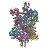 7uzmC  8ekwC  8ekyC  8em2C  8emrC 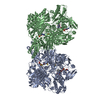 8emtC 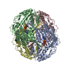 8eneC  8eojC  8eorC  23433 M: このデータのモデリングに利用したマップデータ C: 同じ文献を引用 ( |
|---|---|
| 類似構造データ | |
| 実験データセット #1 | データ参照: 10.6019/EMPIAR-11250 / データの種類: EMPIAR |


-リンク
 PDBj
PDBj


- 集合体
集合体
| 登録構造単位 | 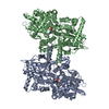
|
|---|---|
| 1 |
|
-要素
| #1: タンパク質 | 分子量: 95376.406 Da / 分子数: 2 / 由来タイプ: 天然 / 由来: (天然) Homo sapiens (ヒト) / 器官: liver / 参照: UniProt: P06737, glycogen phosphorylase#2: 化合物 |   分子量: 247.142 Da / 分子数: 2 / 由来タイプ: 天然 / 式: C8H10NO6P / タイプ: SUBJECT OF INVESTIGATION 分子量: 247.142 Da / 分子数: 2 / 由来タイプ: 天然 / 式: C8H10NO6P / タイプ: SUBJECT OF INVESTIGATION研究の焦点であるリガンドがあるか | Y | |
|---|
-実験情報
-実験
| 実験 | 手法: 電子顕微鏡法 |
|---|---|
| EM実験 | 試料の集合状態: PARTICLE / 3次元再構成法: 単粒子再構成法 |
- 試料調製
試料調製
| 構成要素 | 名称: Glycogen Phosphorylase / タイプ: COMPLEX / Entity ID: #1 / 由来: NATURAL |
|---|---|
| 由来(天然) | 生物種: Homo sapiens (ヒト) |
| 緩衝液 | pH: 7.5 |
| 試料 | 包埋: NO / シャドウイング: NO / 染色: NO / 凍結: YES |
| 急速凍結 | 凍結剤: ETHANE |
- 電子顕微鏡撮影
| 実験機器 |  モデル: Titan Krios / 画像提供: FEI Company |
|---|---|
| 顕微鏡 | モデル: FEI TITAN KRIOS |
| 電子銃 | 電子線源:  FIELD EMISSION GUN / 加速電圧: 300 kV / 照射モード: FLOOD BEAM FIELD EMISSION GUN / 加速電圧: 300 kV / 照射モード: FLOOD BEAM |
| 電子レンズ | モード: BRIGHT FIELD / 最大 デフォーカス(公称値): 2500 nm / 最小 デフォーカス(公称値): 1000 nm |
| 撮影 | 電子線照射量: 41.25 e/Å2 フィルム・検出器のモデル: GATAN K3 BIOQUANTUM (6k x 4k) |
- 解析
解析
| ソフトウェア | 名称: PHENIX / バージョン: 1.20.1_4487: / 分類: 精密化 | ||||||||||||||||||||||||
|---|---|---|---|---|---|---|---|---|---|---|---|---|---|---|---|---|---|---|---|---|---|---|---|---|---|
| CTF補正 | タイプ: PHASE FLIPPING AND AMPLITUDE CORRECTION | ||||||||||||||||||||||||
| 3次元再構成 | 解像度: 2.65 Å / 解像度の算出法: FSC 0.143 CUT-OFF / 粒子像の数: 764806 / 対称性のタイプ: POINT | ||||||||||||||||||||||||
| 拘束条件 |
|
