 Movie
Movie Controller

[English] 日本語
 Yorodumi
- PDB-7qid: tentative model of the human insulin receptor ectodomain bound by... -
+ Open data

- Basic information
| Entry | Database: PDB / ID: 7qid | |||||||||||||||
|---|---|---|---|---|---|---|---|---|---|---|---|---|---|---|---|---|
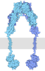
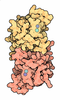
| Title | tentative model of the human insulin receptor ectodomain bound by three insulin | |||||||||||||||
 Components Components |
| |||||||||||||||
 Keywords Keywords | MEMBRANE PROTEIN / cell surface receptor / insulin / tyrosine kinase receptor / glucose homeostasis / hormone / diabetes | |||||||||||||||
| Function / homology |  Function and homology information Function and homology informationregulation of female gonad development / positive regulation of meiotic cell cycle / insulin-like growth factor II binding / positive regulation of developmental growth / insulin receptor complex / insulin-like growth factor I binding / insulin receptor activity / positive regulation of protein-containing complex disassembly / adrenal gland development / dendritic spine maintenance ...regulation of female gonad development / positive regulation of meiotic cell cycle / insulin-like growth factor II binding / positive regulation of developmental growth / insulin receptor complex / insulin-like growth factor I binding / insulin receptor activity / positive regulation of protein-containing complex disassembly / adrenal gland development / dendritic spine maintenance / insulin binding / : / cargo receptor activity / negative regulation of glycogen catabolic process / negative regulation of fatty acid metabolic process / Signaling by Insulin receptor / PTB domain binding / IRS activation / Insulin processing / regulation of protein secretion / negative regulation of feeding behavior / positive regulation of peptide hormone secretion / negative regulation of acute inflammatory response / Regulation of gene expression in beta cells / positive regulation of respiratory burst / neuronal cell body membrane / alpha-beta T cell activation / amyloid-beta clearance / heart morphogenesis / positive regulation of receptor internalization / insulin receptor substrate binding / Synthesis, secretion, and deacylation of Ghrelin / negative regulation of gluconeogenesis / negative regulation of protein secretion / positive regulation of dendritic spine maintenance / fatty acid homeostasis / positive regulation of brown fat cell differentiation / positive regulation of glycogen biosynthetic process / positive regulation of insulin receptor signaling pathway / Signal attenuation / FOXO-mediated transcription of oxidative stress, metabolic and neuronal genes / positive regulation of lipid biosynthetic process / negative regulation of respiratory burst involved in inflammatory response / protein kinase activator activity / negative regulation of lipid catabolic process / negative regulation of oxidative stress-induced intrinsic apoptotic signaling pathway / nitric oxide-cGMP-mediated signaling / regulation of protein localization to plasma membrane / transport vesicle / phosphatidylinositol 3-kinase binding / transport across blood-brain barrier / Insulin receptor recycling / COPI-mediated anterograde transport / positive regulation of nitric-oxide synthase activity / negative regulation of reactive oxygen species biosynthetic process / insulin-like growth factor receptor binding / NPAS4 regulates expression of target genes / positive regulation of D-glucose import across plasma membrane / positive regulation of mitotic nuclear division / neuron projection maintenance / male gonad development / endoplasmic reticulum-Golgi intermediate compartment membrane / receptor-mediated endocytosis / positive regulation of glycolytic process / learning / Insulin receptor signalling cascade / regulation of embryonic development / dendrite membrane / endosome lumen / positive regulation of protein secretion / acute-phase response / positive regulation of cytokine production / wound healing / insulin receptor binding / positive regulation of long-term synaptic potentiation / positive regulation of neuron projection development / glucose metabolic process / positive regulation of cell differentiation / negative regulation of protein catabolic process / Regulation of insulin secretion / memory / regulation of synaptic plasticity / receptor protein-tyrosine kinase / hormone activity / positive regulation of protein localization to nucleus / vasodilation / caveola / Golgi lumen / cognition / insulin receptor signaling pathway / positive regulation of nitric oxide biosynthetic process / glucose homeostasis / cellular response to insulin stimulus / late endosome / regulation of protein localization / cell-cell signaling / protein autophosphorylation / positive regulation of cell growth / amyloid-beta binding / PI5P, PP2A and IER3 Regulate PI3K/AKT Signaling Similarity search - Function | |||||||||||||||
| Biological species |  Homo sapiens (human) Homo sapiens (human) | |||||||||||||||
| Method | ELECTRON MICROSCOPY / single particle reconstruction / cryo EM / Resolution: 5 Å | |||||||||||||||
 Authors Authors | Gutman, T. / Schaefer, I.B. / Poojari, C.S. / Vattulainen, I. / Strauss, M. / Coskun, U. | |||||||||||||||
| Funding support | European Union, 4items
| |||||||||||||||
 Citation Citation | Journal: J Cell Biol / Year: 2020 Title: Cryo-EM structure of the complete and ligand-saturated insulin receptor ectodomain. Authors: Theresia Gutmann / Ingmar B Schäfer / Chetan Poojari / Beate Brankatschk / Ilpo Vattulainen / Mike Strauss / Ünal Coskun /    Abstract: Glucose homeostasis and growth essentially depend on the hormone insulin engaging its receptor. Despite biochemical and structural advances, a fundamental contradiction has persisted in the current ...Glucose homeostasis and growth essentially depend on the hormone insulin engaging its receptor. Despite biochemical and structural advances, a fundamental contradiction has persisted in the current understanding of insulin ligand-receptor interactions. While biochemistry predicts two distinct insulin binding sites, 1 and 2, recent structural analyses have resolved only site 1. Using a combined approach of cryo-EM and atomistic molecular dynamics simulation, we present the structure of the entire dimeric insulin receptor ectodomain saturated with four insulin molecules. Complementing the previously described insulin-site 1 interaction, we present the first view of insulin bound to the discrete insulin receptor site 2. Insulin binding stabilizes the receptor ectodomain in a T-shaped conformation wherein the membrane-proximal domains converge and contact each other. These findings expand the current models of insulin binding to its receptor and of its regulation. In summary, we provide the structural basis for a comprehensive description of ligand-receptor interactions that ultimately will inform new approaches to structure-based drug design. | |||||||||||||||
| History |
|
- Structure visualization
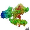
| Movie |
Movie viewer |
|---|---|
| Structure viewer | Molecule: MolmilJmol/JSmol |
- Downloads & links
-Download
| PDBx/mmCIF format | 7qid.cif.gz | 624 KB | Display | PDBx/mmCIF format |
|---|---|---|---|---|
| PDB format | pdb7qid.ent.gz | 522.5 KB | Display | PDB format |
| PDBx/mmJSON format | 7qid.json.gz | Tree view | PDBx/mmJSON format | |
| Others |  Other downloads Other downloads |
-Validation report
| Arichive directory | https://data.pdbj.org/pub/pdb/validation_reports/qi/7qidftp://data.pdbj.org/pub/pdb/validation_reports/qi/7qid | HTTPS FTP |
|---|
-Related structure data
| Related structure data |  10311MC  6sofC M: map data used to model this data C: citing same article ( |
|---|---|
| Similar structure data |




-Links
 PDBj
PDBj







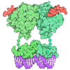
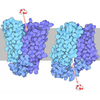
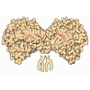
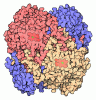


- Assembly
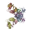
| Deposited unit | 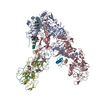
|
|---|---|
| 1 |
|
-Components
| #1: Protein | Mass: 82551.742 Da / Num. of mol.: 2 Source method: isolated from a genetically manipulated source Source: (gene. exp.) Homo sapiens (human) / Gene: INSR / Cell line (production host): HEK293F / Production host: Homo sapiens (human)References: UniProt: P06213, receptor protein-tyrosine kinase #2: Protein | Mass: 21627.248 Da / Num. of mol.: 2 Source method: isolated from a genetically manipulated source Source: (gene. exp.) Homo sapiens (human) / Gene: INSR / Cell line (production host): HEK293F / Production host: Homo sapiens (human)References: UniProt: P06213-2, receptor protein-tyrosine kinase #3: Protein/peptide | Mass: 2383.698 Da / Num. of mol.: 3 Source method: isolated from a genetically manipulated source Source: (gene. exp.) Homo sapiens (human) / Gene: INS / Production host:   Saccharomyces cerevisiae (brewer's yeast) / References: UniProt: P01308 Saccharomyces cerevisiae (brewer's yeast) / References: UniProt: P01308#4: Protein/peptide | Mass: 3433.953 Da / Num. of mol.: 3 Source method: isolated from a genetically manipulated source Source: (gene. exp.) Homo sapiens (human) / Gene: INS / Production host: Saccharomyces cerevisiae (brewer's yeast) / References: UniProt: P01308Has protein modification | Y | |
|---|
-Experimental details
-Experiment
| Experiment | Method: ELECTRON MICROSCOPY |
|---|---|
| EM experiment | Aggregation state: PARTICLE / 3D reconstruction method: single particle reconstruction |
- Sample preparation
| Component | Name: insulin receptor ectodomain with three insulin bound / Type: COMPLEX / Entity ID: all / Source: MULTIPLE SOURCES |
|---|---|
| Molecular weight | Value: 0.37 MDa / Experimental value: YES |
| Buffer solution | pH: 7.5 |
| Specimen | Embedding applied: NO / Shadowing applied: NO / Staining applied: NO / Vitrification applied: YES |
| Vitrification | Cryogen name: ETHANE-PROPANE |
- Electron microscopy imaging
| Experimental equipment |  Model: Titan Krios / Image courtesy: FEI Company |
|---|---|
| Microscopy | Model: FEI TITAN KRIOS |
| Electron gun | Electron source:  FIELD EMISSION GUN / Accelerating voltage: 300 kV / Illumination mode: FLOOD BEAM FIELD EMISSION GUN / Accelerating voltage: 300 kV / Illumination mode: FLOOD BEAM |
| Electron lens | Mode: BRIGHT FIELD / Nominal defocus max: 3500 nm / Nominal defocus min: 500 nm |
| Image recording | Average exposure time: 10.2 sec. / Electron dose: 55 e/Å2 / Detector mode: COUNTING / Film or detector model: GATAN K2 SUMMIT (4k x 4k) / Num. of grids imaged: 1 / Num. of real images: 51 |
- Processing
| Software | Name: PHENIX / Version: 1.19.2_4158: / Classification: refinement | ||||||||||||||||||||||||
|---|---|---|---|---|---|---|---|---|---|---|---|---|---|---|---|---|---|---|---|---|---|---|---|---|---|
| EM software |
| ||||||||||||||||||||||||
| CTF correction | Type: NONE | ||||||||||||||||||||||||
| Symmetry | Point symmetry: C1 (asymmetric) | ||||||||||||||||||||||||
| 3D reconstruction | Resolution: 5 Å / Resolution method: FSC 0.143 CUT-OFF / Num. of particles: 50079 / Symmetry type: POINT | ||||||||||||||||||||||||
| Refine LS restraints |
|
