+ Open data
- Basic information
| Entry | Database: PDB / ID: 7djt | ||||||||||||
|---|---|---|---|---|---|---|---|---|---|---|---|---|---|
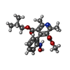
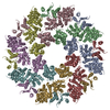
| Title | Human SARM1 inhibitory state bounded with inhibitor dHNN | ||||||||||||
 Components Components | NAD(+) hydrolase SARM1 | ||||||||||||
 Keywords Keywords | HYDROLASE / NAD(+) hydrolase | ||||||||||||
| Function / homology |  Function and homology information Function and homology informationnegative regulation of MyD88-independent toll-like receptor signaling pathway / MyD88-independent TLR4 cascade / Toll Like Receptor 3 (TLR3) Cascade / NADP+ nucleosidase activity / NAD catabolic process / ADP-ribosyl cyclase/cyclic ADP-ribose hydrolase / NAD+ nucleosidase activity / protein localization to mitochondrion / NAD+ nucleotidase, cyclic ADP-ribose generating / nervous system process ...negative regulation of MyD88-independent toll-like receptor signaling pathway / MyD88-independent TLR4 cascade / Toll Like Receptor 3 (TLR3) Cascade / NADP+ nucleosidase activity / NAD catabolic process / ADP-ribosyl cyclase/cyclic ADP-ribose hydrolase / NAD+ nucleosidase activity / protein localization to mitochondrion / NAD+ nucleotidase, cyclic ADP-ribose generating / nervous system process / Hydrolases; Glycosylases; Hydrolysing N-glycosyl compounds / regulation of dendrite morphogenesis / response to axon injury / response to glucose / regulation of neuron apoptotic process / signaling adaptor activity / TRAF6-mediated induction of TAK1 complex within TLR4 complex / Activation of IRF3, IRF7 mediated by TBK1, IKKε (IKBKE) / IKK complex recruitment mediated by RIP1 / nervous system development / mitochondrial outer membrane / microtubule / cell differentiation / axon / innate immune response / dendrite / synapse / cell surface / signal transduction / protein-containing complex / mitochondrion / identical protein binding / cytosol / cytoplasm Similarity search - Function | ||||||||||||
| Biological species |  Homo sapiens (human) Homo sapiens (human) | ||||||||||||
| Method | ELECTRON MICROSCOPY / single particle reconstruction / cryo EM / Resolution: 2.8 Å | ||||||||||||
 Authors Authors | Cai, Y. / Zhang, H. | ||||||||||||
 Citation Citation |  Journal: Elife / Year: 2021 Journal: Elife / Year: 2021Title: Permeant fluorescent probes visualize the activation of SARM1 and uncover an anti-neurodegenerative drug candidate. Authors: Wan Hua Li / Ke Huang / Yang Cai / Qian Wen Wang / Wen Jie Zhu / Yun Nan Hou / Sujing Wang / Sheng Cao / Zhi Ying Zhao / Xu Jie Xie / Yang Du / Chi-Sing Lee / Hon Cheung Lee / Hongmin Zhang / Yong Juan Zhao /  Abstract: SARM1 regulates axonal degeneration through its NAD-metabolizing activity and is a drug target for neurodegenerative disorders. We designed and synthesized fluorescent conjugates of styryl derivative ...SARM1 regulates axonal degeneration through its NAD-metabolizing activity and is a drug target for neurodegenerative disorders. We designed and synthesized fluorescent conjugates of styryl derivative with pyridine to serve as substrates of SARM1, which exhibited large red shifts after conversion. With the conjugates, SARM1 activation was visualized in live cells following elevation of endogenous NMN or treatment with a cell-permeant NMN-analog. In neurons, imaging documented mouse SARM1 activation preceded vincristine-induced axonal degeneration by hours. Library screening identified a derivative of nisoldipine (NSDP) as a covalent inhibitor of SARM1 that reacted with the cysteines, especially Cys311 in its ARM domain and blocked its NMN-activation, protecting axons from degeneration. The Cryo-EM structure showed that SARM1 was locked into an inactive conformation by the inhibitor, uncovering a potential neuroprotective mechanism of dihydropyridines. | ||||||||||||
| History |
|
- Structure visualization
| Movie |
 Movie viewer Movie viewer |
|---|---|
| Structure viewer | Molecule:  Molmil Molmil Jmol/JSmol Jmol/JSmol |
- Downloads & links
- Download
| PDBx/mmCIF format |  7djt.cif.gz 7djt.cif.gz | 849.5 KB | Display |  PDBx/mmCIF format PDBx/mmCIF format |
|---|---|---|---|---|
| PDB format |  pdb7djt.ent.gz pdb7djt.ent.gz | 714.9 KB | Display |  PDB format PDB format |
| PDBx/mmJSON format |  7djt.json.gz 7djt.json.gz | Tree view |  PDBx/mmJSON format PDBx/mmJSON format | |
| Others |  Other downloads Other downloads |
-Validation report
| Summary document |  7djt_validation.pdf.gz 7djt_validation.pdf.gz | 1.4 MB | Display |  wwPDB validaton report wwPDB validaton report |
|---|---|---|---|---|
| Full document |  7djt_full_validation.pdf.gz 7djt_full_validation.pdf.gz | 1.4 MB | Display | |
| Data in XML |  7djt_validation.xml.gz 7djt_validation.xml.gz | 132 KB | Display | |
| Data in CIF |  7djt_validation.cif.gz 7djt_validation.cif.gz | 192 KB | Display | |
| Arichive directory |  https://data.pdbj.org/pub/pdb/validation_reports/dj/7djt https://data.pdbj.org/pub/pdb/validation_reports/dj/7djt ftp://data.pdbj.org/pub/pdb/validation_reports/dj/7djt ftp://data.pdbj.org/pub/pdb/validation_reports/dj/7djt | HTTPS FTP |
-Related structure data
| Related structure data |  30700MC M: map data used to model this data C: citing same article ( |
|---|---|
| Similar structure data |
- Links
Links
- Assembly
| Deposited unit | 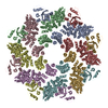
|
|---|---|
| 1 |
|
- Components
| #1: Protein | Mass: 76659.656 Da / Num. of mol.: 8 Source method: isolated from a genetically manipulated source Source: (gene. exp.)  Homo sapiens (human) / Gene: SARM1, KIAA0524, SAMD2, SARM / Cell line (production host): HEK293F / Production host: Homo sapiens (human) / Gene: SARM1, KIAA0524, SAMD2, SARM / Cell line (production host): HEK293F / Production host:  Homo sapiens (human) Homo sapiens (human)References: UniProt: Q6SZW1, ADP-ribosyl cyclase/cyclic ADP-ribose hydrolase, Hydrolases; Glycosylases; Hydrolysing N-glycosyl compounds #2: Chemical | ChemComp-H8L / Has ligand of interest | Y | |
|---|
-Experimental details
-Experiment
| Experiment | Method: ELECTRON MICROSCOPY |
|---|---|
| EM experiment | Aggregation state: PARTICLE / 3D reconstruction method: single particle reconstruction |
- Sample preparation
| Component | Name: Human NAD(+) hydrolysis SARM1 / Type: CELL / Entity ID: #1 / Source: MULTIPLE SOURCES | ||||||||||||||||||||
|---|---|---|---|---|---|---|---|---|---|---|---|---|---|---|---|---|---|---|---|---|---|
| Source (natural) | Organism:  Homo sapiens (human) Homo sapiens (human) | ||||||||||||||||||||
| Source (recombinant) | Organism:  Homo sapiens (human) / Cell: HEK293F Homo sapiens (human) / Cell: HEK293F | ||||||||||||||||||||
| Buffer solution | pH: 8 | ||||||||||||||||||||
| Buffer component |
| ||||||||||||||||||||
| Specimen | Conc.: 3 mg/ml / Embedding applied: NO / Shadowing applied: NO / Staining applied: NO / Vitrification applied: YES | ||||||||||||||||||||
| Vitrification | Cryogen name: ETHANE / Details: BLOT TIME: 4S BLOT FORCE: -2 |
- Electron microscopy imaging
| Experimental equipment |  Model: Titan Krios / Image courtesy: FEI Company |
|---|---|
| Microscopy | Model: FEI TITAN KRIOS |
| Electron gun | Electron source:  FIELD EMISSION GUN / Accelerating voltage: 300 kV / Illumination mode: SPOT SCAN FIELD EMISSION GUN / Accelerating voltage: 300 kV / Illumination mode: SPOT SCAN |
| Electron lens | Mode: BRIGHT FIELD / Nominal magnification: 130000 X / Calibrated magnification: 130000 X / Nominal defocus max: -800 nm / Calibrated defocus min: -2000 nm / Calibrated defocus max: -2500 nm / Cs: 2.7 mm / C2 aperture diameter: 70 µm |
| Specimen holder | Cryogen: NITROGEN / Specimen holder model: FEI TITAN KRIOS AUTOGRID HOLDER |
| Image recording | Electron dose: 1.28 e/Å2 / Film or detector model: GATAN K2 SUMMIT (4k x 4k) / Num. of real images: 2673 |
| Image scans | Movie frames/image: 39 / Used frames/image: 4-36 |
- Processing
| Software | Name: PHENIX / Version: 1.16_3549: / Classification: refinement | ||||||||||||||||||||||||||||||||
|---|---|---|---|---|---|---|---|---|---|---|---|---|---|---|---|---|---|---|---|---|---|---|---|---|---|---|---|---|---|---|---|---|---|
| EM software |
| ||||||||||||||||||||||||||||||||
| CTF correction | Type: PHASE FLIPPING AND AMPLITUDE CORRECTION | ||||||||||||||||||||||||||||||||
| Particle selection | Num. of particles selected: 2655835 | ||||||||||||||||||||||||||||||||
| 3D reconstruction | Resolution: 2.8 Å / Resolution method: FSC 0.143 CUT-OFF / Num. of particles: 247454 / Symmetry type: POINT | ||||||||||||||||||||||||||||||||
| Atomic model building | Protocol: RIGID BODY FIT / Space: REAL | ||||||||||||||||||||||||||||||||
| Refinement | Highest resolution: 2.8 Å | ||||||||||||||||||||||||||||||||
| Refine LS restraints |
|
 Movie
Movie Controller






 PDBj
PDBj


