National Health and Medical Research Council (NHMRC, Australia)
1107804
Australia
National Health and Medical Research Council (NHMRC, Australia)
1160570
Australia
National Health and Medical Research Council (NHMRC, Australia)
1071659
Australia
National Health and Medical Research Council (NHMRC, Australia)
1108859
Australia
Australian Research Council (ARC)
FL180100109
Australia
Australian Research Council (ARC)
DE170100783
Australia
Citation
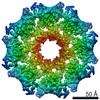
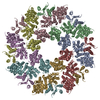
Journal: Neuron / Year: 2021 Title: SARM1 is a metabolic sensor activated by an increased NMN/NAD ratio to trigger axon degeneration. Authors: Matthew D Figley / Weixi Gu / Jeffrey D Nanson / Yun Shi / Yo Sasaki / Katie Cunnea / Alpeshkumar K Malde / Xinying Jia / Zhenyao Luo / Forhad K Saikot / Tamim Mosaiab / Veronika Masic / ...Authors: Matthew D Figley / Weixi Gu / Jeffrey D Nanson / Yun Shi / Yo Sasaki / Katie Cunnea / Alpeshkumar K Malde / Xinying Jia / Zhenyao Luo / Forhad K Saikot / Tamim Mosaiab / Veronika Masic / Stephanie Holt / Lauren Hartley-Tassell / Helen Y McGuinness / Mohammad K Manik / Todd Bosanac / Michael J Landsberg / Philip S Kerry / Mehdi Mobli / Robert O Hughes / Jeffrey Milbrandt / Bostjan Kobe / Aaron DiAntonio / Thomas Ve / Abstract: Axon degeneration is a central pathological feature of many neurodegenerative diseases. Sterile alpha and Toll/interleukin-1 receptor motif-containing 1 (SARM1) is a nicotinamide adenine dinucleotide ...Axon degeneration is a central pathological feature of many neurodegenerative diseases. Sterile alpha and Toll/interleukin-1 receptor motif-containing 1 (SARM1) is a nicotinamide adenine dinucleotide (NAD)-cleaving enzyme whose activation triggers axon destruction. Loss of the biosynthetic enzyme NMNAT2, which converts nicotinamide mononucleotide (NMN) to NAD, activates SARM1 via an unknown mechanism. Using structural, biochemical, biophysical, and cellular assays, we demonstrate that SARM1 is activated by an increase in the ratio of NMN to NAD and show that both metabolites compete for binding to the auto-inhibitory N-terminal armadillo repeat (ARM) domain of SARM1. We report structures of the SARM1 ARM domain bound to NMN and of the homo-octameric SARM1 complex in the absence of ligands. We show that NMN influences the structure of SARM1 and demonstrate via mutagenesis that NMN binding is required for injury-induced SARM1 activation and axon destruction. Hence, SARM1 is a metabolic sensor responding to an increased NMN/NAD ratio by cleaving residual NAD, thereby inducing feedforward metabolic catastrophe and axonal demise.
History
Deposition
Jan 12, 2021
-
Header (metadata) release
Mar 10, 2021
-
Map release
Mar 10, 2021
-
Update
May 14, 2025
-
Current status
May 14, 2025
Processing site: RCSB / Status: Released
-
Structure visualization
Movie
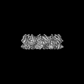
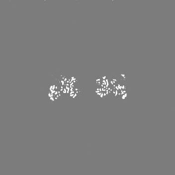
Surface view with section colored by density value
Model: Quantifoil R2/2 / Material: COPPER / Mesh: 300 / Support film - Material: CARBON / Support film - topology: CONTINUOUS / Support film - Film thickness: 6 / Pretreatment - Type: GLOW DISCHARGE
Vitrification
Cryogen name: ETHANE / Instrument: FEI VITROBOT MARK IV
-
Electron microscopy
Microscope
TFS KRIOS
Specialist optics
Energy filter - Slit width: 20 eV
Image recording
Film or detector model: GATAN K3 (6k x 4k) / Average exposure time: 4.99 sec. / Average electron dose: 54.0 e/Å2 Details: exposure rate of 12.88e-/pixel/second super-resolution 0.543 A/pixel 45 frames per image
Electron beam
Acceleration voltage: 300 kV / Electron source: FIELD EMISSION GUN
Electron optics
C2 aperture diameter: 50.0 µm / Illumination mode: FLOOD BEAM / Imaging mode: BRIGHT FIELD / Cs: 2.7 mm
In the structure databanks used in Yorodumi, some data are registered as the other names, "COVID-19 virus" and "2019-nCoV". Here are the details of the virus and the list of structure data.
Jan 31, 2019. EMDB accession codes are about to change! (news from PDBe EMDB page)
EMDB accession codes are about to change! (news from PDBe EMDB page)
The allocation of 4 digits for EMDB accession codes will soon come to an end. Whilst these codes will remain in use, new EMDB accession codes will include an additional digit and will expand incrementally as the available range of codes is exhausted. The current 4-digit format prefixed with “EMD-” (i.e. EMD-XXXX) will advance to a 5-digit format (i.e. EMD-XXXXX), and so on. It is currently estimated that the 4-digit codes will be depleted around Spring 2019, at which point the 5-digit format will come into force.
The EM Navigator/Yorodumi systems omit the EMD- prefix.
Related info.:Q: What is EMD? / ID/Accession-code notation in Yorodumi/EM Navigator
Yorodumi is a browser for structure data from EMDB, PDB, SASBDB, etc.
This page is also the successor to EM Navigator detail page, and also detail information page/front-end page for Omokage search.
The word "yorodu" (or yorozu) is an old Japanese word meaning "ten thousand". "mi" (miru) is to see.
Related info.:EMDB / PDB / SASBDB / Comparison of 3 databanks / Yorodumi Search / Aug 31, 2016. New EM Navigator & Yorodumi / Yorodumi Papers / Jmol/JSmol / Function and homology information / Changes in new EM Navigator and Yorodumi
 Movie
Movie Controller


 Open data
 Basic information
 Map data
 Sample
 Keywords
 Function and homology information
 Homo sapiens (human)
 Authors
 Australia, 6 items
  Citation

 Structure visualization
 Downloads & links
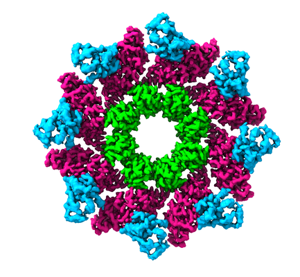 emd_23278.png
 http://ftp.pdbj.org/pub/emdb/structures/EMD-23278


 Z (Sec.)
 Y (Row.)
 X (Col.)


 Sample components
 Processing
 Electron microscopy
 FIELD EMISSION GUN

