 Movie
Movie Controller


+ Open data

- Basic information
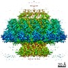
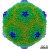
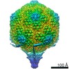
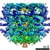
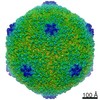
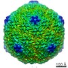
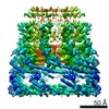
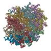
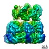
| Entry | Database: PDB / ID: 7bou | |||||||||
|---|---|---|---|---|---|---|---|---|---|---|
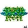
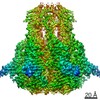
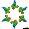
| Title | GP8 of Mature Bacteriophage T7 | |||||||||
 Components Components | Portal protein | |||||||||
 Keywords Keywords | STRUCTURAL PROTEIN / Portal / Bacteriophage / Mature / Transport | |||||||||
| Function / homology | Portal protein, Caudovirales / Head-to-tail connector protein, podovirus-type / Bacteriophage head to tail connecting protein / viral portal complex / symbiont genome ejection through host cell envelope, short tail mechanism / viral DNA genome packaging / Portal protein Function and homology information Function and homology information | |||||||||
| Biological species |   Escherichia phage T7 (virus) Escherichia phage T7 (virus) | |||||||||
| Method | ELECTRON MICROSCOPY / single particle reconstruction / Resolution: 3.6 Å | |||||||||
 Authors Authors | Chen, W.Y. / Xiao, H. | |||||||||
| Funding support |  China, 2items China, 2items
| |||||||||
 Citation Citation | Journal: Protein Cell / Year: 2020 Title: Structural changes of a bacteriophage upon DNA packaging and maturation. Authors: Wenyuan Chen / Hao Xiao / Xurong Wang / Shuanglin Song / Zhen Han / Xiaowu Li / Fan Yang / Li Wang / Jingdong Song / Hongrong Liu / Lingpeng Cheng / | |||||||||
| History |
|
- Structure visualization
| Movie |
Movie viewer |
|---|---|
| Structure viewer | Molecule: MolmilJmol/JSmol |
- Downloads & links
-Download
| PDBx/mmCIF format | 7bou.cif.gz | 1019.3 KB | Display | PDBx/mmCIF format |
|---|---|---|---|---|
| PDB format | pdb7bou.ent.gz | 870.5 KB | Display | PDB format |
| PDBx/mmJSON format | 7bou.json.gz | Tree view | PDBx/mmJSON format | |
| Others |  Other downloads Other downloads |
-Validation report
| Arichive directory | https://data.pdbj.org/pub/pdb/validation_reports/bo/7bouftp://data.pdbj.org/pub/pdb/validation_reports/bo/7bou | HTTPS FTP |
|---|
-Related structure data
| Related structure data | 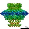 30134MC 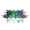 7boxC 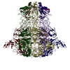 7boyC 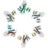 7bozC 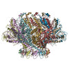 7bp0C M: map data used to model this data C: citing same article ( |
|---|---|
| Similar structure data |


-Links
 PDBj
PDBj- Assembly
| Deposited unit | 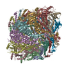
|
|---|---|
| 1 |
|
-Components
| #1: Protein | Mass: 59173.984 Da / Num. of mol.: 12 Source method: isolated from a genetically manipulated source Source: (gene. exp.) Escherichia phage T7 (virus) / Production host:  |
|---|
-Experimental details
-Experiment
| Experiment | Method: ELECTRON MICROSCOPY |
|---|---|
| EM experiment | Aggregation state: PARTICLE / 3D reconstruction method: single particle reconstruction |
- Sample preparation
| Component | Name: Mature GP8 / Type: COMPLEX / Entity ID: all / Source: NATURAL |
|---|---|
| Source (natural) | Organism: Escherichia phage T7 (virus) |
| Buffer solution | pH: 7 |
| Specimen | Embedding applied: NO / Shadowing applied: NO / Staining applied: NO / Vitrification applied: NO |
- Electron microscopy imaging
| Experimental equipment |  Model: Talos Arctica / Image courtesy: FEI Company |
|---|---|
| Microscopy | Model: FEI TECNAI ARCTICA |
| Electron gun | Electron source:  FIELD EMISSION GUN / Accelerating voltage: 200 kV / Illumination mode: FLOOD BEAM FIELD EMISSION GUN / Accelerating voltage: 200 kV / Illumination mode: FLOOD BEAM |
| Electron lens | Mode: BRIGHT FIELD / Cs: 2.7 mm / C2 aperture diameter: 70 µm |
| Image recording | Electron dose: 24 e/Å2 / Film or detector model: FEI FALCON II (4k x 4k) |
- Processing
| CTF correction | Type: PHASE FLIPPING AND AMPLITUDE CORRECTION |
|---|---|
| 3D reconstruction | Resolution: 3.6 Å / Resolution method: FSC 0.143 CUT-OFF / Num. of particles: 50000 / Symmetry type: POINT |
