ムービー
ムービー コントローラー


+ データを開く

- 基本情報
基本情報
| 登録情報 | データベース: PDB / ID: 6yi5 | ||||||||||||
|---|---|---|---|---|---|---|---|---|---|---|---|---|---|
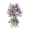
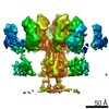
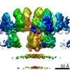
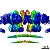
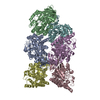
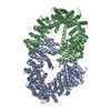
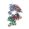
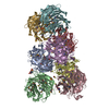
| タイトル | In-situ structure of the trimeric HEF from influenza C by flexible fitting into a cryo-ET map. | ||||||||||||
 要素 要素 | (Hemagglutinin-esterase-fusion glycoprotein) x 2 | ||||||||||||
 キーワード キーワード | VIRAL PROTEIN / Receptor / esterase / fusion / influenza | ||||||||||||
| 機能・相同性 |  機能・相同性情報 機能・相同性情報sialate 9-O-acetylesterase activity / sialate 4-O-acetylesterase activity / sialate O-acetylesterase / viral budding from plasma membrane / endocytosis involved in viral entry into host cell / host cell surface receptor binding / fusion of virus membrane with host plasma membrane / fusion of virus membrane with host endosome membrane / viral envelope / virion attachment to host cell ...sialate 9-O-acetylesterase activity / sialate 4-O-acetylesterase activity / sialate O-acetylesterase / viral budding from plasma membrane / endocytosis involved in viral entry into host cell / host cell surface receptor binding / fusion of virus membrane with host plasma membrane / fusion of virus membrane with host endosome membrane / viral envelope / virion attachment to host cell / host cell plasma membrane / virion membrane / membrane 類似検索 - 分子機能 | ||||||||||||
| 生物種 |  Influenza C virus (インフルエンザウイルス) Influenza C virus (インフルエンザウイルス) | ||||||||||||
| 手法 | 電子顕微鏡法 / サブトモグラム平均法 / クライオ電子顕微鏡法 / 解像度: 9.1 Å | ||||||||||||
 データ登録者 データ登録者 | Halldorsson, S. / Rosenthal, P.B. | ||||||||||||
| 資金援助 |  英国, 3件 英国, 3件
| ||||||||||||
 引用 引用 | ジャーナル: Nat Commun / 年: 2021 タイトル: In situ structure and organization of the influenza C virus surface glycoprotein. 著者: Steinar Halldorsson / Kasim Sader / Jack Turner / Lesley J Calder / Peter B Rosenthal /  要旨: The lipid-enveloped influenza C virus contains a single surface glycoprotein, the haemagglutinin-esterase-fusion (HEF) protein, that mediates receptor binding, receptor destruction, and membrane ...The lipid-enveloped influenza C virus contains a single surface glycoprotein, the haemagglutinin-esterase-fusion (HEF) protein, that mediates receptor binding, receptor destruction, and membrane fusion at the low pH of the endosome. Here we apply electron cryotomography and subtomogram averaging to describe the structural basis for hexagonal lattice formation by HEF on the viral surface. The conformation of the glycoprotein in situ is distinct from the structure of the isolated trimeric ectodomain, showing that a splaying of the membrane distal domains is required to mediate contacts that form the lattice. The splaying of these domains is also coupled to changes in the structure of the stem region which is involved in membrane fusion, thereby linking HEF's membrane fusion conformation with its assembly on the virus surface. The glycoprotein lattice can form independent of other virion components but we show a major role for the matrix layer in particle formation. | ||||||||||||
| 履歴 |
|
- 構造の表示
構造の表示
| ムービー |
ムービービューア |
|---|---|
| 構造ビューア | 分子: MolmilJmol/JSmol |
- ダウンロードとリンク
-ダウンロード
| PDBx/mmCIF形式 | 6yi5.cif.gz | 303.8 KB | 表示 | PDBx/mmCIF形式 |
|---|---|---|---|---|
| PDB形式 | pdb6yi5.ent.gz | 251.5 KB | 表示 | PDB形式 |
| PDBx/mmJSON形式 | 6yi5.json.gz | ツリー表示 | PDBx/mmJSON形式 | |
| その他 |  その他のダウンロード その他のダウンロード |
-検証レポート
| アーカイブディレクトリ | https://data.pdbj.org/pub/pdb/validation_reports/yi/6yi5ftp://data.pdbj.org/pub/pdb/validation_reports/yi/6yi5 | HTTPS FTP |
|---|
-関連構造データ


-リンク
 PDBj
PDBj


- 集合体
集合体
| 登録構造単位 | 
|
|---|---|
| 1 |
|
-要素
| #1: タンパク質 | 分子量: 48230.699 Da / 分子数: 3 / 由来タイプ: 天然 由来: (天然) Influenza C virus (strain C/Johannesburg/1/1966) (インフルエンザウイルス)細胞株: MDCK / 参照: UniProt: P07975, sialate O-acetylesterase #2: タンパク質 | 分子量: 18977.541 Da / 分子数: 3 / 由来タイプ: 天然 由来: (天然) Influenza C virus (strain C/Johannesburg/1/1966) (インフルエンザウイルス)細胞株: MDCK / 参照: UniProt: P07975, sialate O-acetylesterase #3: 多糖 | beta-D-mannopyranose-(1-4)-2-acetamido-2-deoxy-beta-D-glucopyranose-(1-4)-2-acetamido-2-deoxy-beta- ...beta-D-mannopyranose-(1-4)-2-acetamido-2-deoxy-beta-D-glucopyranose-(1-4)-2-acetamido-2-deoxy-beta-D-glucopyranose #4: 多糖 | 研究の焦点であるリガンドがあるか | N | |
|---|
-実験情報
-実験
| 実験 | 手法: 電子顕微鏡法 |
|---|---|
| EM実験 | 試料の集合状態: PARTICLE / 3次元再構成法: サブトモグラム平均法 |
- 試料調製
試料調製
| 構成要素 | 名称: Influenza C virus / タイプ: VIRUS / 詳細: Purified from infected cells. / Entity ID: #1-#2 / 由来: NATURAL | ||||||||||||||||||||
|---|---|---|---|---|---|---|---|---|---|---|---|---|---|---|---|---|---|---|---|---|---|
| 由来(天然) | 生物種: Influenza C virus (インフルエンザウイルス) | ||||||||||||||||||||
| ウイルスについての詳細 | 中空か: NO / エンベロープを持つか: YES / 単離: STRAIN / タイプ: VIRION | ||||||||||||||||||||
| 天然宿主 | 生物種: Homo sapiens | ||||||||||||||||||||
| 緩衝液 | pH: 7.4 | ||||||||||||||||||||
| 緩衝液成分 |
| ||||||||||||||||||||
| 試料 | 濃度: 2 mg/ml / 包埋: NO / シャドウイング: NO / 染色: NO / 凍結: YES 詳細: Heterogeneous mix of viral particles, viral like particles and cellular vesicles | ||||||||||||||||||||
| 試料支持 | グリッドの材料: COPPER / グリッドのタイプ: Quantifoil R2/2 | ||||||||||||||||||||
| 急速凍結 | 装置: FEI VITROBOT MARK III / 凍結剤: ETHANE / 湿度: 100 % / 凍結前の試料温度: 277 K |
- 電子顕微鏡撮影
| 実験機器 |  モデル: Titan Krios / 画像提供: FEI Company |
|---|---|
| 顕微鏡 | モデル: FEI TITAN KRIOS |
| 電子銃 | 電子線源:  FIELD EMISSION GUN / 加速電圧: 300 kV / 照射モード: FLOOD BEAM FIELD EMISSION GUN / 加速電圧: 300 kV / 照射モード: FLOOD BEAM |
| 電子レンズ | モード: BRIGHT FIELD / 倍率(公称値): 64000 X / 最大 デフォーカス(公称値): 4.5 nm / 最小 デフォーカス(公称値): 2.5 nm / Cs: 2.7 mm / C2レンズ絞り径: 100 µm / アライメント法: COMA FREE |
| 試料ホルダ | 凍結剤: NITROGEN 試料ホルダーモデル: FEI TITAN KRIOS AUTOGRID HOLDER |
| 撮影 | 平均露光時間: 1.57 sec. / 電子線照射量: 1.57 e/Å2 / 検出モード: COUNTING フィルム・検出器のモデル: GATAN K2 QUANTUM (4k x 4k) 撮影したグリッド数: 2 |
| 電子光学装置 | エネルギーフィルタースリット幅: 20 eV |
| 画像スキャン | 横: 3838 / 縦: 3710 / 動画フレーム数/画像: 4 |
- 解析
解析
| EMソフトウェア |
| ||||||||||||||||||||||||||||||||||||
|---|---|---|---|---|---|---|---|---|---|---|---|---|---|---|---|---|---|---|---|---|---|---|---|---|---|---|---|---|---|---|---|---|---|---|---|---|---|
| CTF補正 | タイプ: PHASE FLIPPING ONLY | ||||||||||||||||||||||||||||||||||||
| 対称性 | 点対称性: C3 (3回回転対称) | ||||||||||||||||||||||||||||||||||||
| 3次元再構成 | 解像度: 9.1 Å / 解像度の算出法: FSC 0.143 CUT-OFF / 粒子像の数: 14057 / アルゴリズム: BACK PROJECTION / クラス平均像の数: 1 / 対称性のタイプ: POINT | ||||||||||||||||||||||||||||||||||||
| EM volume selection | 手法: Random seeds / 詳細: A vesicle model with random seeds on the surface. / Num. of tomograms: 41 / Num. of volumes extracted: 31947 | ||||||||||||||||||||||||||||||||||||
| 原子モデル構築 | プロトコル: FLEXIBLE FIT / 空間: REAL | ||||||||||||||||||||||||||||||||||||
| 原子モデル構築 | PDB-ID: 1FLC |