 ムービー
ムービー コントローラー


+ データを開く

- 基本情報
基本情報
| 登録情報 | データベース: PDB / ID: 6wqk | |||||||||
|---|---|---|---|---|---|---|---|---|---|---|
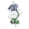
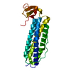
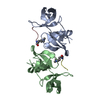
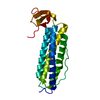
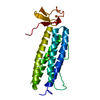
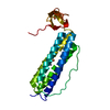
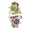
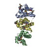
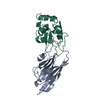
| タイトル | hnRNPA2 Low complexity domain (LCD) determined by cryoEM | |||||||||
 要素 要素 | MCherry fluorescent protein,Heterogeneous nuclear ribonucleoproteins A2/B1 chimera | |||||||||
 キーワード キーワード | PROTEIN FIBRIL / Ribinucleoprotein / RNA binding protein / Low complexity domain | |||||||||
| 機能・相同性 |  機能・相同性情報 機能・相同性情報: / miRNA transport / positive regulation of telomere maintenance via telomere lengthening / RNA transport / G-quadruplex DNA unwinding / primary miRNA processing / single-stranded telomeric DNA binding / N6-methyladenosine-containing RNA reader activity / G-rich strand telomeric DNA binding / miRNA binding ...: / miRNA transport / positive regulation of telomere maintenance via telomere lengthening / RNA transport / G-quadruplex DNA unwinding / primary miRNA processing / single-stranded telomeric DNA binding / N6-methyladenosine-containing RNA reader activity / G-rich strand telomeric DNA binding / miRNA binding / Cajal body / negative regulation of mRNA splicing, via spliceosome / bioluminescence / Processing of Capped Intron-Containing Pre-mRNA / mRNA transport / generation of precursor metabolites and energy / mRNA export from nucleus / pre-mRNA intronic binding / Gene and protein expression by JAK-STAT signaling after Interleukin-12 stimulation / catalytic step 2 spliceosome / mRNA Splicing - Major Pathway / molecular condensate scaffold activity / mRNA 3'-UTR binding / spliceosomal complex / nuclear matrix / mRNA splicing, via spliceosome / mRNA processing / chromosome, telomeric region / ribonucleoprotein complex / negative regulation of transcription by RNA polymerase II / RNA binding / extracellular exosome / nucleoplasm / identical protein binding / membrane / nucleus / cytoplasm 類似検索 - 分子機能 | |||||||||
| 生物種 |  Anaplasma marginale (バクテリア) Anaplasma marginale (バクテリア) Homo sapiens (ヒト) Homo sapiens (ヒト) | |||||||||
| 手法 | 電子顕微鏡法 / らせん対称体再構成法 / クライオ電子顕微鏡法 / 解像度: 3.1 Å | |||||||||
 データ登録者 データ登録者 | Lu, J. / Cao, Q. / Hughes, M.P. / Sawaya, M.R. / Boyer, D.R. / Cascio, D. / Eisenberg, D.S. | |||||||||
| 資金援助 |  米国, 2件 米国, 2件
| |||||||||
 引用 引用 | ジャーナル: Nat Commun / 年: 2020 タイトル: CryoEM structure of the low-complexity domain of hnRNPA2 and its conversion to pathogenic amyloid. 著者: Jiahui Lu / Qin Cao / Michael P Hughes / Michael R Sawaya / David R Boyer / Duilio Cascio / David S Eisenberg / 要旨: hnRNPA2 is a human ribonucleoprotein (RNP) involved in RNA metabolism. It forms fibrils both under cellular stress and in mutated form in neurodegenerative conditions. Previous work established that ...hnRNPA2 is a human ribonucleoprotein (RNP) involved in RNA metabolism. It forms fibrils both under cellular stress and in mutated form in neurodegenerative conditions. Previous work established that the C-terminal low-complexity domain (LCD) of hnRNPA2 fibrillizes under stress, and missense mutations in this domain are found in the disease multisystem proteinopathy (MSP). However, little is known at the atomic level about the hnRNPA2 LCD structure that is involved in those processes and how disease mutations cause structural change. Here we present the cryo-electron microscopy (cryoEM) structure of the hnRNPA2 LCD fibril core and demonstrate its capability to form a reversible hydrogel in vitro containing amyloid-like fibrils. Whereas these fibrils, like pathogenic amyloid, are formed from protein chains stacked into β-sheets by backbone hydrogen bonds, they display distinct structural differences: the chains are kinked, enabling non-covalent cross-linking of fibrils and disfavoring formation of pathogenic steric zippers. Both reversibility and energetic calculations suggest these fibrils are less stable than pathogenic amyloid. Moreover, the crystal structure of the disease-mutation-containing segment (D290V) of hnRNPA2 suggests that the replacement fundamentally alters the fibril structure to a more stable energetic state. These findings illuminate how molecular interactions promote protein fibril networks and how mutation can transform fibril structure from functional to a pathogenic form. | |||||||||
| 履歴 |
|
- 構造の表示
構造の表示
| ムービー |
ムービービューア |
|---|---|
| 構造ビューア | 分子: MolmilJmol/JSmol |
- ダウンロードとリンク
-ダウンロード
| PDBx/mmCIF形式 | 6wqk.cif.gz | 84.1 KB | 表示 | PDBx/mmCIF形式 |
|---|---|---|---|---|
| PDB形式 | pdb6wqk.ent.gz | 47.9 KB | 表示 | PDB形式 |
| PDBx/mmJSON形式 | 6wqk.json.gz | ツリー表示 | PDBx/mmJSON形式 | |
| その他 |  その他のダウンロード その他のダウンロード |
-検証レポート
| 文書・要旨 | 6wqk_validation.pdf.gz | 913.2 KB | 表示 | wwPDB検証レポート |
|---|---|---|---|---|
| 文書・詳細版 | 6wqk_full_validation.pdf.gz | 913 KB | 表示 | |
| XML形式データ | 6wqk_validation.xml.gz | 12.7 KB | 表示 | |
| CIF形式データ | 6wqk_validation.cif.gz | 18 KB | 表示 | |
| アーカイブディレクトリ | https://data.pdbj.org/pub/pdb/validation_reports/wq/6wqkftp://data.pdbj.org/pub/pdb/validation_reports/wq/6wqk | HTTPS FTP |
-関連構造データ
| 関連構造データ |  21871MC  6wpqC C: 同じ文献を引用 ( M: このデータのモデリングに利用したマップデータ |
|---|---|
| 類似構造データ | |
| 電子顕微鏡画像生データ | EMPIAR-10634 (タイトル: CryoEM structure of the LCD of hnRNPA2 amyloid-like fibrils Data size: 1.2 TB Data #1: Unaligned micrographs, 1.15e-/A^2/frame [micrographs - multiframe]) |

-リンク
 PDBj
PDBj


- 集合体
集合体
| 登録構造単位 | 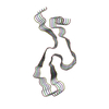
|
|---|---|
| 1 |
|
| 対称性 | らせん対称: (回転対称性: 1 / Dyad axis: no / N subunits divisor: 1 / Num. of operations: 5 / Rise per n subunits: 4.81 Å / Rotation per n subunits: -2.88 °) |
-要素
| #1: タンパク質 | 分子量: 45666.625 Da / 分子数: 5 / 断片: low complexity domain (UNP residues 193-353) / 由来タイプ: 組換発現 由来: (組換発現) Anaplasma marginale (バクテリア), (組換発現) Homo sapiens (ヒト)遺伝子: mCherry, HNRNPA2B1, HNRPA2B1 / 発現宿主: |
|---|
-実験情報
-実験
| 実験 | 手法: 電子顕微鏡法 |
|---|---|
| EM実験 | 試料の集合状態: FILAMENT / 3次元再構成法: らせん対称体再構成法 |
- 試料調製
試料調製
| 構成要素 | 名称: heterogeneous nuclear ribonucleo-protein A2 / タイプ: COMPLEX / Entity ID: all / 由来: RECOMBINANT |
|---|---|
| 由来(天然) | 生物種: Homo sapiens (ヒト) |
| 由来(組換発現) | 生物種: |
| 緩衝液 | pH: 7.5 |
| 試料 | 包埋: NO / シャドウイング: NO / 染色: NO / 凍結: YES |
| 急速凍結 | 凍結剤: ETHANE |
- 電子顕微鏡撮影
| 実験機器 |  モデル: Titan Krios / 画像提供: FEI Company |
|---|---|
| 顕微鏡 | モデル: FEI TITAN KRIOS |
| 電子銃 | 電子線源:  FIELD EMISSION GUN / 加速電圧: 300 kV / 照射モード: OTHER FIELD EMISSION GUN / 加速電圧: 300 kV / 照射モード: OTHER |
| 電子レンズ | モード: BRIGHT FIELD |
| 撮影 | 電子線照射量: 1.15 e/Å2 / フィルム・検出器のモデル: GATAN K2 IS (4k x 4k) |
- 解析
解析
| ソフトウェア | 名称: PHENIX / バージョン: dev_3724: / 分類: 精密化 | ||||||||||||||||||||||||
|---|---|---|---|---|---|---|---|---|---|---|---|---|---|---|---|---|---|---|---|---|---|---|---|---|---|
| EMソフトウェア |
| ||||||||||||||||||||||||
| CTF補正 | タイプ: PHASE FLIPPING AND AMPLITUDE CORRECTION | ||||||||||||||||||||||||
| らせん対称 | 回転角度/サブユニット: -2.88 ° / 軸方向距離/サブユニット: 4.81 Å / らせん対称軸の対称性: C1 | ||||||||||||||||||||||||
| 3次元再構成 | 解像度: 3.1 Å / 解像度の算出法: FSC 0.143 CUT-OFF / 粒子像の数: 132571 / 対称性のタイプ: HELICAL | ||||||||||||||||||||||||
| 拘束条件 |
|
