 Movie
Movie Controller


+ Open data

- Basic information
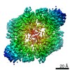
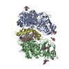
| Entry | Database: PDB / ID: 6sxb | ||||||
|---|---|---|---|---|---|---|---|
| Title | XPF-ERCC1 Cryo-EM Structure, DNA-Bound form | ||||||
 Components Components |
| ||||||
 Keywords Keywords | DNA BINDING PROTEIN / DNA Repair enzyme. Nucleotide excision repair | ||||||
| Function / homology |  Function and homology information Function and homology informationpositive regulation of t-circle formation / telomeric DNA-containing double minutes formation / ERCC4-ERCC1 complex / negative regulation of protection from non-homologous end joining at telomere / : / pyrimidine dimer repair by nucleotide-excision repair / nucleotide-excision repair factor 1 complex / nucleotide-excision repair involved in interstrand cross-link repair / nucleotide-excision repair complex / negative regulation of telomere maintenance ...positive regulation of t-circle formation / telomeric DNA-containing double minutes formation / ERCC4-ERCC1 complex / negative regulation of protection from non-homologous end joining at telomere / : / pyrimidine dimer repair by nucleotide-excision repair / nucleotide-excision repair factor 1 complex / nucleotide-excision repair involved in interstrand cross-link repair / nucleotide-excision repair complex / negative regulation of telomere maintenance / single-stranded DNA endonuclease activity / resolution of meiotic recombination intermediates / t-circle formation / mitotic recombination / post-embryonic hemopoiesis / isotype switching / UV protection / negative regulation of telomere maintenance via telomere lengthening / UV-damage excision repair / HDR through Single Strand Annealing (SSA) / oogenesis / TFIID-class transcription factor complex binding / replicative senescence / positive regulation of transcription initiation by RNA polymerase II / response to X-ray / response to UV / interstrand cross-link repair / insulin-like growth factor receptor signaling pathway / telomere maintenance / determination of adult lifespan / regulation of autophagy / nucleotide-excision repair / DNA endonuclease activity / promoter-specific chromatin binding / Fanconi Anemia Pathway / double-strand break repair via homologous recombination / male gonad development / double-strand break repair via nonhomologous end joining / multicellular organism growth / Dual Incision in GG-NER / Formation of Incision Complex in GG-NER / Dual incision in TC-NER / cellular response to UV / single-stranded DNA binding / response to oxidative stress / spermatogenesis / Hydrolases; Acting on ester bonds / damaged DNA binding / protein-macromolecule adaptor activity / chromosome, telomeric region / cell population proliferation / DNA repair / nucleoplasm / identical protein binding / nucleus / cytoplasm Similarity search - Function | ||||||
| Biological species |  Homo sapiens (human) Homo sapiens (human) | ||||||
| Method | ELECTRON MICROSCOPY / single particle reconstruction / cryo EM / Resolution: 7.9 Å | ||||||
 Authors Authors | Jones, M.L. / Briggs, D.C. / McDonald, N.Q. | ||||||
| Funding support |  United Kingdom, 1items United Kingdom, 1items
| ||||||
 Citation Citation | Journal: Nat Commun / Year: 2020 Title: Cryo-EM structures of the XPF-ERCC1 endonuclease reveal how DNA-junction engagement disrupts an auto-inhibited conformation. Authors: Morgan Jones / Fabienne Beuron / Aaron Borg / Andrea Nans / Christopher P Earl / David C Briggs / Ambrosius P Snijders / Maureen Bowles / Edward P Morris / Mark Linch / Neil Q McDonald / Abstract: The structure-specific endonuclease XPF-ERCC1 participates in multiple DNA damage repair pathways including nucleotide excision repair (NER) and inter-strand crosslink repair (ICLR). How XPF-ERCC1 is ...The structure-specific endonuclease XPF-ERCC1 participates in multiple DNA damage repair pathways including nucleotide excision repair (NER) and inter-strand crosslink repair (ICLR). How XPF-ERCC1 is catalytically activated by DNA junction substrates is not currently understood. Here we report cryo-electron microscopy structures of both DNA-free and DNA-bound human XPF-ERCC1. DNA-free XPF-ERCC1 adopts an auto-inhibited conformation in which the XPF helical domain masks the ERCC1 (HhH) domain and restricts access to the XPF catalytic site. DNA junction engagement releases the ERCC1 (HhH) domain to couple with the XPF-ERCC1 nuclease/nuclease-like domains. Structure-function data indicate xeroderma pigmentosum patient mutations frequently compromise the structural integrity of XPF-ERCC1. Fanconi anaemia patient mutations in XPF often display substantial in-vitro activity but are resistant to activation by ICLR recruitment factor SLX4. Our data provide insights into XPF-ERCC1 architecture and catalytic activation. | ||||||
| History |
|
- Structure visualization
| Movie |
Movie viewer |
|---|---|
| Structure viewer | Molecule: MolmilJmol/JSmol |
- Downloads & links
-Download
| PDBx/mmCIF format | 6sxb.cif.gz | 184.7 KB | Display | PDBx/mmCIF format |
|---|---|---|---|---|
| PDB format | pdb6sxb.ent.gz | 138.1 KB | Display | PDB format |
| PDBx/mmJSON format | 6sxb.json.gz | Tree view | PDBx/mmJSON format | |
| Others |  Other downloads Other downloads |
-Validation report
| Arichive directory | https://data.pdbj.org/pub/pdb/validation_reports/sx/6sxbftp://data.pdbj.org/pub/pdb/validation_reports/sx/6sxb | HTTPS FTP |
|---|
-Related structure data
| Related structure data | 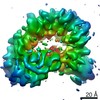 10338MC 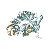 6sxaC M: map data used to model this data C: citing same article ( |
|---|---|
| Similar structure data |


-Links
 PDBj
PDBj


- Assembly
| Deposited unit | 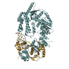
|
|---|---|
| 1 |
|
-Components
| #1: Protein | Mass: 104636.156 Da / Num. of mol.: 1 Source method: isolated from a genetically manipulated source Details: Some residues have been modelled as Alanine where sidechains were not visible. Source: (gene. exp.) Homo sapiens (human) / Gene: ERCC4, ERCC11, XPF / Production host:   Spodoptera frugiperda (fall armyworm) Spodoptera frugiperda (fall armyworm)References: UniProt: Q92889, Hydrolases; Acting on ester bonds |
|---|---|
| #2: Protein | Mass: 32598.301 Da / Num. of mol.: 1 Source method: isolated from a genetically manipulated source Details: Some residues modelled as Alanine where no sidechain information available. Source: (gene. exp.) Homo sapiens (human) / Gene: ERCC1 / Production host: Spodoptera frugiperda (fall armyworm) / References: UniProt: P07992 |
| #3: DNA chain | Mass: 3069.030 Da / Num. of mol.: 1 / Source method: obtained synthetically / Source: (synth.) Homo sapiens (human) |
| #4: DNA chain | Mass: 3019.992 Da / Num. of mol.: 1 / Source method: obtained synthetically / Source: (synth.) Homo sapiens (human) |
-Experimental details
-Experiment
| Experiment | Method: ELECTRON MICROSCOPY |
|---|---|
| EM experiment | Aggregation state: 2D ARRAY / 3D reconstruction method: single particle reconstruction |
- Sample preparation
| Component |
| ||||||||||||||||||||||||
|---|---|---|---|---|---|---|---|---|---|---|---|---|---|---|---|---|---|---|---|---|---|---|---|---|---|
| Molecular weight | Experimental value: NO | ||||||||||||||||||||||||
| Source (natural) |
| ||||||||||||||||||||||||
| Source (recombinant) |
| ||||||||||||||||||||||||
| Buffer solution | pH: 7.8 Details: 20 mM HEPES pH 7.8, 150 mM NaCl, 1 mM TCEP, 0.01% CHAPS | ||||||||||||||||||||||||
| Specimen | Embedding applied: NO / Shadowing applied: NO / Staining applied: NO / Vitrification applied: YES | ||||||||||||||||||||||||
| Vitrification | Instrument: FEI VITROBOT MARK IV / Cryogen name: ETHANE / Humidity: 100 % |
- Electron microscopy imaging
| Experimental equipment |  Model: Titan Krios / Image courtesy: FEI Company |
|---|---|
| Microscopy | Model: FEI TITAN KRIOS |
| Electron gun | Electron source:  FIELD EMISSION GUN / Accelerating voltage: 300 kV / Illumination mode: FLOOD BEAM FIELD EMISSION GUN / Accelerating voltage: 300 kV / Illumination mode: FLOOD BEAM |
| Electron lens | Mode: BRIGHT FIELD / Nominal defocus max: 4200 nm / Nominal defocus min: 1200 nm / Calibrated defocus min: 900 nm / Calibrated defocus max: 4500 nm / Cs: 2.7 mm / Alignment procedure: BASIC |
| Specimen holder | Specimen holder model: FEI TITAN KRIOS AUTOGRID HOLDER |
| Image recording | Electron dose: 63 e/Å2 / Detector mode: COUNTING / Film or detector model: GATAN K2 SUMMIT (4k x 4k) / Num. of real images: 15315 |
| Image scans | Movie frames/image: 20 / Used frames/image: 2-17 |
- Processing
| EM software | Name: cryoSPARC / Version: 2 / Category: 3D reconstruction |
|---|---|
| CTF correction | Type: PHASE FLIPPING AND AMPLITUDE CORRECTION |
| 3D reconstruction | Resolution: 7.9 Å / Resolution method: FSC 0.143 CUT-OFF / Num. of particles: 198212 / Symmetry type: POINT |
