 Movie
Movie Controller


+ Open data

- Basic information
| Entry | Database: EMDB / ID: EMD-10337 | |||||||||
|---|---|---|---|---|---|---|---|---|---|---|
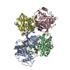
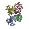
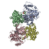
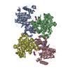
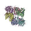
| Title | XPF-ERCC1 Cryo-EM Structure, Apo-form | |||||||||
 Map data Map data | Unsharpened Map | |||||||||
 Sample Sample |
| |||||||||
 Keywords Keywords | DNA Repair enzyme. Nucleotide excision repair / DNA BINDING PROTEIN | |||||||||
| Function / homology |  Function and homology information Function and homology informationpositive regulation of t-circle formation / telomeric DNA-containing double minutes formation / ERCC4-ERCC1 complex / negative regulation of protection from non-homologous end joining at telomere / : / pyrimidine dimer repair by nucleotide-excision repair / nucleotide-excision repair factor 1 complex / nucleotide-excision repair involved in interstrand cross-link repair / nucleotide-excision repair complex / negative regulation of telomere maintenance ...positive regulation of t-circle formation / telomeric DNA-containing double minutes formation / ERCC4-ERCC1 complex / negative regulation of protection from non-homologous end joining at telomere / : / pyrimidine dimer repair by nucleotide-excision repair / nucleotide-excision repair factor 1 complex / nucleotide-excision repair involved in interstrand cross-link repair / nucleotide-excision repair complex / negative regulation of telomere maintenance / single-stranded DNA endonuclease activity / resolution of meiotic recombination intermediates / t-circle formation / mitotic recombination / post-embryonic hemopoiesis / UV protection / isotype switching / negative regulation of telomere maintenance via telomere lengthening / UV-damage excision repair / HDR through Single Strand Annealing (SSA) / oogenesis / TFIID-class transcription factor complex binding / replicative senescence / positive regulation of transcription initiation by RNA polymerase II / response to X-ray / response to UV / interstrand cross-link repair / insulin-like growth factor receptor signaling pathway / telomere maintenance / determination of adult lifespan / regulation of autophagy / nucleotide-excision repair / DNA endonuclease activity / promoter-specific chromatin binding / Fanconi Anemia Pathway / double-strand break repair via homologous recombination / male gonad development / double-strand break repair via nonhomologous end joining / multicellular organism growth / Dual Incision in GG-NER / Formation of Incision Complex in GG-NER / Dual incision in TC-NER / cellular response to UV / single-stranded DNA binding / response to oxidative stress / spermatogenesis / Hydrolases; Acting on ester bonds / damaged DNA binding / protein-macromolecule adaptor activity / chromosome, telomeric region / cell population proliferation / DNA repair / nucleoplasm / identical protein binding / nucleus / cytoplasm Similarity search - Function | |||||||||
| Biological species |  Homo sapiens (human) Homo sapiens (human) | |||||||||
| Method | single particle reconstruction / cryo EM / Resolution: 3.6 Å | |||||||||
 Authors Authors | Jones ML / Briggs DC | |||||||||
| Funding support |  United Kingdom, 1 items United Kingdom, 1 items
| |||||||||
 Citation Citation | Journal: Nat Commun / Year: 2020 Title: Cryo-EM structures of the XPF-ERCC1 endonuclease reveal how DNA-junction engagement disrupts an auto-inhibited conformation. Authors: Morgan Jones / Fabienne Beuron / Aaron Borg / Andrea Nans / Christopher P Earl / David C Briggs / Ambrosius P Snijders / Maureen Bowles / Edward P Morris / Mark Linch / Neil Q McDonald / Abstract: The structure-specific endonuclease XPF-ERCC1 participates in multiple DNA damage repair pathways including nucleotide excision repair (NER) and inter-strand crosslink repair (ICLR). How XPF-ERCC1 is ...The structure-specific endonuclease XPF-ERCC1 participates in multiple DNA damage repair pathways including nucleotide excision repair (NER) and inter-strand crosslink repair (ICLR). How XPF-ERCC1 is catalytically activated by DNA junction substrates is not currently understood. Here we report cryo-electron microscopy structures of both DNA-free and DNA-bound human XPF-ERCC1. DNA-free XPF-ERCC1 adopts an auto-inhibited conformation in which the XPF helical domain masks the ERCC1 (HhH) domain and restricts access to the XPF catalytic site. DNA junction engagement releases the ERCC1 (HhH) domain to couple with the XPF-ERCC1 nuclease/nuclease-like domains. Structure-function data indicate xeroderma pigmentosum patient mutations frequently compromise the structural integrity of XPF-ERCC1. Fanconi anaemia patient mutations in XPF often display substantial in-vitro activity but are resistant to activation by ICLR recruitment factor SLX4. Our data provide insights into XPF-ERCC1 architecture and catalytic activation. | |||||||||
| History |
|
- Structure visualization
| Movie |
Movie viewer |
|---|---|
| Structure viewer | EM map: SurfViewMolmilJmol/JSmol |
| Supplemental images |

- Downloads & links
-EMDB archive
| Map data | emd_10337.map.gz | 15.4 MB | EMDB map data format | |
|---|---|---|---|---|
| Header (meta data) | emd-10337-v30.xmlemd-10337.xml | 19.3 KB 19.3 KB | Display Display | EMDB header |
| Images | 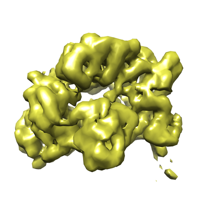 emd_10337.png emd_10337.png | 104.5 KB | ||
| Masks | emd_10337_msk_1.map | 30.5 MB | Mask map | |
| Filedesc metadata | emd-10337.cif.gz | 6.4 KB | ||
| Others | emd_10337_additional.map.gzemd_10337_half_map_1.map.gzemd_10337_half_map_2.map.gz | 28.7 MB 28.3 MB 28.3 MB | ||
| Archive directory |  http://ftp.pdbj.org/pub/emdb/structures/EMD-10337ftp://ftp.pdbj.org/pub/emdb/structures/EMD-10337 http://ftp.pdbj.org/pub/emdb/structures/EMD-10337ftp://ftp.pdbj.org/pub/emdb/structures/EMD-10337 | HTTPS FTP |
-Related structure data
| Related structure data |  6sxaMC  6sxbC M: atomic model generated by this map C: citing same article ( |
|---|---|
| Similar structure data |


-Links
| EMDB pages | EMDB (EBI/PDBe) / EMDataResource |
|---|---|
| Related items in Molecule of the Month |


-Map
| File | Download / File: emd_10337.map.gz / Format: CCP4 / Size: 30.5 MB / Type: IMAGE STORED AS FLOATING POINT NUMBER (4 BYTES) | ||||||||||||||||||||||||||||||||||||||||||||||||||||||||||||||||||||
|---|---|---|---|---|---|---|---|---|---|---|---|---|---|---|---|---|---|---|---|---|---|---|---|---|---|---|---|---|---|---|---|---|---|---|---|---|---|---|---|---|---|---|---|---|---|---|---|---|---|---|---|---|---|---|---|---|---|---|---|---|---|---|---|---|---|---|---|---|---|
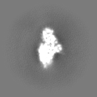
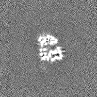
| Annotation | Unsharpened Map | ||||||||||||||||||||||||||||||||||||||||||||||||||||||||||||||||||||
| Projections & slices | Image control
Images are generated by Spider. | ||||||||||||||||||||||||||||||||||||||||||||||||||||||||||||||||||||
| Voxel size | X=Y=Z: 1.38 Å | ||||||||||||||||||||||||||||||||||||||||||||||||||||||||||||||||||||
| Density |
| ||||||||||||||||||||||||||||||||||||||||||||||||||||||||||||||||||||
| Symmetry | Space group: 1 | ||||||||||||||||||||||||||||||||||||||||||||||||||||||||||||||||||||
| Details | EMDB XML:
CCP4 map header:
| ||||||||||||||||||||||||||||||||||||||||||||||||||||||||||||||||||||
 Z (Sec.)
 Y (Row.)
 X (Col.)


-Supplemental data
-Mask #1
| File | emd_10337_msk_1.map | ||||||||||||
|---|---|---|---|---|---|---|---|---|---|---|---|---|---|
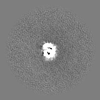
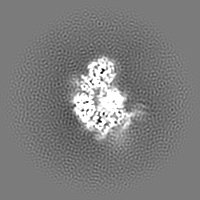
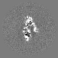
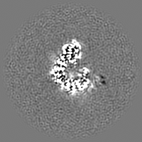
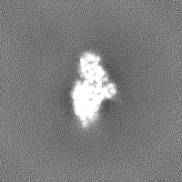
| Projections & Slices |
| ||||||||||||
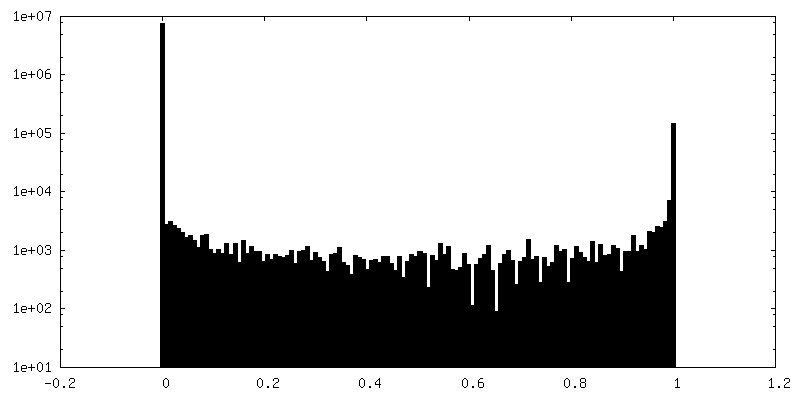
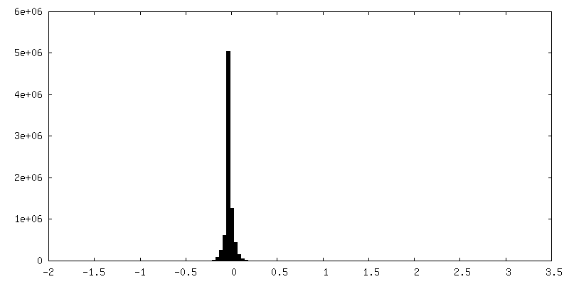
| Density Histograms |


-Additional map: Sharpened Map
| File | emd_10337_additional.map | ||||||||||||
|---|---|---|---|---|---|---|---|---|---|---|---|---|---|
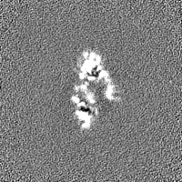
| Annotation | Sharpened Map | ||||||||||||
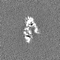
| Projections & Slices |
| ||||||||||||
| Density Histograms |


-Half map: Half Map A
| File | emd_10337_half_map_1.map | ||||||||||||
|---|---|---|---|---|---|---|---|---|---|---|---|---|---|
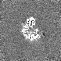
| Annotation | Half Map A | ||||||||||||
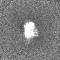
| Projections & Slices |
| ||||||||||||
| Density Histograms |


-Half map: Half Map B
| File | emd_10337_half_map_2.map | ||||||||||||
|---|---|---|---|---|---|---|---|---|---|---|---|---|---|
| Annotation | Half Map B | ||||||||||||
| Projections & Slices |
| ||||||||||||
| Density Histograms |


- Sample components
-Entire : ERCC1/XPF complex
| Entire | Name: ERCC1/XPF complex |
|---|---|
| Components |
|
-Supramolecule #1: ERCC1/XPF complex
| Supramolecule | Name: ERCC1/XPF complex / type: complex / ID: 1 / Parent: 0 / Macromolecule list: all |
|---|---|
| Source (natural) | Organism: Homo sapiens (human) |
-Macromolecule #1: DNA repair endonuclease XPF
| Macromolecule | Name: DNA repair endonuclease XPF / type: protein_or_peptide / ID: 1 / Number of copies: 1 / Enantiomer: LEVO / EC number: Hydrolases; Acting on ester bonds |
|---|---|
| Source (natural) | Organism: Homo sapiens (human) |
| Molecular weight | Theoretical: 104.636156 KDa |
| Recombinant expression | Organism:   Spodoptera frugiperda (fall armyworm) Spodoptera frugiperda (fall armyworm) |
| Sequence | String: MESGQPARRI AMAPLLEYER QLVLELLDTD GLVVCARGLG ADRLLYHFLQ LHCHPACLVL VLNTQPAEEE YFINQLKIEG VEHLPRRVT NEITSNSRYE VYTQGGVIFA TSRILVVDFL TDRIPSDLIT GILVYRAHRI IESCQEAFIL RLFRQKNKRG F IKAFTDNA ...String: MESGQPARRI AMAPLLEYER QLVLELLDTD GLVVCARGLG ADRLLYHFLQ LHCHPACLVL VLNTQPAEEE YFINQLKIEG VEHLPRRVT NEITSNSRYE VYTQGGVIFA TSRILVVDFL TDRIPSDLIT GILVYRAHRI IESCQEAFIL RLFRQKNKRG F IKAFTDNA VAFDTGFCHV ERVMRNLFVR KLYLWPRFHV AVNSFLEQHK PEVVEIHVSM TPTMLAIQTA ILDILNACLK EL KCHNPSL EVEDLSLENA IGKPFDKTIR HYLDPLWHQL GAKTKSLVQD LKILRTLLQY LSQYDCVTFL NLLESLRATE KAF GQNSGW LFLDSSTSMF INARARVYHL PDAKMSKKEK ISEKMEIKEG EETKKELVLE SNPKWEALTE VLKEIEAENK ESEA LGGPG QVLICASDDR TCSQLRDYIT LGAEAFLLRL YRKTFEKDSK AEEVWMKFRK EDSSKRIRKS HKRPKDPQNK ERAST KERT LKKKKRKLTL TQMVGKPEEL EEEGDVEEGY RREISSSPES CPEEIKHEEF DVNLSSDAAF GILKEPLTII HPLLGC SDP YALTRVLHEV EPRYVVLYDA ELTFVRQLEI YRASRPGKPL RVYFLIYGGS TEEQRYLTAL RKEKEAFEKL IREKASM VV PEEREGRDET NLDLVRGTAS ADVSTDTRKA GGQEQNGTQQ SIVVDMREFR SELPSLIHRR GIDIEPVTLE VGDYILTP E MCVERKSISD LIGSLNNGRL YSQCISMSRY YKRPVLLIEF DPSKPFSLTS RGALFQEISS NDISSKLTLL TLHFPRLRI LWCPSPHATA ELFEELKQSK PQPDAATALA ITADSETLPE SEKYNPGPQD FLLKMPGVNA KNCRSLMHHV KNIAELAALS QDELTSILG NAANAKQLYD FIHTSFAEVV SKGKGKK UniProtKB: DNA repair endonuclease XPF |
-Macromolecule #2: DNA excision repair protein ERCC-1
| Macromolecule | Name: DNA excision repair protein ERCC-1 / type: protein_or_peptide / ID: 2 / Number of copies: 1 / Enantiomer: LEVO |
|---|---|
| Source (natural) | Organism: Homo sapiens (human) |
| Molecular weight | Theoretical: 32.598301 KDa |
| Recombinant expression | Organism: Spodoptera frugiperda (fall armyworm) |
| Sequence | String: MDPGKDKEGV PQPSGPPARK KFVIPLDEDE VPPGVAKPLF RSTQSLPTVD TSAQAAPQTY AEYAISQPLE GAGATCPTGS EPLAGETPN QALKPGAKSN SIIVSPRQRG NPVLKFVRNV PWEFGDVIPD YVLGQSTCAL FLSLRYHNLH PDYIHGRLQS L GKNFALRV ...String: MDPGKDKEGV PQPSGPPARK KFVIPLDEDE VPPGVAKPLF RSTQSLPTVD TSAQAAPQTY AEYAISQPLE GAGATCPTGS EPLAGETPN QALKPGAKSN SIIVSPRQRG NPVLKFVRNV PWEFGDVIPD YVLGQSTCAL FLSLRYHNLH PDYIHGRLQS L GKNFALRV LLVQVDVKDP QQALKELAKM CILADCTLIL AWSPEEAGRY LETYKAYEQK PADLLMEKLE QDFVSRVTEC LT TVKSVNK TDSQTLLTTF GSLEQLIAAS REDLALCPGL GPQKARRLFD VLHEPFLKVP UniProtKB: DNA excision repair protein ERCC-1 |
-Experimental details
-Structure determination
| Method | cryo EM |
|---|---|
 Processing Processing | single particle reconstruction |
| Aggregation state | 2D array |
-Sample preparation
| Buffer | pH: 7.8 Details: 20 mM HEPES pH 7.8, 150 mM NaCl, 1 mM TCEP, 0.01% CHAPS |
|---|---|
| Grid | Model: Quantifoil R1.2/1.3 / Mesh: 400 |
| Vitrification | Cryogen name: ETHANE / Chamber humidity: 100 % / Instrument: FEI VITROBOT MARK IV |
- Electron microscopy
| Microscope | FEI TITAN KRIOS |
|---|---|
| Image recording | Film or detector model: GATAN K2 SUMMIT (4k x 4k) / Detector mode: COUNTING / Digitization - Frames/image: 2-17 / Number real images: 15315 / Average electron dose: 63.0 e/Å2 |
| Electron beam | Acceleration voltage: 300 kV / Electron source:  FIELD EMISSION GUN FIELD EMISSION GUN |
| Electron optics | Calibrated defocus max: 4.5 µm / Calibrated defocus min: 0.9 µm / Illumination mode: FLOOD BEAM / Imaging mode: BRIGHT FIELD / Cs: 2.7 mm / Nominal defocus max: 4.2 µm / Nominal defocus min: 1.2 µm |
| Sample stage | Specimen holder model: FEI TITAN KRIOS AUTOGRID HOLDER |
| Experimental equipment |  Model: Titan Krios / Image courtesy: FEI Company |
-Image processing
| Startup model | Type of model: INSILICO MODEL |
|---|---|
| Final reconstruction | Number classes used: 1 / Applied symmetry - Point group: C1 (asymmetric) / Resolution.type: BY AUTHOR / Resolution: 3.6 Å / Resolution method: FSC 0.143 CUT-OFF / Software - Name: cryoSPARC (ver. 2) / Number images used: 405339 |
| Initial angle assignment | Type: ANGULAR RECONSTITUTION |
| Final angle assignment | Type: ANGULAR RECONSTITUTION |
