 Movie
Movie Controller


+ Open data

- Basic information
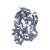
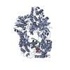
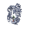
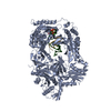
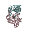
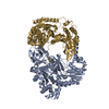
| Entry | Database: PDB / ID: 3avv | ||||||
|---|---|---|---|---|---|---|---|
| Title | Structure of viral RNA polymerase complex 3 | ||||||
 Components Components |
| ||||||
 Keywords Keywords | TRANSLATION / TRANSFERASE/RNA / RNA polymerase / TRANSFERASE-RNA complex | ||||||
| Function / homology |  Function and homology information Function and homology informationguanyl-nucleotide exchange factor complex / protein-synthesizing GTPase / guanosine tetraphosphate binding / translation elongation factor activity / RNA-directed RNA polymerase / response to antibiotic / viral RNA genome replication / nucleotide binding / RNA-directed RNA polymerase activity / GTPase activity ...guanyl-nucleotide exchange factor complex / protein-synthesizing GTPase / guanosine tetraphosphate binding / translation elongation factor activity / RNA-directed RNA polymerase / response to antibiotic / viral RNA genome replication / nucleotide binding / RNA-directed RNA polymerase activity / GTPase activity / GTP binding / magnesium ion binding / RNA binding / metal ion binding / plasma membrane / cytosol / cytoplasm Similarity search - Function | ||||||
| Biological species |  synthetic construct (others)  Escherichia phage Qbeta (virus) Escherichia phage Qbeta (virus) | ||||||
| Method |  X-RAY DIFFRACTION / SYNCHROTRON / MOLECULAR REPLACEMENT / Resolution: 3.119 Å X-RAY DIFFRACTION / SYNCHROTRON / MOLECULAR REPLACEMENT / Resolution: 3.119 Å | ||||||
 Authors Authors | Takeshita, D. / Tomita, K. | ||||||
 Citation Citation | Journal: Nat.Struct.Mol.Biol. / Year: 2012 Title: Molecular basis for RNA polymerization by Q beta replicase Authors: Takeshita, D. / Tomita, K. | ||||||
| History |
|
- Structure visualization
| Structure viewer | Molecule: MolmilJmol/JSmol |
|---|
- Downloads & links
-Download
| PDBx/mmCIF format | 3avv.cif.gz | 259.2 KB | Display | PDBx/mmCIF format |
|---|---|---|---|---|
| PDB format | pdb3avv.ent.gz | 200.2 KB | Display | PDB format |
| PDBx/mmJSON format | 3avv.json.gz | Tree view | PDBx/mmJSON format | |
| Others |  Other downloads Other downloads |
-Validation report
| Arichive directory | https://data.pdbj.org/pub/pdb/validation_reports/av/3avvftp://data.pdbj.org/pub/pdb/validation_reports/av/3avv | HTTPS FTP |
|---|
-Related structure data
| Related structure data | 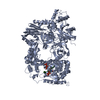 3avtC 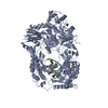 3avuC  3avwC  3avxC 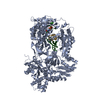 3avyC 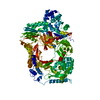 3agpS C: citing same article ( S: Starting model for refinement |
|---|---|
| Similar structure data |


-Links
 PDBj
PDBj


- Assembly
| Deposited unit | 
| ||||||||
|---|---|---|---|---|---|---|---|---|---|
| 1 |
| ||||||||
| Unit cell |
|
-Components
| #1: Protein | Mass: 141417.688 Da / Num. of mol.: 1 Source method: isolated from a genetically manipulated source Details: chimera of Elongation factor Ts, Elongation factor Tu and Q beta replicase Source: (gene. exp.) Escherichia phage Qbeta (virus)Production host: References: UniProt: P0A6P3, UniProt: P0A6N3, UniProt: Q8LTE0 | ||
|---|---|---|---|
| #2: RNA chain | Mass: 2542.561 Da / Num. of mol.: 1 / Source method: obtained synthetically | ||
| #3: RNA chain | Mass: 4163.582 Da / Num. of mol.: 1 / Source method: obtained synthetically | ||
| #4: Chemical |   Mass: 40.078 Da / Num. of mol.: 2 / Source method: obtained synthetically / Formula: Ca Mass: 40.078 Da / Num. of mol.: 2 / Source method: obtained synthetically / Formula: Ca#5: Water | ChemComp-HOH / |  Mass: 18.015 Da / Num. of mol.: 10 / Source method: isolated from a natural source / Formula: H2O Mass: 18.015 Da / Num. of mol.: 10 / Source method: isolated from a natural source / Formula: H2O |
-Experimental details
-Experiment
| Experiment | Method: X-RAY DIFFRACTION / Number of used crystals: 1 |
|---|
- Sample preparation
| Crystal | Density Matthews: 3.02 Å3/Da / Density % sol: 59.32 % |
|---|---|
| Crystal grow | Method: vapor diffusion, sitting drop / Details: VAPOR DIFFUSION, SITTING DROP |
-Data collection
| Diffraction source | Source: SYNCHROTRON / Site: Photon Factory  / Beamline: BL-17A / Beamline: BL-17A |
|---|---|
| Detector | Date: Oct 1, 2010 |
| Radiation | Protocol: SINGLE WAVELENGTH / Monochromatic (M) / Laue (L): M / Scattering type: x-ray |
| Radiation wavelength | Relative weight: 1 |
| Reflection | Resolution: 3.1→50 Å / Num. obs: 31085 / Biso Wilson estimate: 60.23 Å2 |
- Processing
| Software |
| ||||||||||||||||||||||||||||||||||||||||||||||||||||||||||||||||||||||||||||||||||||
|---|---|---|---|---|---|---|---|---|---|---|---|---|---|---|---|---|---|---|---|---|---|---|---|---|---|---|---|---|---|---|---|---|---|---|---|---|---|---|---|---|---|---|---|---|---|---|---|---|---|---|---|---|---|---|---|---|---|---|---|---|---|---|---|---|---|---|---|---|---|---|---|---|---|---|---|---|---|---|---|---|---|---|---|---|---|
| Refinement | Method to determine structure: MOLECULAR REPLACEMENT Starting model: 3AGP Resolution: 3.119→19.996 Å / Occupancy max: 1 / Occupancy min: 1 / FOM work R set: 0.8052 / SU ML: 0.44 / σ(F): 1.45 / Phase error: 26.9 / Stereochemistry target values: ML
| ||||||||||||||||||||||||||||||||||||||||||||||||||||||||||||||||||||||||||||||||||||
| Solvent computation | Shrinkage radii: 0.27 Å / VDW probe radii: 0.6 Å / Solvent model: FLAT BULK SOLVENT MODEL / Bsol: 0.001 Å2 / ksol: 0.209 e/Å3 | ||||||||||||||||||||||||||||||||||||||||||||||||||||||||||||||||||||||||||||||||||||
| Displacement parameters | Biso max: 164.5 Å2 / Biso mean: 68.4012 Å2 / Biso min: 19.87 Å2
| ||||||||||||||||||||||||||||||||||||||||||||||||||||||||||||||||||||||||||||||||||||
| Refinement step | Cycle: LAST / Resolution: 3.119→19.996 Å
| ||||||||||||||||||||||||||||||||||||||||||||||||||||||||||||||||||||||||||||||||||||
| Refine LS restraints |
| ||||||||||||||||||||||||||||||||||||||||||||||||||||||||||||||||||||||||||||||||||||
| LS refinement shell | Refine-ID: X-RAY DIFFRACTION / Total num. of bins used: 11
|
