[English] 日本語
 Yorodumi
- PDB-6gzv: Identification of a druggable VP1-VP3 interprotomer pocket in the... -
+ Open data
- Basic information
| Entry | Database: PDB / ID: 6gzv | ||||||
|---|---|---|---|---|---|---|---|
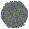
| Title | Identification of a druggable VP1-VP3 interprotomer pocket in the capsid of enteroviruses | ||||||
 Components Components |
| ||||||
 Keywords Keywords | VIRUS / Inhibitor / complex | ||||||
| Function / homology |  Function and homology information Function and homology informationsymbiont-mediated perturbation of host transcription / symbiont-mediated suppression of host cytoplasmic pattern recognition receptor signaling pathway via inhibition of RIG-I activity / symbiont-mediated suppression of host cytoplasmic pattern recognition receptor signaling pathway via inhibition of MDA-5 activity / symbiont-mediated suppression of host cytoplasmic pattern recognition receptor signaling pathway via inhibition of MAVS activity / picornain 2A / symbiont-mediated suppression of host mRNA export from nucleus / symbiont genome entry into host cell via pore formation in plasma membrane / picornain 3C / T=pseudo3 icosahedral viral capsid / host cell cytoplasmic vesicle membrane ...symbiont-mediated perturbation of host transcription / symbiont-mediated suppression of host cytoplasmic pattern recognition receptor signaling pathway via inhibition of RIG-I activity / symbiont-mediated suppression of host cytoplasmic pattern recognition receptor signaling pathway via inhibition of MDA-5 activity / symbiont-mediated suppression of host cytoplasmic pattern recognition receptor signaling pathway via inhibition of MAVS activity / picornain 2A / symbiont-mediated suppression of host mRNA export from nucleus / symbiont genome entry into host cell via pore formation in plasma membrane / picornain 3C / T=pseudo3 icosahedral viral capsid / host cell cytoplasmic vesicle membrane / endocytosis involved in viral entry into host cell / nucleoside-triphosphate phosphatase / channel activity / symbiont-mediated suppression of host NF-kappaB cascade / monoatomic ion transmembrane transport / DNA replication / RNA helicase activity / induction by virus of host autophagy / RNA-directed RNA polymerase / viral RNA genome replication / cysteine-type endopeptidase activity / RNA-dependent RNA polymerase activity / virus-mediated perturbation of host defense response / DNA-templated transcription / host cell nucleus / virion attachment to host cell / structural molecule activity / ATP hydrolysis activity / proteolysis / RNA binding / ATP binding / membrane / metal ion binding Similarity search - Function | ||||||
| Biological species |   Coxsackievirus B3 Coxsackievirus B3 | ||||||
| Method | ELECTRON MICROSCOPY / single particle reconstruction / cryo EM / Resolution: 4 Å | ||||||
 Authors Authors | Geraets, J.A. / Flatt, J.W. / Domanska, A. / Butcher, S.J. | ||||||
| Funding support |  Finland, 1items Finland, 1items
| ||||||
 Citation Citation |  Journal: PLoS Biol / Year: 2019 Journal: PLoS Biol / Year: 2019Title: A novel druggable interprotomer pocket in the capsid of rhino- and enteroviruses. Authors: Rana Abdelnabi / James A Geraets / Yipeng Ma / Carmen Mirabelli / Justin W Flatt / Aušra Domanska / Leen Delang / Dirk Jochmans / Timiri Ajay Kumar / Venkatesan Jayaprakash / Barij Nayan ...Authors: Rana Abdelnabi / James A Geraets / Yipeng Ma / Carmen Mirabelli / Justin W Flatt / Aušra Domanska / Leen Delang / Dirk Jochmans / Timiri Ajay Kumar / Venkatesan Jayaprakash / Barij Nayan Sinha / Pieter Leyssen / Sarah J Butcher / Johan Neyts /    Abstract: Rhino- and enteroviruses are important human pathogens, against which no antivirals are available. The best-studied inhibitors are "capsid binders" that fit in a hydrophobic pocket of the viral ...Rhino- and enteroviruses are important human pathogens, against which no antivirals are available. The best-studied inhibitors are "capsid binders" that fit in a hydrophobic pocket of the viral capsid. Employing a new class of entero-/rhinovirus inhibitors and by means of cryo-electron microscopy (EM), followed by resistance selection and reverse genetics, we discovered a hitherto unknown druggable pocket that is formed by viral proteins VP1 and VP3 and that is conserved across entero-/rhinovirus species. We propose that these inhibitors stabilize a key region of the virion, thereby preventing the conformational expansion needed for viral RNA release. A medicinal chemistry effort resulted in the identification of analogues targeting this pocket with broad-spectrum activity against Coxsackieviruses B (CVBs) and compounds with activity against enteroviruses (EV) of groups C and D, and even rhinoviruses (RV). Our findings provide novel insights in the biology of the entry of entero-/rhinoviruses and open new avenues for the design of broad-spectrum antivirals against these pathogens. | ||||||
| History |
|
- Structure visualization
| Movie |
 Movie viewer Movie viewer |
|---|---|
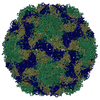
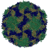
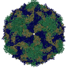
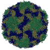
| Structure viewer | Molecule:  Molmil Molmil Jmol/JSmol Jmol/JSmol |
- Downloads & links
- Download
| PDBx/mmCIF format |  6gzv.cif.gz 6gzv.cif.gz | 269.4 KB | Display |  PDBx/mmCIF format PDBx/mmCIF format |
|---|---|---|---|---|
| PDB format |  pdb6gzv.ent.gz pdb6gzv.ent.gz | 216 KB | Display |  PDB format PDB format |
| PDBx/mmJSON format |  6gzv.json.gz 6gzv.json.gz | Tree view |  PDBx/mmJSON format PDBx/mmJSON format | |
| Others |  Other downloads Other downloads |
-Validation report
| Summary document |  6gzv_validation.pdf.gz 6gzv_validation.pdf.gz | 1.2 MB | Display |  wwPDB validaton report wwPDB validaton report |
|---|---|---|---|---|
| Full document |  6gzv_full_validation.pdf.gz 6gzv_full_validation.pdf.gz | 1.2 MB | Display | |
| Data in XML |  6gzv_validation.xml.gz 6gzv_validation.xml.gz | 39.8 KB | Display | |
| Data in CIF |  6gzv_validation.cif.gz 6gzv_validation.cif.gz | 59.2 KB | Display | |
| Arichive directory |  https://data.pdbj.org/pub/pdb/validation_reports/gz/6gzv https://data.pdbj.org/pub/pdb/validation_reports/gz/6gzv ftp://data.pdbj.org/pub/pdb/validation_reports/gz/6gzv ftp://data.pdbj.org/pub/pdb/validation_reports/gz/6gzv | HTTPS FTP |
-Related structure data
| Related structure data |  0103MC M: map data used to model this data C: citing same article ( |
|---|---|
| Similar structure data | |
| EM raw data |  EMPIAR-10199 (Title: Identification of a druggable VP1-VP3 interprotomer pocket in the capsid of enteroviruses EMPIAR-10199 (Title: Identification of a druggable VP1-VP3 interprotomer pocket in the capsid of enterovirusesData size: 2.2 TB Data #1: Unaligned multi-frame micrographs of CVB3 in complex with a small molecular inhibitor [micrographs - single frame]) |
- Links
Links
- Assembly
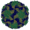
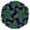
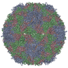
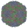
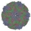
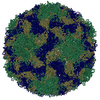
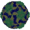
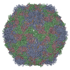
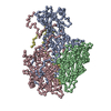
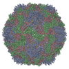
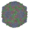
| Deposited unit | 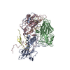
|
|---|---|
| 1 | x 60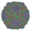
|
| Symmetry | Point symmetry: (Schoenflies symbol: I (icosahedral)) |
- Components
| #1: Protein | Mass: 31639.438 Da / Num. of mol.: 1 / Source method: isolated from a natural source / Source: (natural)  Coxsackievirus B3 (strain Nancy) / Strain: Nancy Coxsackievirus B3 (strain Nancy) / Strain: NancyReferences: UniProt: P03313, picornain 2A, nucleoside-triphosphate phosphatase, picornain 3C, RNA-directed RNA polymerase |
|---|---|
| #2: Protein | Mass: 28836.502 Da / Num. of mol.: 1 / Source method: isolated from a natural source / Source: (natural)  Coxsackievirus B3 (strain Nancy) / Strain: Nancy Coxsackievirus B3 (strain Nancy) / Strain: NancyReferences: UniProt: P03313, picornain 2A, nucleoside-triphosphate phosphatase, picornain 3C, RNA-directed RNA polymerase |
| #3: Protein | Mass: 26195.727 Da / Num. of mol.: 1 / Source method: isolated from a natural source / Source: (natural)  Coxsackievirus B3 (strain Nancy) / Strain: Nancy Coxsackievirus B3 (strain Nancy) / Strain: NancyReferences: UniProt: P03313, picornain 2A, nucleoside-triphosphate phosphatase, picornain 3C, RNA-directed RNA polymerase |
| #4: Protein | Mass: 7580.377 Da / Num. of mol.: 1 / Source method: isolated from a natural source / Source: (natural)  Coxsackievirus B3 (strain Nancy) / Strain: Nancy Coxsackievirus B3 (strain Nancy) / Strain: NancyReferences: UniProt: P03313, picornain 2A, nucleoside-triphosphate phosphatase, picornain 3C, RNA-directed RNA polymerase |
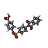
| #5: Chemical | ChemComp-FHK / |
-Experimental details
-Experiment
| Experiment | Method: ELECTRON MICROSCOPY |
|---|---|
| EM experiment | Aggregation state: PARTICLE / 3D reconstruction method: single particle reconstruction |
- Sample preparation
| Component | Name: Coxsackievirus B3 (strain Nancy) / Type: VIRUS Details: Coxsackievirus B3 (strain Nancy) NCBI Taxonomy ID: 103903 Full, non-enveloped virion from natural source Diameter: ~314A Triangulation: 1 (pT3) Benzene sulfonamide derivative Molecular ...Details: Coxsackievirus B3 (strain Nancy) NCBI Taxonomy ID: 103903 Full, non-enveloped virion from natural source Diameter: ~314A Triangulation: 1 (pT3) Benzene sulfonamide derivative Molecular weight: 422Da Binding sites on capsid: 60 Entity ID: #1-#4 / Source: NATURAL | |||||||||||||||
|---|---|---|---|---|---|---|---|---|---|---|---|---|---|---|---|---|
| Source (natural) | Organism:  Coxsackievirus B3 (strain Nancy) Coxsackievirus B3 (strain Nancy) | |||||||||||||||
| Details of virus | Empty: NO / Enveloped: NO / Isolate: STRAIN / Type: VIRION | |||||||||||||||
| Natural host | Organism: Homo sapiens | |||||||||||||||
| Virus shell | Diameter: 314 nm / Triangulation number (T number): 1 | |||||||||||||||
| Buffer solution | pH: 7.5 | |||||||||||||||
| Buffer component |
| |||||||||||||||
| Specimen | Embedding applied: NO / Shadowing applied: NO / Staining applied: NO / Vitrification applied: YES Details: Inhibitor was added at 2500:1 molar ratio to purified CVB3 and incubated for 1h at 37C | |||||||||||||||
| Vitrification | Instrument: HOMEMADE PLUNGER / Cryogen name: ETHANE |
- Electron microscopy imaging
| Experimental equipment |  Model: Titan Krios / Image courtesy: FEI Company |
|---|---|
| Microscopy | Model: FEI TITAN KRIOS |
| Electron gun | Electron source:  FIELD EMISSION GUN / Accelerating voltage: 300 kV / Illumination mode: FLOOD BEAM FIELD EMISSION GUN / Accelerating voltage: 300 kV / Illumination mode: FLOOD BEAM |
| Electron lens | Mode: BRIGHT FIELD / Cs: 2.7 mm |
| Specimen holder | Specimen holder model: FEI TITAN KRIOS AUTOGRID HOLDER |
| Image recording | Electron dose: 47 e/Å2 / Detector mode: COUNTING / Film or detector model: GATAN K2 SUMMIT (4k x 4k) |
| Image scans | Sampling size: 5 µm / Movie frames/image: 20 |
- Processing
| EM software |
| ||||||||||||||||||||||||||||||||||||
|---|---|---|---|---|---|---|---|---|---|---|---|---|---|---|---|---|---|---|---|---|---|---|---|---|---|---|---|---|---|---|---|---|---|---|---|---|---|
| CTF correction | Type: PHASE FLIPPING AND AMPLITUDE CORRECTION | ||||||||||||||||||||||||||||||||||||
| Symmetry | Point symmetry: I (icosahedral) | ||||||||||||||||||||||||||||||||||||
| 3D reconstruction | Resolution: 4 Å / Resolution method: FSC 0.143 CUT-OFF / Num. of particles: 4891 / Symmetry type: POINT | ||||||||||||||||||||||||||||||||||||
| Atomic model building | Protocol: FLEXIBLE FIT / Space: REAL |
 Movie
Movie Controller



 PDBj
PDBj

