 Movie
Movie Controller


+ Open data

- Basic information
| Entry | Database: PDB / ID: 5xxb | ||||||
|---|---|---|---|---|---|---|---|
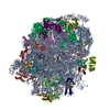
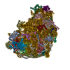
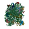
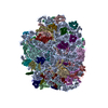
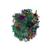
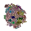
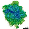
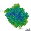
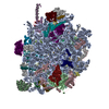
| Title | Large subunit of Toxoplasma gondii ribosome | ||||||
 Components Components |
| ||||||
 Keywords Keywords | RIBOSOME / parasitic ribosome / rRNA / rproteins | ||||||
| Function / homology | RNA / RNA (> 10) / RNA (> 100) / RNA (> 1000) Function and homology information Function and homology information | ||||||
| Biological species |  | ||||||
| Method | ELECTRON MICROSCOPY / single particle reconstruction / cryo EM / Resolution: 3.17 Å | ||||||
 Authors Authors | Li, Z. / Guo, Q. / Zheng, L. / Ji, Y. / Xie, Y. / Lai, D. / Lun, Z. / Suo, X. / Gao, N. | ||||||
 Citation Citation | Journal: Cell Res / Year: 2017 Title: Cryo-EM structures of the 80S ribosomes from human parasites Trichomonas vaginalis and Toxoplasma gondii. Authors: Zhifei Li / Qiang Guo / Lvqin Zheng / Yongsheng Ji / Yi-Ting Xie / De-Hua Lai / Zhao-Rong Lun / Xun Suo / Ning Gao /  Abstract: As an indispensable molecular machine universal in all living organisms, the ribosome has been selected by evolution to be the natural target of many antibiotics and small-molecule inhibitors. High- ...As an indispensable molecular machine universal in all living organisms, the ribosome has been selected by evolution to be the natural target of many antibiotics and small-molecule inhibitors. High-resolution structures of pathogen ribosomes are crucial for understanding the general and unique aspects of translation control in disease-causing microbes. With cryo-electron microscopy technique, we have determined structures of the cytosolic ribosomes from two human parasites, Trichomonas vaginalis and Toxoplasma gondii, at resolution of 3.2-3.4 Å. Although the ribosomal proteins from both pathogens are typical members of eukaryotic families, with a co-evolution pattern between certain species-specific insertions/extensions and neighboring ribosomal RNA (rRNA) expansion segments, the sizes of their rRNAs are sharply different. Very interestingly, rRNAs of T. vaginalis are in size comparable to prokaryotic counterparts, with nearly all the eukaryote-specific rRNA expansion segments missing. These structures facilitate the dissection of evolution path for ribosomal proteins and RNAs, and may aid in design of novel translation inhibitors. | ||||||
| History |
|
- Structure visualization
| Movie |
Movie viewer |
|---|---|
| Structure viewer | Molecule: MolmilJmol/JSmol |
- Downloads & links
-Download
| PDBx/mmCIF format | 5xxb.cif.gz | 3 MB | Display | PDBx/mmCIF format |
|---|---|---|---|---|
| PDB format | pdb5xxb.ent.gz | Display | PDB format | |
| PDBx/mmJSON format | 5xxb.json.gz | Tree view | PDBx/mmJSON format | |
| Others |  Other downloads Other downloads |
-Validation report
| Arichive directory | https://data.pdbj.org/pub/pdb/validation_reports/xx/5xxbftp://data.pdbj.org/pub/pdb/validation_reports/xx/5xxb | HTTPS FTP |
|---|
-Related structure data
| Related structure data | 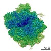 6778MC  6780C 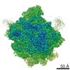 6784C 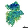 6788C 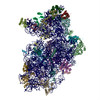 5xxuC 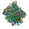 5xy3C 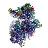 5xyiC M: map data used to model this data C: citing same article ( |
|---|---|
| Similar structure data |

-Links
 PDBj
PDBj


- Assembly
| Deposited unit | 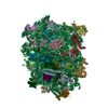
|
|---|---|
| 1 |
|
-Components
-RNA chain , 3 types, 3 molecules 134
| #1: RNA chain | Mass: 1120153.125 Da / Num. of mol.: 1 / Source method: isolated from a natural source / Source: (natural) |
|---|---|
| #2: RNA chain | Mass: 39921.641 Da / Num. of mol.: 1 / Source method: isolated from a natural source / Source: (natural) |
| #3: RNA chain | Mass: 50787.980 Da / Num. of mol.: 1 / Source method: isolated from a natural source / Source: (natural) |
+Ribosomal protein ... , 41 types, 41 molecules ABCDEFGHIJKLMNOPQRSTUVWXYZabcd...
-Details
| Has protein modification | Y |
|---|---|
| Sequence details | Sequences used in this study were derived from Toxoplasma gondii RH strain, which were not ...Sequences used in this study were derived from Toxoplasma gondii RH strain, which were not available at UNIPROT at the time of data processing. |
-Experimental details
-Experiment
| Experiment | Method: ELECTRON MICROSCOPY |
|---|---|
| EM experiment | Aggregation state: PARTICLE / 3D reconstruction method: single particle reconstruction |
- Sample preparation
| Component | Name: Large subunit of Toxoplasma gondii ribosome / Type: RIBOSOME / Entity ID: all / Source: NATURAL |
|---|---|
| Source (natural) | Organism: |
| Buffer solution | pH: 7.4 |
| Specimen | Embedding applied: NO / Shadowing applied: NO / Staining applied: NO / Vitrification applied: YES |
| Vitrification | Cryogen name: ETHANE |
- Electron microscopy imaging
| Microscopy | Model: FEI TITAN |
|---|---|
| Electron gun | Electron source:  FIELD EMISSION GUN / Accelerating voltage: 300 kV / Illumination mode: FLOOD BEAM FIELD EMISSION GUN / Accelerating voltage: 300 kV / Illumination mode: FLOOD BEAM |
| Electron lens | Mode: BRIGHT FIELD |
| Image recording | Electron dose: 2 e/Å2 / Film or detector model: GATAN K2 SUMMIT (4k x 4k) |
- Processing
| CTF correction | Type: PHASE FLIPPING AND AMPLITUDE CORRECTION |
|---|---|
| 3D reconstruction | Resolution: 3.17 Å / Resolution method: FSC 0.143 CUT-OFF / Num. of particles: 108162 / Symmetry type: POINT |
