 Movie
Movie Controller


+ Open data

- Basic information
| Entry | Database: PDB / ID: 3j7r | |||||||||||||||
|---|---|---|---|---|---|---|---|---|---|---|---|---|---|---|---|---|
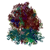
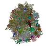
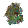
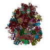
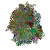
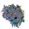
| Title | Structure of the translating mammalian ribosome-Sec61 complex | |||||||||||||||
 Components Components |
| |||||||||||||||
 Keywords Keywords | RIBOSOME / mammalian / Sec61 / translocation / translation | |||||||||||||||
| Function / homology |  Function and homology information Function and homology informationregulation of cell cycle => GO:0051726 / regulation of cell cycle => GO:0051726 / TNFR1-mediated ceramide production / Translation initiation complex formation / Formation of the ternary complex, and subsequently, the 43S complex / Ribosomal scanning and start codon recognition / Degradation of CDH1 / Regulation of TNFR1 signaling / TNFR1-induced NF-kappa-B signaling pathway / : ...regulation of cell cycle => GO:0051726 / regulation of cell cycle => GO:0051726 / TNFR1-mediated ceramide production / Translation initiation complex formation / Formation of the ternary complex, and subsequently, the 43S complex / Ribosomal scanning and start codon recognition / Degradation of CDH1 / Regulation of TNFR1 signaling / TNFR1-induced NF-kappa-B signaling pathway / : / L13a-mediated translational silencing of Ceruloplasmin expression / SRP-dependent cotranslational protein targeting to membrane / Major pathway of rRNA processing in the nucleolus and cytosol / Formation of a pool of free 40S subunits / GTP hydrolysis and joining of the 60S ribosomal subunit / Nonsense Mediated Decay (NMD) independent of the Exon Junction Complex (EJC) / Nonsense Mediated Decay (NMD) enhanced by the Exon Junction Complex (EJC) / pronephric nephron development / Sec61 translocon complex / protein insertion into ER membrane / (S)-malate dehydrogenase (NAD+, oxaloacetate-forming) / protein-transporting ATPase activity / L-malate dehydrogenase (NAD+) activity / carboxylic acid metabolic process / SRP-dependent cotranslational protein targeting to membrane / embryonic brain development / post-translational protein targeting to membrane, translocation / translation at presynapse / cellular response to actinomycin D / protein-DNA complex disassembly / positive regulation of gastrulation / protein tyrosine kinase inhibitor activity / IRE1-RACK1-PP2A complex / positive regulation of Golgi to plasma membrane protein transport / alpha-beta T cell differentiation / negative regulation of RNA splicing / neural crest cell differentiation / cysteine-type endopeptidase activator activity involved in apoptotic process / negative regulation of intrinsic apoptotic signaling pathway in response to hydrogen peroxide / regulation of establishment of cell polarity / negative regulation of phagocytosis / cytoplasmic side of rough endoplasmic reticulum membrane / organelle membrane / laminin receptor activity / positive regulation of mitochondrial depolarization / negative regulation of Wnt signaling pathway / BH3 domain binding / negative regulation of translational frameshifting / regulation of adenylate cyclase-activating G protein-coupled receptor signaling pathway / positive regulation of GTPase activity / membrane => GO:0016020 / regulation of cell division / protein serine/threonine kinase inhibitor activity / positive regulation of intrinsic apoptotic signaling pathway by p53 class mediator / ubiquitin ligase inhibitor activity / endonucleolytic cleavage to generate mature 3'-end of SSU-rRNA from (SSU-rRNA, 5.8S rRNA, LSU-rRNA) / positive regulation of signal transduction by p53 class mediator / protein localization to nucleus / negative regulation of ubiquitin-dependent protein catabolic process / protein targeting / phagocytic cup / tricarboxylic acid cycle / positive regulation of intrinsic apoptotic signaling pathway / translation regulator activity / endonucleolytic cleavage in ITS1 to separate SSU-rRNA from 5.8S rRNA and LSU-rRNA from tricistronic rRNA transcript (SSU-rRNA, 5.8S rRNA, LSU-rRNA) / rough endoplasmic reticulum / laminin binding / gastrulation / signaling adaptor activity / negative regulation of proteasomal ubiquitin-dependent protein catabolic process / SH2 domain binding / rescue of stalled cytosolic ribosome / class I DNA-(apurinic or apyrimidinic site) endonuclease activity / cytosolic ribosome / cyclin binding / negative regulation of phosphatidylinositol 3-kinase/protein kinase B signal transduction / DNA-(apurinic or apyrimidinic site) lyase / protein kinase C binding / ribosomal large subunit biogenesis / maturation of LSU-rRNA from tricistronic rRNA transcript (SSU-rRNA, 5.8S rRNA, LSU-rRNA) / maturation of SSU-rRNA from tricistronic rRNA transcript (SSU-rRNA, 5.8S rRNA, LSU-rRNA) / positive regulation of apoptotic signaling pathway / mRNA 3'-UTR binding / maturation of SSU-rRNA / negative regulation of smoothened signaling pathway / neural tube closure / cellular response to glucose stimulus / small-subunit processome / positive regulation of protein-containing complex assembly / negative regulation of cell growth / positive regulation of protein phosphorylation / receptor tyrosine kinase binding / maintenance of translational fidelity / cellular response to growth factor stimulus / modification-dependent protein catabolic process / spindle / transcription coactivator binding / mRNA 5'-UTR binding / calcium channel activity / protein tag activity Similarity search - Function | |||||||||||||||
| Biological species |  | |||||||||||||||
| Method | ELECTRON MICROSCOPY / single particle reconstruction / cryo EM / Resolution: 3.9 Å | |||||||||||||||
 Authors Authors | Voorhees, R.M. / Fernandez, I.S. / Scheres, S.H.W. / Hegde, R.S. | |||||||||||||||
 Citation Citation | Journal: Cell / Year: 2014 Title: Structure of the mammalian ribosome-Sec61 complex to 3.4 Å resolution. Authors: Rebecca M Voorhees / Israel S Fernández / Sjors H W Scheres / Ramanujan S Hegde /  Abstract: Cotranslational protein translocation is a universally conserved process for secretory and membrane protein biosynthesis. Nascent polypeptides emerging from a translating ribosome are either ...Cotranslational protein translocation is a universally conserved process for secretory and membrane protein biosynthesis. Nascent polypeptides emerging from a translating ribosome are either transported across or inserted into the membrane via the ribosome-bound Sec61 channel. Here, we report structures of a mammalian ribosome-Sec61 complex in both idle and translating states, determined to 3.4 and 3.9 Å resolution. The data sets permit building of a near-complete atomic model of the mammalian ribosome, visualization of A/P and P/E hybrid-state tRNAs, and analysis of a nascent polypeptide in the exit tunnel. Unprecedented chemical detail is observed for both the ribosome-Sec61 interaction and the conformational state of Sec61 upon ribosome binding. Comparison of the maps from idle and translating complexes suggests how conformational changes to the Sec61 channel could facilitate translocation of a secreted polypeptide. The high-resolution structure of the mammalian ribosome-Sec61 complex provides a valuable reference for future functional and structural studies. | |||||||||||||||
| History |
|
- Structure visualization
| Movie |
Movie viewer |
|---|---|
| Structure viewer | Molecule: MolmilJmol/JSmol |
- Downloads & links
-Download
| PDBx/mmCIF format | 3j7r.cif.gz | 5.5 MB | Display | PDBx/mmCIF format |
|---|---|---|---|---|
| PDB format | pdb3j7r.ent.gz | Display | PDB format | |
| PDBx/mmJSON format | 3j7r.json.gz | Tree view | PDBx/mmJSON format | |
| Others |  Other downloads Other downloads |
-Validation report
| Arichive directory | https://data.pdbj.org/pub/pdb/validation_reports/j7/3j7rftp://data.pdbj.org/pub/pdb/validation_reports/j7/3j7r | HTTPS FTP |
|---|
-Related structure data
| Related structure data | 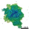 2644MC 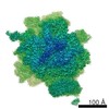 2646C 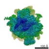 2649C 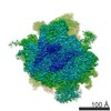 2650C  3j7oC  3j7pC  3j7qC M: map data used to model this data C: citing same article ( |
|---|---|
| Similar structure data |


-Links
 PDBj
PDBj


- Assembly
| Deposited unit | 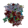
|
|---|---|
| 1 |
|
-Components
-RNA chain , 7 types, 7 molecules 578S2S4S5S6
| #1: RNA chain | Mass: 1206101.875 Da / Num. of mol.: 1 / Source method: isolated from a natural source / Source: (natural) |
|---|---|
| #2: RNA chain | Mass: 38691.914 Da / Num. of mol.: 1 / Source method: isolated from a natural source / Source: (natural) |
| #3: RNA chain | Mass: 50143.648 Da / Num. of mol.: 1 / Source method: isolated from a natural source / Source: (natural) |
| #50: RNA chain | Mass: 561958.812 Da / Num. of mol.: 1 / Source method: isolated from a natural source / Source: (natural) |
| #84: RNA chain | Mass: 3039.740 Da / Num. of mol.: 1 / Source method: isolated from a natural source / Source: (natural) |
| #85: RNA chain | Mass: 23874.191 Da / Num. of mol.: 1 / Source method: isolated from a natural source / Source: (natural) |
| #86: RNA chain | Mass: 24484.555 Da / Num. of mol.: 1 / Source method: isolated from a natural source / Source: (natural) |
+Ribosomal protein ... , 76 types, 76 molecules ABCDEFGHIJLMNOPQRSTUVWXYZabcde...
-Protein , 2 types, 2 molecules 12
| #47: Protein | Mass: 43496.219 Da / Num. of mol.: 1 / Source method: isolated from a natural source / Source: (natural) |
|---|---|
| #48: Protein | Mass: 7752.325 Da / Num. of mol.: 1 / Source method: isolated from a natural source / Source: (natural) |
-Protein/peptide , 1 types, 1 molecules 3
| #49: Protein/peptide | Mass: 3081.790 Da / Num. of mol.: 1 / Source method: isolated from a natural source / Source: (natural) |
|---|
-Non-polymers , 2 types, 170 molecules 

| #87: Chemical | ChemComp-MG /  Mass: 24.305 Da / Num. of mol.: 166 / Source method: obtained synthetically / Formula: Mg Mass: 24.305 Da / Num. of mol.: 166 / Source method: obtained synthetically / Formula: Mg#88: Chemical | ChemComp-ZN /  Mass: 65.409 Da / Num. of mol.: 4 / Source method: obtained synthetically / Formula: Zn Mass: 65.409 Da / Num. of mol.: 4 / Source method: obtained synthetically / Formula: Zn |
|---|
-Details
| Has protein modification | Y |
|---|
-Experimental details
-Experiment
| Experiment | Method: ELECTRON MICROSCOPY |
|---|---|
| EM experiment | Aggregation state: PARTICLE / 3D reconstruction method: single particle reconstruction |
- Sample preparation
| Component | Name: The 80S-Sec61 complex purified from porcine pancreas / Type: RIBOSOME |
|---|---|
| Buffer solution | Name: 50 mM HEPES, 200 mM potassium acetate, 15 mM magnesium acetate, 1 mM DTT, 0.25% Digitonin pH: 7.5 Details: 50 mM HEPES, 200 mM potassium acetate, 15 mM magnesium acetate, 1 mM DTT, 0.25% Digitonin |
| Specimen | Embedding applied: NO / Shadowing applied: NO / Staining applied: NO / Vitrification applied: YES |
| Specimen support | Details: Quantifoil R2/2 400 mesh copper grids |
| Vitrification | Instrument: FEI VITROBOT MARK IV / Cryogen name: ETHANE / Temp: 120 K Details: 3 uL sample was incubated on the grid for 30 seconds and blotted for 9 seconds before being plunged into liquid ethane (FEI VITROBOT MARK IV). |
- Electron microscopy imaging
| Experimental equipment |  Model: Titan Krios / Image courtesy: FEI Company |
|---|---|
| Microscopy | Model: FEI TITAN KRIOS / Date: Apr 7, 2014 |
| Electron gun | Electron source:  FIELD EMISSION GUN / Accelerating voltage: 300 kV / Illumination mode: OTHER FIELD EMISSION GUN / Accelerating voltage: 300 kV / Illumination mode: OTHER |
| Electron lens | Mode: BRIGHT FIELD / Nominal magnification: 59000 X / Calibrated magnification: 104478 X / Nominal defocus max: 3500 nm / Nominal defocus min: 2500 nm / Cs: 2.7 mm |
| Specimen holder | Specimen holder type: FEI TITAN KRIOS AUTOGRID HOLDER / Temperature: 70 K / Tilt angle max: 0 ° / Tilt angle min: 0 ° |
| Image recording | Electron dose: 27 e/Å2 / Film or detector model: FEI FALCON II (4k x 4k) / Details: Back-thinned |
| Radiation | Protocol: SINGLE WAVELENGTH / Monochromatic (M) / Laue (L): M / Scattering type: x-ray |
| Radiation wavelength | Relative weight: 1 |
- Processing
| EM software |
| |||||||||||||||||||||||||||||||||||
|---|---|---|---|---|---|---|---|---|---|---|---|---|---|---|---|---|---|---|---|---|---|---|---|---|---|---|---|---|---|---|---|---|---|---|---|---|
| CTF correction | Details: Each particle | |||||||||||||||||||||||||||||||||||
| Symmetry | Point symmetry: C1 (asymmetric) | |||||||||||||||||||||||||||||||||||
| 3D reconstruction | Method: Single particle / Resolution: 3.9 Å / Resolution method: FSC 0.143 CUT-OFF / Num. of particles: 14723 / Nominal pixel size: 1.34 Å / Symmetry type: POINT | |||||||||||||||||||||||||||||||||||
| Atomic model building | B value: 37 / Protocol: OTHER / Space: RECIPROCAL / Target criteria: R-factor and FSC / Details: METHOD--Maximum likelihood | |||||||||||||||||||||||||||||||||||
| Atomic model building |
| |||||||||||||||||||||||||||||||||||
| Refinement step | Cycle: LAST
|

