 Movie
Movie Controller


+ Open data

- Basic information
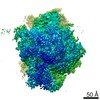
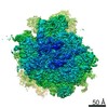
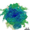
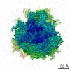
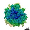
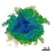
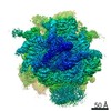
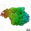
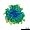
| Entry | Database: EMDB / ID: EMD-2649 | |||||||||
|---|---|---|---|---|---|---|---|---|---|---|
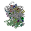
| Title | Structure of the mammalian ribosome-Sec61 complex | |||||||||
 Map data Map data | The final map is 60S_masked.mrc | |||||||||
 Sample Sample |
| |||||||||
 Keywords Keywords | translation / ribosome / mammalian / sec61 | |||||||||
| Function / homology |  Function and homology information Function and homology informationProtein hydroxylation / Ribosome Quality Control (RQC) complex extracts and degrades nascent peptide / PELO:HBS1L and ABCE1 dissociate a ribosome on a non-stop mRNA / ZNF598 and the Ribosome-associated Quality Trigger (RQT) complex dissociate a ribosome stalled on a no-go mRNA / L13a-mediated translational silencing of Ceruloplasmin expression / SRP-dependent cotranslational protein targeting to membrane / Major pathway of rRNA processing in the nucleolus and cytosol / Formation of a pool of free 40S subunits / GTP hydrolysis and joining of the 60S ribosomal subunit / Nonsense Mediated Decay (NMD) independent of the Exon Junction Complex (EJC) ...Protein hydroxylation / Ribosome Quality Control (RQC) complex extracts and degrades nascent peptide / PELO:HBS1L and ABCE1 dissociate a ribosome on a non-stop mRNA / ZNF598 and the Ribosome-associated Quality Trigger (RQT) complex dissociate a ribosome stalled on a no-go mRNA / L13a-mediated translational silencing of Ceruloplasmin expression / SRP-dependent cotranslational protein targeting to membrane / Major pathway of rRNA processing in the nucleolus and cytosol / Formation of a pool of free 40S subunits / GTP hydrolysis and joining of the 60S ribosomal subunit / Nonsense Mediated Decay (NMD) independent of the Exon Junction Complex (EJC) / Nonsense Mediated Decay (NMD) enhanced by the Exon Junction Complex (EJC) / translation at presynapse / alpha-beta T cell differentiation / protein-DNA complex disassembly / cytoplasmic side of rough endoplasmic reticulum membrane / organelle membrane / ubiquitin ligase inhibitor activity / protein localization to nucleus / positive regulation of signal transduction by p53 class mediator / negative regulation of ubiquitin-dependent protein catabolic process / rough endoplasmic reticulum / negative regulation of proteasomal ubiquitin-dependent protein catabolic process / cytosolic ribosome / ribosomal large subunit biogenesis / maturation of LSU-rRNA from tricistronic rRNA transcript (SSU-rRNA, 5.8S rRNA, LSU-rRNA) / modification-dependent protein catabolic process / transcription coactivator binding / protein tag activity / rRNA processing / heparin binding / large ribosomal subunit / presynapse / 5S rRNA binding / large ribosomal subunit rRNA binding / cytosolic large ribosomal subunit / cytoplasmic translation / negative regulation of translation / protein stabilization / rRNA binding / structural constituent of ribosome / protein ubiquitination / ribosome / translation / ribonucleoprotein complex / mRNA binding / positive regulation of cell population proliferation / ubiquitin protein ligase binding / synapse / nucleolus / glutamatergic synapse / negative regulation of transcription by RNA polymerase II / endoplasmic reticulum / RNA binding / nucleoplasm / nucleus / cytoplasm Similarity search - Function | |||||||||
| Biological species |  | |||||||||
| Method | single particle reconstruction / cryo EM / Resolution: 3.35 Å | |||||||||
 Authors Authors | Voorhees RM / Fernandez IS / Scheres SHW / Hegde R | |||||||||
 Citation Citation | Journal: Cell / Year: 2014 Title: Structure of the mammalian ribosome-Sec61 complex to 3.4 Å resolution. Authors: Rebecca M Voorhees / Israel S Fernández / Sjors H W Scheres / Ramanujan S Hegde /  Abstract: Cotranslational protein translocation is a universally conserved process for secretory and membrane protein biosynthesis. Nascent polypeptides emerging from a translating ribosome are either ...Cotranslational protein translocation is a universally conserved process for secretory and membrane protein biosynthesis. Nascent polypeptides emerging from a translating ribosome are either transported across or inserted into the membrane via the ribosome-bound Sec61 channel. Here, we report structures of a mammalian ribosome-Sec61 complex in both idle and translating states, determined to 3.4 and 3.9 Å resolution. The data sets permit building of a near-complete atomic model of the mammalian ribosome, visualization of A/P and P/E hybrid-state tRNAs, and analysis of a nascent polypeptide in the exit tunnel. Unprecedented chemical detail is observed for both the ribosome-Sec61 interaction and the conformational state of Sec61 upon ribosome binding. Comparison of the maps from idle and translating complexes suggests how conformational changes to the Sec61 channel could facilitate translocation of a secreted polypeptide. The high-resolution structure of the mammalian ribosome-Sec61 complex provides a valuable reference for future functional and structural studies. | |||||||||
| History |
|
- Structure visualization
| Movie |
Movie viewer |
|---|---|
| Structure viewer | EM map: SurfViewMolmilJmol/JSmol |
| Supplemental images |

- Downloads & links
-EMDB archive
| Map data | emd_2649.map.gz | 30.5 MB | EMDB map data format | |
|---|---|---|---|---|
| Header (meta data) | emd-2649-v30.xmlemd-2649.xml | 8.4 KB 8.4 KB | Display Display | EMDB header |
| Images | 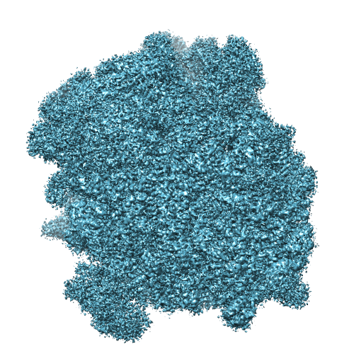 EMD-2649.png EMD-2649.png | 358.4 KB | ||
| Others | emd_2649_half_map_1.map.gzemd_2649_half_map_2.map.gz | 209.7 MB 209.7 MB | ||
| Archive directory |  http://ftp.pdbj.org/pub/emdb/structures/EMD-2649ftp://ftp.pdbj.org/pub/emdb/structures/EMD-2649 http://ftp.pdbj.org/pub/emdb/structures/EMD-2649ftp://ftp.pdbj.org/pub/emdb/structures/EMD-2649 | HTTPS FTP |
-Related structure data
| Related structure data |  3j7oMC 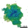 2644C 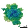 2646C 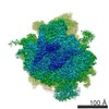 2650C  3j7pC  3j7qC  3j7rC M: atomic model generated by this map C: citing same article ( |
|---|---|
| Similar structure data |
-Links
| EMDB pages | EMDB (EBI/PDBe) / EMDataResource |
|---|---|
| Related items in Molecule of the Month |


-Map
| File | Download / File: emd_2649.map.gz / Format: CCP4 / Size: 256.8 MB / Type: IMAGE STORED AS FLOATING POINT NUMBER (4 BYTES) | ||||||||||||||||||||||||||||||||||||||||||||||||||||||||||||
|---|---|---|---|---|---|---|---|---|---|---|---|---|---|---|---|---|---|---|---|---|---|---|---|---|---|---|---|---|---|---|---|---|---|---|---|---|---|---|---|---|---|---|---|---|---|---|---|---|---|---|---|---|---|---|---|---|---|---|---|---|---|
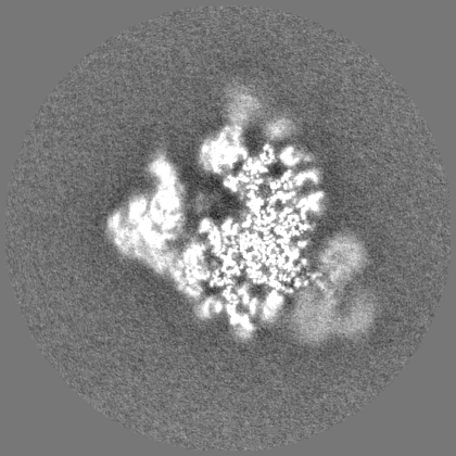
| Annotation | The final map is 60S_masked.mrc | ||||||||||||||||||||||||||||||||||||||||||||||||||||||||||||
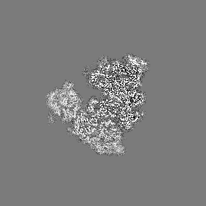
| Projections & slices | Image control
Images are generated by Spider. | ||||||||||||||||||||||||||||||||||||||||||||||||||||||||||||
| Voxel size | X=Y=Z: 1.34 Å | ||||||||||||||||||||||||||||||||||||||||||||||||||||||||||||
| Density |
| ||||||||||||||||||||||||||||||||||||||||||||||||||||||||||||
| Symmetry | Space group: 1 | ||||||||||||||||||||||||||||||||||||||||||||||||||||||||||||
| Details | EMDB XML:
CCP4 map header:
| ||||||||||||||||||||||||||||||||||||||||||||||||||||||||||||
 Z (Sec.)
 Y (Row.)
 X (Col.)


-Supplemental data
-Supplemental map: emd 2649 half map 1.map
| File | emd_2649_half_map_1.map | ||||||||||||
|---|---|---|---|---|---|---|---|---|---|---|---|---|---|
| Projections & Slices |
| ||||||||||||
| Density Histograms |


-Supplemental map: emd 2649 half map 2.map
| File | emd_2649_half_map_2.map | ||||||||||||
|---|---|---|---|---|---|---|---|---|---|---|---|---|---|
| Projections & Slices |
| ||||||||||||
| Density Histograms |


- Sample components
-Entire : Mammalian ribosome in complex with Sec61 with the large subunit (...
| Entire | Name: Mammalian ribosome in complex with Sec61 with the large subunit (60S) masked during processing. |
|---|---|
| Components |
|
-Supramolecule #1000: Mammalian ribosome in complex with Sec61 with the large subunit (...
| Supramolecule | Name: Mammalian ribosome in complex with Sec61 with the large subunit (60S) masked during processing. type: sample / ID: 1000 / Number unique components: 2 |
|---|
-Supramolecule #1: Mammalian ribosome
| Supramolecule | Name: Mammalian ribosome / type: complex / ID: 1 / Recombinant expression: No / Ribosome-details: ribosome-eukaryote: ALL |
|---|---|
| Source (natural) | Organism: |
-Macromolecule #1: Sec61
| Macromolecule | Name: Sec61 / type: protein_or_peptide / ID: 1 / Recombinant expression: No |
|---|---|
| Source (natural) | Organism: |
-Experimental details
-Structure determination
| Method | cryo EM |
|---|---|
 Processing Processing | single particle reconstruction |
| Aggregation state | particle |
-Sample preparation
| Buffer | pH: 7.5 Details: 50mM HEPES, 200mM K-acetate, 15mM Mg-acetate, 1mM DTT |
|---|---|
| Grid | Details: Quantifoil R2/2 400 mesh copper grids. |
| Vitrification | Cryogen name: ETHANE / Chamber humidity: 90 % / Chamber temperature: 120 K / Instrument: FEI VITROBOT MARK IV Method: 3uL of sampled was incubated on the grid for 30 seconds before blotting for 9 second |
- Electron microscopy
| Microscope | FEI TITAN KRIOS |
|---|---|
| Date | Apr 7, 2014 |
| Image recording | Category: CCD / Film or detector model: FEI FALCON II (4k x 4k) / Number real images: 1900 / Average electron dose: 25 e/Å2 |
| Electron beam | Acceleration voltage: 300 kV / Electron source:  FIELD EMISSION GUN FIELD EMISSION GUN |
| Electron optics | Illumination mode: OTHER / Imaging mode: BRIGHT FIELD / Nominal defocus max: 0.003 µm / Nominal defocus min: 0.001 µm / Nominal magnification: 47000 |
| Sample stage | Specimen holder model: FEI TITAN KRIOS AUTOGRID HOLDER |
| Experimental equipment |  Model: Titan Krios / Image courtesy: FEI Company |
-Image processing
| CTF correction | Details: Each particle |
|---|---|
| Final reconstruction | Applied symmetry - Point group: C1 (asymmetric) / Resolution.type: BY AUTHOR / Resolution: 3.35 Å / Resolution method: OTHER / Software - Name: Relion / Number images used: 80019 |
