5.8S rRNA binding / Protein hydroxylation / negative regulation of protein neddylation / Formation of a pool of free 40S subunits / SRP-dependent cotranslational protein targeting to membrane / Major pathway of rRNA processing in the nucleolus and cytosol / Nonsense Mediated Decay (NMD) independent of the Exon Junction Complex (EJC) / Nonsense Mediated Decay (NMD) enhanced by the Exon Junction Complex (EJC) / selenocysteine insertion sequence binding / L13a-mediated translational silencing of Ceruloplasmin expression ...5.8S rRNA binding / Protein hydroxylation / negative regulation of protein neddylation / Formation of a pool of free 40S subunits / SRP-dependent cotranslational protein targeting to membrane / Major pathway of rRNA processing in the nucleolus and cytosol / Nonsense Mediated Decay (NMD) independent of the Exon Junction Complex (EJC) / Nonsense Mediated Decay (NMD) enhanced by the Exon Junction Complex (EJC) / selenocysteine insertion sequence binding / L13a-mediated translational silencing of Ceruloplasmin expression / GTP hydrolysis and joining of the 60S ribosomal subunit / translation at postsynapse / A band / aminoacyl-tRNA synthetase multienzyme complex / embryonic brain development / translation at presynapse / response to aldosterone / alpha-beta T cell differentiation / exit from mitosis / optic nerve development / peroxisome proliferator activated receptor binding / eukaryotic 80S initiation complex / cellular response to actinomycin D / regulation of G1 to G0 transition / axial mesoderm development / retinal ganglion cell axon guidance / protein-DNA complex disassembly / negative regulation of formation of translation preinitiation complex / 90S preribosome assembly / middle ear morphogenesis / GAIT complex / TORC2 complex binding / G1 to G0 transition / growth factor binding / cell-substrate adhesion / homeostatic process / lung morphogenesis / macrophage chemotaxis / positive regulation of natural killer cell proliferation / cellular response to dexamethasone stimulus / protein targeting / ribonucleoprotein complex binding / ubiquitin ligase inhibitor activity / protein localization to nucleus / positive regulation of signal transduction by p53 class mediator / positive regulation of G1/S transition of mitotic cell cycle / negative regulation of ubiquitin-dependent protein catabolic process / maturation of LSU-rRNA / protein-RNA complex assembly / cellular response to interleukin-4 / Neutrophil degranulation / translation regulator activity / rough endoplasmic reticulum / negative regulation of proteasomal ubiquitin-dependent protein catabolic process / MDM2/MDM4 family protein binding / liver regeneration / ossification / regulation of signal transduction by p53 class mediator / cellular response to amino acid starvation / cytosolic ribosome / multicellular organism growth / ribosomal large subunit biogenesis / skeletal system development / maturation of LSU-rRNA from tricistronic rRNA transcript (SSU-rRNA, 5.8S rRNA, LSU-rRNA) / positive regulation of translation / innate immune response in mucosa / positive regulation of cell differentiation / mRNA 3'-UTR binding / sensory perception of sound / bone development / cellular response to type II interferon / mRNA 5'-UTR binding / transcription coactivator binding / cytoplasmic ribonucleoprotein granule / rRNA processing / transcription corepressor activity / antimicrobial humoral immune response mediated by antimicrobial peptide / heparin binding / regulation of translation / large ribosomal subunit / antibacterial humoral response / presynapse / retina development in camera-type eye / 5S rRNA binding / cell body / ribosomal large subunit assembly / large ribosomal subunit rRNA binding / response to lipopolysaccharide / killing of cells of another organism / defense response to Gram-negative bacterium / cytosolic large ribosomal subunit / nucleic acid binding / cytoplasmic translation / cell population proliferation / tRNA binding / postsynapse / negative regulation of translation / postsynaptic density / nuclear body / protein stabilization Similarity search - Function
PA2G4 family / : / Peptidase M24A, methionine aminopeptidase, subfamily 2, binding site / Methionine aminopeptidase subfamily 2 signature. / Peptidase M24 / Metallopeptidase family M24 / Creatinase/aminopeptidase-like / Ribosomal protein L6, N-terminal / Ribosomal protein L6, N-terminal domain / Ribosomal protein L30e ...PA2G4 family / : / Peptidase M24A, methionine aminopeptidase, subfamily 2, binding site / Methionine aminopeptidase subfamily 2 signature. / Peptidase M24 / Metallopeptidase family M24 / Creatinase/aminopeptidase-like / Ribosomal protein L6, N-terminal / Ribosomal protein L6, N-terminal domain / Ribosomal protein L30e / Ribosomal L15/L27a, N-terminal / Ribosomal protein L28e / : / Ribosomal L28e/Mak16 / Ribosomal L28e protein family / Ribosomal protein L23 / Ribosomal protein L2, archaeal-type / metallochaperone-like domain / TRASH domain / Ribosomal protein L41 / Ribosomal protein L41 / Ribosomal protein L13e, conserved site / Ribosomal protein L13e signature. / Ribosomal protein L29e / Ribosomal L29e protein family / Ribosomal protein L22e / Ribosomal protein L22e superfamily / Ribosomal L22e protein family / Ribosomal protein L27e, conserved site / Ribosomal protein L27e signature. / Ribosomal protein L13e / Ribosomal protein L13e / : / Ribosomal protein L38e / Ribosomal protein L38e superfamily / Ribosomal L38e protein family / Ribosomal protein L6e signature. / Ribosomal protein L19, eukaryotic / 60S ribosomal protein L18a/ L20, eukaryotes / Ribosomal protein L10e, conserved site / Ribosomal protein L10e signature. / Ribosomal protein L18/L18-A/B/e, conserved site / Ribosomal protein L18e signature. / Ribosomal protein L19/L19e conserved site / Ribosomal protein L19e signature. / Ribosomal protein L44e signature. / Ribosomal protein L24e, conserved site / Ribosomal protein L24e signature. / Ribosomal protein L10e / Ribosomal protein L34e, conserved site / Ribosomal protein L34e signature. / Ribosomal protein L5 eukaryotic, C-terminal / Ribosomal L18 C-terminal region / Ribosomal protein L23/L25, N-terminal / Ribosomal protein L23, N-terminal domain / Ribosomal protein L30e signature 1. / Ribosomal L40e family / Ribosomal protein L36e signature. / 50S ribosomal protein L18Ae/60S ribosomal protein L20 and L18a / Ribosomal protein L35Ae, conserved site / Ribosomal protein L35Ae signature. / Eukaryotic Ribosomal Protein L27, KOW domain / : / Ribosomal protein 50S-L18Ae/60S-L20/60S-L18A / Ribosomal proteins 50S-L18Ae/60S-L20/60S-L18A / : / Ribosomal_L40e / Ribosomal protein L27e / Ribosomal protein L40e / Ribosomal protein L40e superfamily / Ribosomal protein L27e superfamily / Ribosomal L27e protein family / Ribosomal protein L44e / Ribosomal Protein L6, KOW domain / Ribosomal protein L44 / Ribosomal protein 60S L18 and 50S L18e / 60S ribosomal protein L35 / Ribosomal protein L30e signature 2. / Ribosomal protein L30e, conserved site / Ribosomal protein L6e / : / Ribosomal protein L7A/L8 / Ribosomal protein L39e, conserved site / Ribosomal protein L39e signature. / Ribosomal protein L34Ae / Ribosomal protein L34e / 60S ribosomal protein L6E / Ribosomal protein L7, eukaryotic / Ribosomal protein L30, N-terminal / Ribosomal L30 N-terminal domain / Ribosomal protein L30/YlxQ / 60S ribosomal protein L19 / 60S ribosomal protein L4, C-terminal domain / 60S ribosomal protein L4 C-terminal domain / Ribosomal protein L13, eukaryotic/archaeal / Ribosomal protein L31e, conserved site / : / Ribosomal protein L31e signature. / Ribosomal protein L36e / Ribosomal protein L14e domain Similarity search - Domain/homology
Large ribosomal subunit protein eL21 / Large ribosomal subunit protein eL33 / Large ribosomal subunit protein eL8 / Large ribosomal subunit protein uL15 / Large ribosomal subunit protein uL30 / Large ribosomal subunit protein uL13 / Large ribosomal subunit protein uL3 / Large ribosomal subunit protein eL18 / Large ribosomal subunit protein eL28 / Large ribosomal subunit protein eL6 ...Large ribosomal subunit protein eL21 / Large ribosomal subunit protein eL33 / Large ribosomal subunit protein eL8 / Large ribosomal subunit protein uL15 / Large ribosomal subunit protein uL30 / Large ribosomal subunit protein uL13 / Large ribosomal subunit protein uL3 / Large ribosomal subunit protein eL18 / Large ribosomal subunit protein eL28 / Large ribosomal subunit protein eL6 / Large ribosomal subunit protein eL29 / Large ribosomal subunit protein uL18 / Large ribosomal subunit protein eL13 / Large ribosomal subunit protein eL36 / Proliferation-associated protein 2G4 / Large ribosomal subunit protein uL6 / Large ribosomal subunit protein eL20 / Large ribosomal subunit protein uL23 / Large ribosomal subunit protein uL14 / Large ribosomal subunit protein eL30 / Large ribosomal subunit protein eL31 / Large ribosomal subunit protein eL32 / Large ribosomal subunit protein uL2 / Small ribosomal subunit protein eS32 / Large ribosomal subunit protein eL22 / Large ribosomal subunit protein eL19 / Large ribosomal subunit protein uL24 / Large ribosomal subunit protein eL43 / Large ribosomal subunit protein eL39 / 60S ribosomal protein L27 / Ubiquitin-ribosomal protein eL40 fusion protein / Ribosomal protein L36A / Large ribosomal subunit protein uL16 / Large ribosomal subunit protein uL29 / Large ribosomal subunit protein eL24 / Large ribosomal subunit protein uL22 / Large ribosomal subunit protein eL14 / Large ribosomal subunit protein uL5 / Large ribosomal subunit protein eL15 / Large ribosomal subunit protein eL34 / Large ribosomal subunit protein eL37 / Large ribosomal subunit protein uL4 / Large ribosomal subunit protein eL38 Similarity search - Component
Biological species
Mus musculus (house mouse)
Method
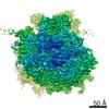
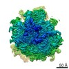
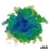
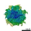
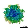
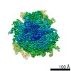
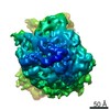
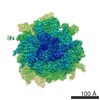
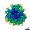
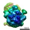
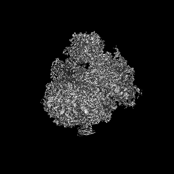
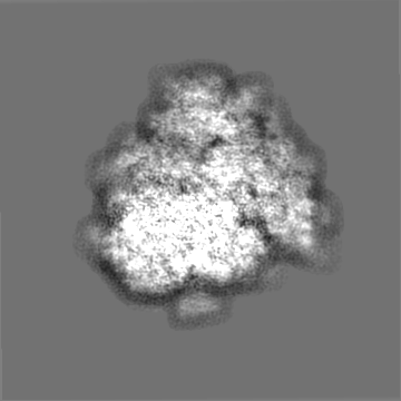
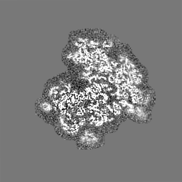
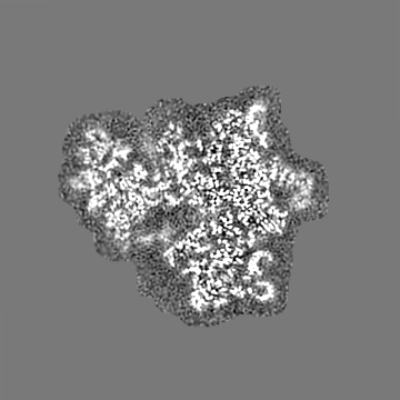
single particle reconstruction / cryo EM / Resolution: 3.1 Å
Journal: Mol Cell / Year: 2021 Title: Protein Synthesis in the Developing Neocortex at Near-Atomic Resolution Reveals Ebp1-Mediated Neuronal Proteostasis at the 60S Tunnel Exit. Authors: Matthew L Kraushar / Ferdinand Krupp / Dermot Harnett / Paul Turko / Mateusz C Ambrozkiewicz / Thiemo Sprink / Koshi Imami / Manuel Günnigmann / Ulrike Zinnall / Carlos H Vieira-Vieira / ...Authors: Matthew L Kraushar / Ferdinand Krupp / Dermot Harnett / Paul Turko / Mateusz C Ambrozkiewicz / Thiemo Sprink / Koshi Imami / Manuel Günnigmann / Ulrike Zinnall / Carlos H Vieira-Vieira / Theres Schaub / Agnieszka Münster-Wandowski / Jörg Bürger / Ekaterina Borisova / Hiroshi Yamamoto / Mladen-Roko Rasin / Uwe Ohler / Dieter Beule / Thorsten Mielke / Victor Tarabykin / Markus Landthaler / Günter Kramer / Imre Vida / Matthias Selbach / Christian M T Spahn / Abstract: Protein synthesis must be finely tuned in the developing nervous system as the final essential step of gene expression. This study investigates the architecture of ribosomes from the neocortex during ...Protein synthesis must be finely tuned in the developing nervous system as the final essential step of gene expression. This study investigates the architecture of ribosomes from the neocortex during neurogenesis, revealing Ebp1 as a high-occupancy 60S peptide tunnel exit (TE) factor during protein synthesis at near-atomic resolution by cryoelectron microscopy (cryo-EM). Ribosome profiling demonstrated Ebp1-60S binding is highest during start codon initiation and N-terminal peptide elongation, regulating ribosome occupancy of these codons. Membrane-targeting domains emerging from the 60S tunnel, which recruit SRP/Sec61 to the shared binding site, displace Ebp1. Ebp1 is particularly abundant in the early-born neural stem cell (NSC) lineage and regulates neuronal morphology. Ebp1 especially impacts the synthesis of membrane-targeted cell adhesion molecules (CAMs), measured by pulsed stable isotope labeling by amino acids in cell culture (pSILAC)/bioorthogonal noncanonical amino acid tagging (BONCAT) mass spectrometry (MS). Therefore, Ebp1 is a central component of protein synthesis, and the ribosome TE is a focal point of gene expression control in the molecular specification of neuronal morphology during development.
History
Deposition
Sep 20, 2019
-
Header (metadata) release
Sep 30, 2020
-
Map release
Sep 30, 2020
-
Update
Nov 13, 2024
-
Current status
Nov 13, 2024
Processing site: PDBe / Status: Released
-
Structure visualization
Movie
Surface view with section colored by density value
Name: 28S ribosomal RNA / type: rna / ID: 43 Details: We used the Oryctolagus cuniculus sequence to model the Mus musculus 28S ribosomal RNA Number of copies: 1
Name: 5.8S ribosomal RNA / type: rna / ID: 44 Details: We used the Oryctolagus cuniculus sequence to model the Mus musculus 5.8S ribosomal RNA Number of copies: 1
Name: 5S ribosomal RNA / type: rna / ID: 45 Details: We used the Oryctolagus cuniculus sequence to model the Mus musculus 5S ribosomal RNA Number of copies: 1
Name: MAGNESIUM ION / type: ligand / ID: 47 / Number of copies: 247 / Formula: MG
Molecular weight
Theoretical: 24.305 Da
+
Macromolecule #48: ZINC ION
Macromolecule
Name: ZINC ION / type: ligand / ID: 48 / Number of copies: 3 / Formula: ZN
Molecular weight
Theoretical: 65.409 Da
-
Experimental details
-
Structure determination
Method
cryo EM
Processing
single particle reconstruction
Aggregation state
particle
-
Sample preparation
Buffer
pH: 7.4 Component:
Concentration
Name
Formula
20.0 mM
HEPES
100.0 mM
potassium chloride
KCl
10.0 mM
magnesium chloride
MgCl2
20.0 mM
Dithiothreitol
0.04 mM
Spermine
0.5 mM
Spermidine
0.1 mg/ml
cycloheximide
Grid
Model: Quantifoil R3/3 / Material: COPPER / Mesh: 100 / Support film - Material: CARBON / Support film - topology: HOLEY / Support film - Film thickness: 2 / Pretreatment - Type: GLOW DISCHARGE
Vitrification
Cryogen name: ETHANE / Chamber humidity: 100 % / Chamber temperature: 277 K / Instrument: FEI VITROBOT MARK II
-
Electron microscopy
Microscope
FEI TITAN KRIOS
Specialist optics
Energy filter - Name: GIF Quantum LS
Image recording
Film or detector model: GATAN K2 SUMMIT (4k x 4k) / Number real images: 5379 / Average exposure time: 20.0 sec. / Average electron dose: 31.78 e/Å2
Electron beam
Acceleration voltage: 300 kV / Electron source: FIELD EMISSION GUN
In the structure databanks used in Yorodumi, some data are registered as the other names, "COVID-19 virus" and "2019-nCoV". Here are the details of the virus and the list of structure data.
Jan 31, 2019. EMDB accession codes are about to change! (news from PDBe EMDB page)
EMDB accession codes are about to change! (news from PDBe EMDB page)
The allocation of 4 digits for EMDB accession codes will soon come to an end. Whilst these codes will remain in use, new EMDB accession codes will include an additional digit and will expand incrementally as the available range of codes is exhausted. The current 4-digit format prefixed with “EMD-” (i.e. EMD-XXXX) will advance to a 5-digit format (i.e. EMD-XXXXX), and so on. It is currently estimated that the 4-digit codes will be depleted around Spring 2019, at which point the 5-digit format will come into force.
The EM Navigator/Yorodumi systems omit the EMD- prefix.
Related info.:Q: What is EMD? / ID/Accession-code notation in Yorodumi/EM Navigator
Yorodumi is a browser for structure data from EMDB, PDB, SASBDB, etc.
This page is also the successor to EM Navigator detail page, and also detail information page/front-end page for Omokage search.
The word "yorodu" (or yorozu) is an old Japanese word meaning "ten thousand". "mi" (miru) is to see.
Related info.:EMDB / PDB / SASBDB / Comparison of 3 databanks / Yorodumi Search / Aug 31, 2016. New EM Navigator & Yorodumi / Yorodumi Papers / Jmol/JSmol / Function and homology information / Changes in new EM Navigator and Yorodumi
 Movie
Movie Controller


 Open data
 Basic information
 Map data
 Sample
 Keywords
 Function and homology information
 Authors
 Germany, 4 items
  Citation

 Structure visualization
 Downloads & links
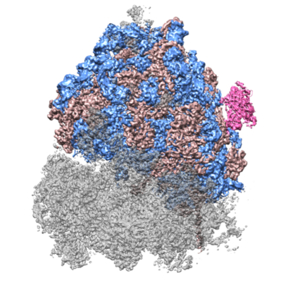 emd_10321.png
 http://ftp.pdbj.org/pub/emdb/structures/EMD-10321


 Z (Sec.)
 Y (Row.)
 X (Col.)


 Sample components
 Processing
 Electron microscopy
 FIELD EMISSION GUN
