 Movie
Movie Controller

[English] 日本語
 Yorodumi
- PDB-3j5s: EttA binds to ribosome exit site and regulates translation by res... -
+ Open data

- Basic information
| Entry | Database: PDB / ID: 3j5s | ||||||
|---|---|---|---|---|---|---|---|
| Title | EttA binds to ribosome exit site and regulates translation by restricting ribosome and tRNA dynamics | ||||||
 Components Components |
| ||||||
 Keywords Keywords | RIBOSOME/TRANSLATION / protein translation regulation / ABC-F protein family / single-molecule FRET / YjjK / RIBOSOME-TRANSLATION complex | ||||||
| Function / homology |  Function and homology information Function and homology informationnegative regulation of translational elongation / translation release factor activity / Hydrolases; Acting on acid anhydrides; In phosphorus-containing anhydrides / negative regulation of translational initiation / ribosomal small subunit assembly / ribosome binding / 5S rRNA binding / ribosomal large subunit assembly / cytosolic small ribosomal subunit / cytosolic large ribosomal subunit ...negative regulation of translational elongation / translation release factor activity / Hydrolases; Acting on acid anhydrides; In phosphorus-containing anhydrides / negative regulation of translational initiation / ribosomal small subunit assembly / ribosome binding / 5S rRNA binding / ribosomal large subunit assembly / cytosolic small ribosomal subunit / cytosolic large ribosomal subunit / cytoplasmic translation / tRNA binding / negative regulation of translation / rRNA binding / structural constituent of ribosome / ribosome / translation / response to antibiotic / mRNA binding / ATP hydrolysis activity / RNA binding / ATP binding / membrane / cytoplasm / cytosol Similarity search - Function | ||||||
| Biological species |  | ||||||
| Method | ELECTRON MICROSCOPY / single particle reconstruction / cryo EM / Resolution: 7.5 Å | ||||||
 Authors Authors | Hashem, Y. | ||||||
 Citation Citation | Journal: Nat Struct Mol Biol / Year: 2014 Title: EttA regulates translation by binding the ribosomal E site and restricting ribosome-tRNA dynamics. Authors: Bo Chen / Grégory Boël / Yaser Hashem / Wei Ning / Jingyi Fei / Chi Wang / Ruben L Gonzalez / John F Hunt / Joachim Frank /  Abstract: Cells express many ribosome-interacting factors whose functions and molecular mechanisms remain unknown. Here, we elucidate the mechanism of a newly characterized regulatory translation factor, ...Cells express many ribosome-interacting factors whose functions and molecular mechanisms remain unknown. Here, we elucidate the mechanism of a newly characterized regulatory translation factor, energy-dependent translational throttle A (EttA), which is an Escherichia coli representative of the ATP-binding cassette F (ABC-F) protein family. Using cryo-EM, we demonstrate that the ATP-bound form of EttA binds to the ribosomal tRNA-exit site, where it forms bridging interactions between the ribosomal L1 stalk and the tRNA bound in the peptidyl-tRNA-binding site. Using single-molecule fluorescence resonance energy transfer, we show that the ATP-bound form of EttA restricts ribosome and tRNA dynamics required for protein synthesis. This work represents the first example, to our knowledge, in which the detailed molecular mechanism of any ABC-F family protein has been determined and establishes a framework for elucidating the mechanisms of other regulatory translation factors. #1: Journal: Nat Struct Mol Biol / Year: 2014Title: The ABC-F protein EttA gates ribosome entry into the translation elongation cycle. Authors: Grégory Boël / Paul C Smith / Wei Ning / Michael T Englander / Bo Chen / Yaser Hashem / Anthony J Testa / Jeffrey J Fischer / Hans-Joachim Wieden / Joachim Frank / Ruben L Gonzalez / John F Hunt /  Abstract: ABC-F proteins have evaded functional characterization even though they compose one of the most widely distributed branches of the ATP-binding cassette (ABC) superfamily. Herein, we demonstrate that ...ABC-F proteins have evaded functional characterization even though they compose one of the most widely distributed branches of the ATP-binding cassette (ABC) superfamily. Herein, we demonstrate that YjjK, the most prevalent eubacterial ABC-F protein, gates ribosome entry into the translation elongation cycle through a nucleotide-dependent interaction sensitive to ATP/ADP ratio. Accordingly, we rename this protein energy-dependent translational throttle A (EttA). We determined the crystal structure of Escherichia coli EttA and used it to design mutants for biochemical studies including enzymological assays of the initial steps of protein synthesis. These studies suggest that EttA may regulate protein synthesis in energy-depleted cells, which have a low ATP/ADP ratio. Consistently with this inference, EttA-deleted cells exhibit a severe fitness defect in long-term stationary phase. These studies demonstrate that an ABC-F protein regulates protein synthesis via a new mechanism sensitive to cellular energy status. | ||||||
| History |
|
- Structure visualization
| Movie |
Movie viewer |
|---|---|
| Structure viewer | Molecule: MolmilJmol/JSmol |
- Downloads & links
-Download
| PDBx/mmCIF format | 3j5s.cif.gz | 383 KB | Display | PDBx/mmCIF format |
|---|---|---|---|---|
| PDB format | pdb3j5s.ent.gz | 262.8 KB | Display | PDB format |
| PDBx/mmJSON format | 3j5s.json.gz | Tree view | PDBx/mmJSON format | |
| Others |  Other downloads Other downloads |
-Validation report
| Arichive directory | https://data.pdbj.org/pub/pdb/validation_reports/j5/3j5sftp://data.pdbj.org/pub/pdb/validation_reports/j5/3j5s | HTTPS FTP |
|---|
-Related structure data
| Related structure data | 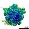 5784MC 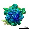 5785C 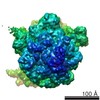 5786C 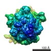 5841C  5842C  5843C M: map data used to model this data C: citing same article ( |
|---|---|
| Similar structure data |


-Links
 PDBj
PDBj


- Assembly
| Deposited unit | 
|
|---|---|
| 1 |
|
-Components
-RNA chain , 3 types, 3 molecules BAE
| #1: RNA chain | Mass: 32636.531 Da / Num. of mol.: 1 / Fragment: SEE REMARK 999 / Source method: isolated from a natural source / Source: (natural) |
|---|---|
| #2: RNA chain | Mass: 116242.516 Da / Num. of mol.: 1 / Fragment: SEE REMARK 999 / Source method: isolated from a natural source / Source: (natural) |
| #3: RNA chain | Mass: 24802.785 Da / Num. of mol.: 1 / Source method: isolated from a natural source / Source: (natural) |
-Protein , 2 types, 2 molecules DI
| #4: Protein | Mass: 63357.668 Da / Num. of mol.: 1 Source method: isolated from a genetically manipulated source Source: (gene. exp.) |
|---|---|
| #8: Protein | Mass: 16861.523 Da / Num. of mol.: 1 / Source method: isolated from a natural source / Source: (natural) |
-50S ribosomal protein ... , 3 types, 3 molecules FGH
| #5: Protein | Mass: 24765.660 Da / Num. of mol.: 1 / Source method: isolated from a natural source / Source: (natural) |
|---|---|
| #6: Protein | Mass: 20202.416 Da / Num. of mol.: 1 / Source method: isolated from a natural source / Source: (natural) |
| #7: Protein/peptide | Mass: 5814.842 Da / Num. of mol.: 1 / Source method: isolated from a natural source / Source: (natural) |
-Details
| Sequence details | THE FULL RIBOSOME WAS IMAGED, BUT ONLY A SUBSET OF THE 16S AND 23S RIBOSOMAL RNA WAS MODELED IN THIS ENTRY. |
|---|
-Experimental details
-Experiment
| Experiment | Method: ELECTRON MICROSCOPY |
|---|---|
| EM experiment | Aggregation state: PARTICLE / 3D reconstruction method: single particle reconstruction |
- Sample preparation
| Component |
| ||||||||||||||||||||||||
|---|---|---|---|---|---|---|---|---|---|---|---|---|---|---|---|---|---|---|---|---|---|---|---|---|---|
| Buffer solution | Name: 50 mM Tris acetate, 100 mM KCl, 5 mM NH4OAc, 3.5 mM Mg(OAc)2, 0.5 mM Ca(OAc)2, 0.1 mM EDTA, 1 mM spermidine, 5 mM putrescine, 6 mM 2-mercaptoethanol, 0.5 mM Mg-ATP pH: 6.9 Details: 50 mM Tris acetate, 100 mM KCl, 5 mM NH4OAc, 3.5 mM Mg(OAc)2, 0.5 mM Ca(OAc)2, 0.1 mM EDTA, 1 mM spermidine, 5 mM putrescine, 6 mM 2-mercaptoethanol, 0.5 mM Mg-ATP | ||||||||||||||||||||||||
| Specimen | Conc.: 1.2 mg/ml / Embedding applied: NO / Shadowing applied: NO / Staining applied: NO / Vitrification applied: YES | ||||||||||||||||||||||||
| Specimen support | Details: Quantifoil R2/4 300 mesh Cu EM grid, coated with thin carbon film, glow discharged in H2/O2 | ||||||||||||||||||||||||
| Vitrification | Instrument: FEI VITROBOT MARK IV / Cryogen name: ETHANE / Temp: 80 K / Humidity: 100 % Details: Wait time 30 sec, blot time 8 sec (4 C), plunge into liquid ethane (FEI VITROBOT MARK IV) Method: Wait time 30 sec, blot time 8 sec, at 4 degrees Celsius |
- Electron microscopy imaging
| Experimental equipment |  Model: Tecnai F20 / Image courtesy: FEI Company |
|---|---|
| Microscopy | Model: FEI TECNAI F20 / Date: Apr 5, 2013 |
| Electron gun | Electron source:  FIELD EMISSION GUN / Accelerating voltage: 200 kV / Illumination mode: FLOOD BEAM FIELD EMISSION GUN / Accelerating voltage: 200 kV / Illumination mode: FLOOD BEAM |
| Electron lens | Mode: BRIGHT FIELD / Nominal magnification: 80000 X / Calibrated magnification: 110637 X / Nominal defocus max: 2500 nm / Nominal defocus min: 1200 nm / Cs: 2 mm |
| Specimen holder | Specimen holder model: GATAN LIQUID NITROGEN Specimen holder type: Single tilt cryoholder, liquid Nitrogen cooled Temperature: 80 K |
| Image recording | Electron dose: 17 e/Å2 / Film or detector model: GATAN ULTRASCAN 4000 (4k x 4k) / Details: Low dose |
| Image scans | Num. digital images: 1816 |
| Radiation | Protocol: SINGLE WAVELENGTH / Monochromatic (M) / Laue (L): M / Scattering type: x-ray |
| Radiation wavelength | Relative weight: 1 |
- Processing
| EM software |
| ||||||||||||||||||||||||||||||||||||
|---|---|---|---|---|---|---|---|---|---|---|---|---|---|---|---|---|---|---|---|---|---|---|---|---|---|---|---|---|---|---|---|---|---|---|---|---|---|
| CTF correction | Details: Each micrograph | ||||||||||||||||||||||||||||||||||||
| Symmetry | Point symmetry: C1 (asymmetric) | ||||||||||||||||||||||||||||||||||||
| 3D reconstruction | Resolution: 7.5 Å / Resolution method: FSC 0.143 CUT-OFF / Num. of particles: 39316 / Nominal pixel size: 2.7116 Å / Actual pixel size: 2.7116 Å / Details: Subset after RELION 3D classification / Refinement type: HALF-MAPS REFINED INDEPENDENTLY / Symmetry type: POINT | ||||||||||||||||||||||||||||||||||||
| Atomic model building |
| ||||||||||||||||||||||||||||||||||||
| Atomic model building | Source name: PDB / Type: experimental model
| ||||||||||||||||||||||||||||||||||||
| Refinement step | Cycle: LAST
|


