[English] 日本語
Yorodumi
- PDB-4fin: Crystal Structure of EttA (formerly YjjK) - an E. coli ABC-type ATPase -

+
Open data


ID or keywords:

Loading...

-
Basic information

Entry
Database: PDB / ID: 4fin
TitleCrystal Structure of EttA (formerly YjjK) - an E. coli ABC-type ATPase
ComponentsEttA (YjjK) ABCF family protein
KeywordsATP-BINDING PROTEIN / ABC protein / ABCF / mechanoenzyme / Gating of ribosomal elongation / Ribosome / Cytosol/Ribosome / Northeast Structural Genomics Consortium / NESG / PSI-Biology / structural genomics
Function / homology
Function and homology information


negative regulation of translational elongation / Hydrolases; Acting on acid anhydrides; In phosphorus-containing anhydrides / ribosome binding / tRNA binding / rRNA binding / translation / ATP hydrolysis activity / ATP binding / cytoplasm
Similarity search - Function
Energy-dependent translational throttle protein EttA / ABC-transporter extension domain / ABC transporter / ABC transporter-like, conserved site / ABC transporters family signature. / ABC transporter / ABC transporter-like, ATP-binding domain / ATP-binding cassette, ABC transporter-type domain profile. / P-loop containing nucleotide triphosphate hydrolases / ATPases associated with a variety of cellular activities ...Energy-dependent translational throttle protein EttA / ABC-transporter extension domain / ABC transporter / ABC transporter-like, conserved site / ABC transporters family signature. / ABC transporter / ABC transporter-like, ATP-binding domain / ATP-binding cassette, ABC transporter-type domain profile. / P-loop containing nucleotide triphosphate hydrolases / ATPases associated with a variety of cellular activities / AAA+ ATPase domain / Rossmann fold / P-loop containing nucleoside triphosphate hydrolase / 3-Layer(aba) Sandwich / Alpha Beta
Similarity search - Domain/homology
CITRIC ACID / TRIETHYLENE GLYCOL / Energy-dependent translational throttle protein EttA
Similarity search - Component
Biological speciesEscherichia coli (E. coli)
MethodX-RAY DIFFRACTION / SYNCHROTRON / SAD / Resolution: 2.4 Å
AuthorsSmith, P. / Yuan, Y. / Hunt, J.F. / Northeast Structural Genomics Consortium (NESG)
CitationJournal: Nat Struct Mol Biol / Year: 2014
Title: The ABC-F protein EttA gates ribosome entry into the translation elongation cycle.
Authors: Grégory Boël / Paul C Smith / Wei Ning / Michael T Englander / Bo Chen / Yaser Hashem / Anthony J Testa / Jeffrey J Fischer / Hans-Joachim Wieden / Joachim Frank / Ruben L Gonzalez / John F Hunt /
Abstract: ABC-F proteins have evaded functional characterization even though they compose one of the most widely distributed branches of the ATP-binding cassette (ABC) superfamily. Herein, we demonstrate that ...ABC-F proteins have evaded functional characterization even though they compose one of the most widely distributed branches of the ATP-binding cassette (ABC) superfamily. Herein, we demonstrate that YjjK, the most prevalent eubacterial ABC-F protein, gates ribosome entry into the translation elongation cycle through a nucleotide-dependent interaction sensitive to ATP/ADP ratio. Accordingly, we rename this protein energy-dependent translational throttle A (EttA). We determined the crystal structure of Escherichia coli EttA and used it to design mutants for biochemical studies including enzymological assays of the initial steps of protein synthesis. These studies suggest that EttA may regulate protein synthesis in energy-depleted cells, which have a low ATP/ADP ratio. Consistently with this inference, EttA-deleted cells exhibit a severe fitness defect in long-term stationary phase. These studies demonstrate that an ABC-F protein regulates protein synthesis via a new mechanism sensitive to cellular energy status.
History
DepositionJun 9, 2012Deposition site: RCSB / Processing site: RCSB
Revision 1.0Jul 3, 2013Provider: repository / Type: Initial release
Revision 1.1Dec 4, 2013Group: Structure summary
Revision 1.2Jan 8, 2014Group: Database references
Revision 1.3Mar 5, 2014Group: Database references

-
Structure visualization

Structure viewerMolecule:
MolmilJmol/JSmol

Downloads & links

-
Assembly

Deposited unit
A: EttA (YjjK) ABCF family protein
B: EttA (YjjK) ABCF family protein
hetero molecules


Theoretical massNumber of molelcules
Total (without water)128,22324
Polymers125,9952
Non-polymers2,22822
Water7,134396
1


  • Idetical with deposited unit
  • defined by author&software
TypeNameSymmetry operationNumber
identity operation1_555x,y,z1
Buried area12480 Å2
ΔGint-199 kcal/mol
Surface area48340 Å2
MethodPISA
2
A: EttA (YjjK) ABCF family protein
hetero molecules


Theoretical massNumber of molelcules
Total (without water)64,18912
Polymers62,9981
Non-polymers1,19111
Water181
TypeNameSymmetry operationNumber
identity operation1_555x,y,z1
3
B: EttA (YjjK) ABCF family protein
hetero molecules


Theoretical massNumber of molelcules
Total (without water)64,03512
Polymers62,9981
Non-polymers1,03711
Water181
TypeNameSymmetry operationNumber
identity operation1_555x,y,z1
Unit cell
Length a, b, c (Å)45.363, 233.474, 54.089
Angle α, β, γ (deg.)90.00, 91.29, 90.00
Int Tables number4
Space group name H-MP1211
Details
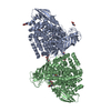
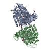
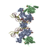
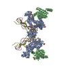
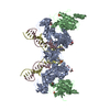
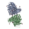
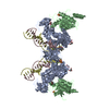
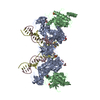
Protein exists in monomer/dimer equilibrium as seen in hydrodynamic studies. Crystal structure here represents the dimer formation. Cryo-EM reconstruction (see accompanying publications) reveal the monomeric ribosomally assocaited form. Both dimer and monomer exhibit Rad50-like head/tail ABC cassette dimer interfaces.

-
Components

-
Protein , 1 types, 2 molecules AB

#1: Protein EttA (YjjK) ABCF family protein


Mass: 62997.727 Da / Num. of mol.: 2
Source method: isolated from a genetically manipulated source
Details: Cloned NdeI/XhoI to express wt full-length protein without any additional tags or amino acids
Source: (gene. exp.) Escherichia coli (E. coli) / Strain: K-12 / Gene: b4391, JW4354, yjjK, YjjK / Plasmid: pET24 / Production host: Escherichia coli (E. coli) / Strain (production host): B834(DE3) / References: UniProt: P0A9W3

-
Non-polymers , 5 types, 418 molecules

#2: Chemical
ChemComp-SO4 / SULFATE ION


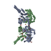
Mass: 96.063 Da / Num. of mol.: 11 / Source method: obtained synthetically / Formula: SO4
#3: Chemical ChemComp-CIT / CITRIC ACID


Mass: 192.124 Da / Num. of mol.: 1 / Source method: obtained synthetically / Formula: C6H8O7
#4: Chemical ChemComp-PGE / TRIETHYLENE GLYCOL


Mass: 150.173 Da / Num. of mol.: 1 / Source method: obtained synthetically / Formula: C6H14O4
#5: Chemical
ChemComp-GOL / GLYCEROL / GLYCERIN / PROPANE-1,2,3-TRIOL


Mass: 92.094 Da / Num. of mol.: 9 / Source method: obtained synthetically / Formula: C3H8O3
#6: Water ChemComp-HOH / water


Mass: 18.015 Da / Num. of mol.: 396 / Source method: isolated from a natural source / Formula: H2O

-
Experimental details

-
Experiment

ExperimentMethod: X-RAY DIFFRACTION / Number of used crystals: 1

-
Sample preparation

CrystalDensity Matthews: 2.27 Å3/Da / Density % sol: 45.88 %
Crystal growTemperature: 300 K / Method: vapor diffusion, hanging drop / pH: 5.6
Details: 200mM Li2SO4, 100mM NaCitrate pH 5.6, 10-20% Polyethylene Glycol (MW 8000), VAPOR DIFFUSION, HANGING DROP, temperature 300KK

-
Data collection

DiffractionMean temperature: 130 K
Diffraction sourceSource: SYNCHROTRON / Site: NSLS / Beamline: X12C / Wavelength: 0.9798 Å
DetectorType: Brandeis-4K / Detector: CCD / Date: Mar 1, 2000 / Details: Si(111) Mono
RadiationMonochromator: Si 111 / Protocol: SINGLE WAVELENGTH / Monochromatic (M) / Laue (L): M / Scattering type: x-ray
Radiation wavelengthWavelength: 0.9798 Å / Relative weight: 1
ReflectionResolution: 2.4→50 Å / Num. all: 41262 / Num. obs: 44036 / % possible obs: 93.7 % / Observed criterion σ(F): -999 / Observed criterion σ(I): -1 / Redundancy: 6.5 % / Rsym value: 0.143 / Net I/σ(I): 14.1
Reflection shell
Resolution (Å)Redundancy (%)Mean I/σ(I) obsRsym valueDiffraction-ID% possible all
2.4-2.443.22.040.577183.1
6.51-508.3440.056199.6

-
Processing

Software
NameVersionClassification
CBASSdata collection
SOLVEphasing
PHENIX(phenix.refine: 1.7.3_928)refinement
DENZOdata reduction
SCALEPACKdata scaling
RefinementMethod to determine structure: SAD / Resolution: 2.4→49.067 Å / SU ML: 0.3 / σ(F): 1.41 / Phase error: 23.36 / Stereochemistry target values: ML
RfactorNum. reflection% reflection
Rfree0.2429 2077 5.05 %
Rwork0.1833 --
obs0.1862 41106 94.03 %
all-41262 -
Solvent computationShrinkage radii: 0.98 Å / VDW probe radii: 1.2 Å / Solvent model: FLAT BULK SOLVENT MODEL / Bsol: 35.005 Å2 / ksol: 0.358 e/Å3
Displacement parameters
Baniso -1Baniso -2Baniso -3
1-1.1892 Å20 Å20.4195 Å2
2--2.8858 Å2-0 Å2
3----3.4889 Å2
Refinement stepCycle: LAST / Resolution: 2.4→49.067 Å
ProteinNucleic acidLigandSolventTotal
Num. atoms8384 0 132 396 8912
Refine LS restraints
Refine-IDTypeDev idealNumber
X-RAY DIFFRACTIONf_bond_d0.0068682
X-RAY DIFFRACTIONf_angle_d0.98411718
X-RAY DIFFRACTIONf_dihedral_angle_d14.3873290
X-RAY DIFFRACTIONf_chiral_restr0.0621264
X-RAY DIFFRACTIONf_plane_restr0.0041536
LS refinement shell
Resolution (Å)Rfactor RfreeNum. reflection RfreeRfactor RworkNum. reflection RworkRefine-ID% reflection obs (%)
2.4-2.45590.31951220.24042328X-RAY DIFFRACTION84
2.4559-2.51730.27621170.2232391X-RAY DIFFRACTION87
2.5173-2.58530.2921130.21512381X-RAY DIFFRACTION85
2.5853-2.66140.2541330.20362425X-RAY DIFFRACTION88
2.6614-2.74730.24191260.20622408X-RAY DIFFRACTION88
2.7473-2.84550.29941350.20032510X-RAY DIFFRACTION90
2.8455-2.95940.27061360.20892505X-RAY DIFFRACTION93
2.9594-3.09410.26031320.19562701X-RAY DIFFRACTION98
3.0941-3.25710.26721760.1912779X-RAY DIFFRACTION100
3.2571-3.46120.23491420.17162755X-RAY DIFFRACTION100
3.4612-3.72830.20221340.16422775X-RAY DIFFRACTION100
3.7283-4.10330.22231650.1542762X-RAY DIFFRACTION100
4.1033-4.69670.2231510.15552740X-RAY DIFFRACTION100
4.6967-5.91570.23211500.18282778X-RAY DIFFRACTION100
5.9157-49.07690.22591450.18862791X-RAY DIFFRACTION100

+
About Yorodumi

-
News

-
Feb 9, 2022. New format data for meta-information of EMDB entries

New format data for meta-information of EMDB entries

  • Version 3 of the EMDB header file is now the official format.
  • The previous official version 1.9 will be removed from the archive.

Related info.:EMDB header

External links:wwPDB to switch to version 3 of the EMDB data model

-
Aug 12, 2020. Covid-19 info

Covid-19 info

URL: https://pdbjlvh1.pdbj.org/emnavi/covid19.php

New page: Covid-19 featured information page in EM Navigator.

Related info.:Covid-19 info / Mar 5, 2020. Novel coronavirus structure data

+
Mar 5, 2020. Novel coronavirus structure data

Novel coronavirus structure data

Related info.:Yorodumi Speices / Aug 12, 2020. Covid-19 info

External links:COVID-19 featured content - PDBj / Molecule of the Month (242):Coronavirus Proteases

+
Jan 31, 2019. EMDB accession codes are about to change! (news from PDBe EMDB page)

EMDB accession codes are about to change! (news from PDBe EMDB page)

  • The allocation of 4 digits for EMDB accession codes will soon come to an end. Whilst these codes will remain in use, new EMDB accession codes will include an additional digit and will expand incrementally as the available range of codes is exhausted. The current 4-digit format prefixed with “EMD-” (i.e. EMD-XXXX) will advance to a 5-digit format (i.e. EMD-XXXXX), and so on. It is currently estimated that the 4-digit codes will be depleted around Spring 2019, at which point the 5-digit format will come into force.
  • The EM Navigator/Yorodumi systems omit the EMD- prefix.

Related info.:Q: What is EMD? / ID/Accession-code notation in Yorodumi/EM Navigator

External links:EMDB Accession Codes are Changing Soon! / Contact to PDBj

+
Jul 12, 2017. Major update of PDB

Major update of PDB

  • wwPDB released updated PDB data conforming to the new PDBx/mmCIF dictionary.
  • This is a major update changing the version number from 4 to 5, and with Remediation, in which all the entries are updated.
  • In this update, many items about electron microscopy experimental information are reorganized (e.g. em_software).
  • Now, EM Navigator and Yorodumi are based on the updated data.

External links:wwPDB Remediation / Enriched Model Files Conforming to OneDep Data Standards Now Available in the PDB FTP Archive

-
Yorodumi

Thousand views of thousand structures

  • Yorodumi is a browser for structure data from EMDB, PDB, SASBDB, etc.
  • This page is also the successor to EM Navigator detail page, and also detail information page/front-end page for Omokage search.
  • The word "yorodu" (or yorozu) is an old Japanese word meaning "ten thousand". "mi" (miru) is to see.

Related info.:EMDB / PDB / SASBDB / Comparison of 3 databanks / Yorodumi Search / Aug 31, 2016. New EM Navigator & Yorodumi / Yorodumi Papers / Jmol/JSmol / Function and homology information / Changes in new EM Navigator and Yorodumi

Read more