Movie
Movie Controller


+ Open data

- Basic information
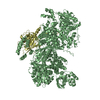
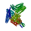
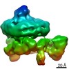
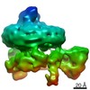
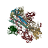
| Entry | Database: EMDB / ID: EMD-4772 | |||||||||
|---|---|---|---|---|---|---|---|---|---|---|
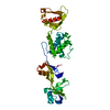
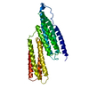
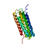
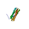
| Title | Cryo-EM structure of autoinhibited human talin-1 | |||||||||
 Map data Map data | best map | |||||||||
 Sample Sample |
| |||||||||
 Keywords Keywords | focal adhesion / signaling / actin / vinculin / CELL ADHESION | |||||||||
| Function / homology |  Function and homology information Function and homology informationLIM domain binding / vinculin binding / cortical microtubule organization / SEMA3A-Plexin repulsion signaling by inhibiting Integrin adhesion / XBP1(S) activates chaperone genes / integrin activation / cell-substrate junction assembly / cell-cell junction assembly / cortical actin cytoskeleton organization / regulation of focal adhesion assembly ...LIM domain binding / vinculin binding / cortical microtubule organization / SEMA3A-Plexin repulsion signaling by inhibiting Integrin adhesion / XBP1(S) activates chaperone genes / integrin activation / cell-substrate junction assembly / cell-cell junction assembly / cortical actin cytoskeleton organization / regulation of focal adhesion assembly / p130Cas linkage to MAPK signaling for integrins / phosphatidylserine binding / GRB2:SOS provides linkage to MAPK signaling for Integrins / Smooth Muscle Contraction / ruffle / Integrin signaling / phosphatidylinositol binding / integrin-mediated signaling pathway / adherens junction / cell-cell adhesion / Signaling by high-kinase activity BRAF mutants / MAP2K and MAPK activation / structural constituent of cytoskeleton / platelet aggregation / integrin binding / ruffle membrane / Signaling by RAF1 mutants / Signaling by moderate kinase activity BRAF mutants / Paradoxical activation of RAF signaling by kinase inactive BRAF / Signaling downstream of RAS mutants / actin filament binding / Signaling by BRAF and RAF1 fusions / Platelet degranulation / actin cytoskeleton organization / High laminar flow shear stress activates signaling by PIEZO1 and PECAM1:CDH5:KDR in endothelial cells / cytoskeleton / cadherin binding / focal adhesion / cell surface / extracellular exosome / extracellular region / plasma membrane / cytosol / cytoplasm Similarity search - Function | |||||||||
| Biological species |  Homo sapiens (human) Homo sapiens (human) | |||||||||
| Method | single particle reconstruction / cryo EM / Resolution: 6.2 Å | |||||||||
 Authors Authors | Dedden D / Schumacher S | |||||||||
| Funding support |  Germany, 2 items Germany, 2 items
| |||||||||
 Citation Citation | Journal: Cell / Year: 2019 Title: The Architecture of Talin1 Reveals an Autoinhibition Mechanism. Authors: Dirk Dedden / Stephanie Schumacher / Charlotte F Kelley / Martin Zacharias / Christian Biertümpfel / Reinhard Fässler / Naoko Mizuno / Abstract: Focal adhesions (FAs) are protein machineries essential for cell adhesion, migration, and differentiation. Talin is an integrin-activating and tension-sensing FA component directly connecting ...Focal adhesions (FAs) are protein machineries essential for cell adhesion, migration, and differentiation. Talin is an integrin-activating and tension-sensing FA component directly connecting integrins in the plasma membrane with the actomyosin cytoskeleton. To understand how talin function is regulated, we determined a cryoelectron microscopy (cryo-EM) structure of full-length talin1 revealing a two-way mode of autoinhibition. The actin-binding rod domains fold into a 15-nm globular arrangement that is interlocked by the integrin-binding FERM head. In turn, the rod domains R9 and R12 shield access of the FERM domain to integrin and the phospholipid PIP at the membrane. This mechanism likely ensures synchronous inhibition of integrin, membrane, and cytoskeleton binding. We also demonstrate that compacted talin1 reversibly unfolds to an ∼60-nm string-like conformation, revealing interaction sites for vinculin and actin. Our data explain how fast switching between active and inactive conformations of talin could regulate FA turnover, a process critical for cell adhesion and signaling. | |||||||||
| History |
|
- Structure visualization
| Movie |
Movie viewer |
|---|---|
| Structure viewer | EM map: SurfViewMolmilJmol/JSmol |
| Supplemental images |

- Downloads & links
-EMDB archive
| Map data | emd_4772.map.gz | 3.5 MB | EMDB map data format | |
|---|---|---|---|---|
| Header (meta data) | emd-4772-v30.xmlemd-4772.xml | 23.1 KB 23.1 KB | Display Display | EMDB header |
| Images | 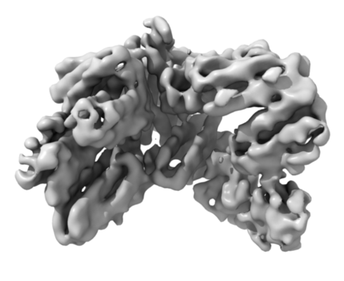 emd_4772.png emd_4772.png | 99.8 KB | ||
| Filedesc metadata | emd-4772.cif.gz | 8 KB | ||
| Others | emd_4772_additional.map.gz | 3.8 MB | ||
| Archive directory |  http://ftp.pdbj.org/pub/emdb/structures/EMD-4772ftp://ftp.pdbj.org/pub/emdb/structures/EMD-4772 http://ftp.pdbj.org/pub/emdb/structures/EMD-4772ftp://ftp.pdbj.org/pub/emdb/structures/EMD-4772 | HTTPS FTP |
-Related structure data
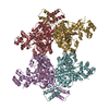
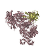
| Related structure data |  6r9tMC M: atomic model generated by this map C: citing same article ( |
|---|---|
| Similar structure data |

-Links
| EMDB pages | EMDB (EBI/PDBe) / EMDataResource |
|---|---|
| Related items in Molecule of the Month |


-Map
| File | Download / File: emd_4772.map.gz / Format: CCP4 / Size: 42.9 MB / Type: IMAGE STORED AS FLOATING POINT NUMBER (4 BYTES) | ||||||||||||||||||||||||||||||||||||||||||||||||||||||||||||
|---|---|---|---|---|---|---|---|---|---|---|---|---|---|---|---|---|---|---|---|---|---|---|---|---|---|---|---|---|---|---|---|---|---|---|---|---|---|---|---|---|---|---|---|---|---|---|---|---|---|---|---|---|---|---|---|---|---|---|---|---|---|
| Annotation | best map | ||||||||||||||||||||||||||||||||||||||||||||||||||||||||||||
| Projections & slices | Image control
Images are generated by Spider. | ||||||||||||||||||||||||||||||||||||||||||||||||||||||||||||
| Voxel size | X=Y=Z: 1.06 Å | ||||||||||||||||||||||||||||||||||||||||||||||||||||||||||||
| Density |
| ||||||||||||||||||||||||||||||||||||||||||||||||||||||||||||
| Symmetry | Space group: 1 | ||||||||||||||||||||||||||||||||||||||||||||||||||||||||||||
| Details | EMDB XML:
CCP4 map header:
| ||||||||||||||||||||||||||||||||||||||||||||||||||||||||||||
 Z (Sec.)
 Y (Row.)
 X (Col.)


-Supplemental data
-Additional map: map with additional density
| File | emd_4772_additional.map | ||||||||||||
|---|---|---|---|---|---|---|---|---|---|---|---|---|---|
| Annotation | map with additional density | ||||||||||||
| Projections & Slices |
| ||||||||||||
| Density Histograms |


- Sample components
-Entire : Talin-1
| Entire | Name: Talin-1 |
|---|---|
| Components |
|
-Supramolecule #1: Talin-1
| Supramolecule | Name: Talin-1 / type: complex / ID: 1 / Parent: 0 / Macromolecule list: all / Details: autoinhibited |
|---|---|
| Source (natural) | Organism: Homo sapiens (human) |
-Macromolecule #1: Talin-1
| Macromolecule | Name: Talin-1 / type: protein_or_peptide / ID: 1 / Number of copies: 1 / Enantiomer: LEVO |
|---|---|
| Source (natural) | Organism: Homo sapiens (human) |
| Molecular weight | Theoretical: 270.773719 KDa |
| Recombinant expression | Organism:  |
| Sequence | String: MVALSLKISI GNVVKTMQFE PSTMVYDACR IIRERIPEAP AGPPSDFGLF LSDDDPKKGI WLEAGKALDY YMLRNGDTME YRKKQRPLK IRMLDGTVKT IMVDDSKTVT DMLMTICARI GITNHDEYSL VRELMEEKKE EITGTLRKDK TLLRDEKKME K LKQKLHTD ...String: MVALSLKISI GNVVKTMQFE PSTMVYDACR IIRERIPEAP AGPPSDFGLF LSDDDPKKGI WLEAGKALDY YMLRNGDTME YRKKQRPLK IRMLDGTVKT IMVDDSKTVT DMLMTICARI GITNHDEYSL VRELMEEKKE EITGTLRKDK TLLRDEKKME K LKQKLHTD DELNWLDHGR TLREQGVEEH ETLLLRRKFF YSDQNVDSRD PVQLNLLYVQ ARDDILNGSH PVSFDKACEF AG FQCQIQF GPHNEQKHKA GFLDLKDFLP KEYVKQKGER KIFQAHKNCG QMSEIEAKVR YVKLARSLKT YGVSFFLVKE KMK GKNKLV PRLLGITKEC VMRVDEKTKE VIQEWNLTNI KRWAASPKSF TLDFGDYQDG YYSVQTTEGE QIAQLIAGYI DIIL KKKKS KDHFGLEGDE ESTMLEDSVS PKKSTVLQQQ YNRVGKVEHG SVALPAIMRS GASGPENFQV GSMPPAQQQI TSGQM HRGH MPPLTSAQQA LTGTINSSMQ AVQAAQATLD DFDTLPPLGQ DAASKAWRKN KMDESKHEIH SQVDAITAGT ASVVNL TAG DPAETDYTAV GCAVTTISSN LTEMSRGVKL LAALLEDEGG SGRPLLQAAK GLAGAVSELL RSAQPASAEP RQNLLQA AG NVGQASGELL QQIGESDTDP HFQDALMQLA KAVASAAAAL VLKAKSVAQR TEDSGLQTQV IAAATQCALS TSQLVACT K VVAPTISSPV CQEQLVEAGR LVAKAVEGCV SASQAATEDG QLLRGVGAAA TAVTQALNEL LQHVKAHATG AGPAGRYDQ ATDTILTVTE NIFSSMGDAG EMVRQARILA QATSDLVNAI KADAEGESDL ENSRKLLSAA KILADATAKM VEAAKGAAAH PDSEEQQQR LREAAEGLRM ATNAAAQNAI KKKLVQRLEH AAKQAAASAT QTIAAAQHAA STPKASAGPQ PLLVQSCKAV A EQIPLLVQ GVRGSQAQPD SPSAQLALIA ASQSFLQPGG KMVAAAKASV PTIQDQASAM QLSQCAKNLG TALAELRTAA QK AQEACGP LEMDSALSVV QNLEKDLQEV KAAARDGKLK PLPGETMEKC TQDLGNSTKA VSSAIAQLLG EVAQGNENYA GIA ARDVAG GLRSLAQAAR GVAALTSDPA VQAIVLDTAS DVLDKASSLI EEAKKAAGHP GDPESQQRLA QVAKAVTQAL NRCV SCLPG QRDVDNALRA VGDASKRLLS DSLPPSTGTF QEAQSRLNEA AAGLNQAATE LVQASRGTPQ DLARASGRFG QDFST FLEA GVEMAGQAPS QEDRAQVVSN LKGISMSSSK LLLAAKALST DPAAPNLKSQ LAAAARAVTD SINQLITMCT QQAPGQ KEC DNALRELETV RELLENPVQP INDMSYFGCL DSVMENSKVL GEAMTGISQN AKNGNLPEFG DAISTASKAL CGFTEAA AQ AAYLVGVSDP NSQAGQQGLV EPTQFARANQ AIQMACQSLG EPGCTQAQVL SAATIVAKHT SALCNSCRLA SARTTNPT A KRQFVQSAKE VANSTANLVK TIKALDGAFT EENRAQCRAA TAPLLEAVDN LSAFASNPEF SSIPAQISPE GRAAMEPIV ISAKTMLESA GGLIQTARAL AVNPRDPPSW SVLAGHSRTV SDSIKKLITS MRDKAPGQLE CETAIAALNS CLRDLDQASL AAVSQQLAP REGISQEALH TQMLTAVQEI SHLIEPLANA ARAEASQLGH KVSQMAQYFE PLTLAAVGAA SKTLSHPQQM A LLDQTKTL AESALQLLYT AKEAGGNPKQ AAHTQEALEE AVQMMTEAVE DLTTTLNEAA SAAGVVGGMV DSITQAINQL DE GPMGEPE GSFVDYQTTM VRTAKAIAVT VQEMVTKSNT SPEELGPLAN QLTSDYGRLA SEAKPAAVAA ENEEIGSHIK HRV QELGHG CAALVTKAGA LQCSPSDAYT KKELIECARR VSEKVSHVLA ALQAGNRGTQ ACITAASAVS GIIADLDTTI MFAT AGTLN REGTETFADH REGILKTAKV LVEDTKVLVQ NAAGSQEKLA QAAQSSVATI TRLADVVKLG AASLGAEDPE TQVVL INAV KDVAKALGDL ISATKAAAGK VGDDPAVWQL KNSAKVMVTN VTSLLKTVKA VEDEATKGTR ALEATTEHIR QELAVF CSP EPPAKTSTPE DFIRMTKGIT MATAKAVAAG NSCRQEDVIA TANLSRRAIA DMLRACKEAA YHPEVAPDVR LRALHYG RE CANGYLELLD HVLLTLQKPS PELKQQLTGH SKRVAGSVTE LIQAAEAMKG TEWVDPEDPT VIAENELLGA AAAIEAAA K KLEQLKPRAK PKEADESLNF EEQILEAAKS IAAATSALVK AASAAQRELV AQGKVGAIPA NALDDGQWSQ GLISAARMV AAATNNLCEA ANAAVQGHAS QEKLISSAKQ VAASTAQLLV ACKVKADQDS EAMKRLQAAG NAVKRASDNL VKAAQKAAAF EEQENETVV VKEKMVGGIA QIIAAQEEML RKERELEEAR KKLAQIRQQQ YKFLPSELRD EHLVVLFQ UniProtKB: Talin-1 |
-Experimental details
-Structure determination
| Method | cryo EM |
|---|---|
 Processing Processing | single particle reconstruction |
| Aggregation state | particle |
-Sample preparation
| Concentration | 0.3 mg/mL | |||||||||||||||
|---|---|---|---|---|---|---|---|---|---|---|---|---|---|---|---|---|
| Buffer | pH: 7.5 Component:
| |||||||||||||||
| Grid | Model: Quantifoil R1.2/1.3 / Material: COPPER / Mesh: 200 / Support film - Material: CARBON / Support film - topology: HOLEY / Pretreatment - Type: GLOW DISCHARGE / Pretreatment - Time: 30 sec. / Pretreatment - Atmosphere: AIR | |||||||||||||||
| Vitrification | Cryogen name: ETHANE-PROPANE / Chamber humidity: 95 % / Chamber temperature: 277 K / Instrument: FEI VITROBOT MARK IV / Details: Blotted 4 seconds before plunging, blot force 4. | |||||||||||||||
| Details | GraFix |
- Electron microscopy
| Microscope | FEI TITAN KRIOS |
|---|---|
| Specialist optics | Energy filter - Name: GIF Quantum LS / Energy filter - Slit width: 20 eV |
| Image recording | Film or detector model: GATAN K2 SUMMIT (4k x 4k) / Detector mode: COUNTING / Digitization - Dimensions - Width: 4096 pixel / Digitization - Dimensions - Height: 4096 pixel / Digitization - Frames/image: 1-40 / Number grids imaged: 1 / Number real images: 11007 / Average exposure time: 10.0 sec. / Average electron dose: 76.8 e/Å2 |
| Electron beam | Acceleration voltage: 300 kV / Electron source:  FIELD EMISSION GUN FIELD EMISSION GUN |
| Electron optics | C2 aperture diameter: 70.0 µm / Illumination mode: FLOOD BEAM / Imaging mode: BRIGHT FIELD / Cs: 2.62 mm / Nominal defocus max: 3.0 µm / Nominal defocus min: 1.0 µm / Nominal magnification: 130000 |
| Sample stage | Specimen holder model: FEI TITAN KRIOS AUTOGRID HOLDER / Cooling holder cryogen: NITROGEN |
| Experimental equipment |  Model: Titan Krios / Image courtesy: FEI Company |
+Image processing
-Atomic model buiding 1
| Initial model |
| ||||||||||||||||||||||
|---|---|---|---|---|---|---|---|---|---|---|---|---|---|---|---|---|---|---|---|---|---|---|---|
| Refinement | Space: REAL / Protocol: FLEXIBLE FIT / Overall B value: 377 | ||||||||||||||||||||||
| Output model | PDB-6r9t: |