Resolution: 1.85→20 Å / Num. obs: 52439 / % possible obs: 98.1 % / Rsym value: 0.085 / Net I/σ(I): 16.8
Reflection shell
Resolution: 1.85→1.92 Å / Mean I/σ(I) obs: 1.7 / Rsym value: 0.449 / % possible all: 87.5
-
Processing
Software
Name
Version
Classification
SOLVE
phasing
REFMAC
5.2.0019
refinement
ADSC
datacollection
DENZO
datareduction
Refinement
Method to determine structure: SAD / Resolution: 1.85→19.57 Å / Cor.coef. Fo:Fc: 0.948 / Cor.coef. Fo:Fc free: 0.933 / SU B: 3.774 / SU ML: 0.115 / Cross valid method: THROUGHOUT / ESU R: 0.166 / ESU R Free: 0.155 / Stereochemistry target values: MAXIMUM LIKELIHOOD / Details: HYDROGENS HAVE BEEN ADDED IN THE RIDING POSITIONS
Rfactor
Num. reflection
% reflection
Selection details
Rfree
0.25989
1300
2.6 %
RANDOM
Rwork
0.2138
-
-
-
obs
0.215
49457
98.3 %
-
Solvent computation
Ion probe radii: 0.8 Å / Shrinkage radii: 0.8 Å / VDW probe radii: 1.2 Å / Solvent model: MASK
Displacement parameters
Biso mean: 30.897 Å2
Baniso -1
Baniso -2
Baniso -3
1-
0 Å2
0 Å2
-0.27 Å2
2-
-
0.18 Å2
0 Å2
3-
-
-
-0.31 Å2
Refinement step
Cycle: LAST / Resolution: 1.85→19.57 Å
Protein
Nucleic acid
Ligand
Solvent
Total
Num. atoms
4678
0
0
332
5010
Refine LS restraints
Refine-ID
Type
Dev ideal
Dev ideal target
Number
X-RAY DIFFRACTION
r_bond_refined_d
0.016
0.022
4726
X-RAY DIFFRACTION
r_bond_other_d
X-RAY DIFFRACTION
r_angle_refined_deg
1.454
1.971
6410
X-RAY DIFFRACTION
r_angle_other_deg
X-RAY DIFFRACTION
r_dihedral_angle_1_deg
4.839
5
632
X-RAY DIFFRACTION
r_dihedral_angle_2_deg
40.945
25.227
176
X-RAY DIFFRACTION
r_dihedral_angle_3_deg
15.849
15
840
X-RAY DIFFRACTION
r_dihedral_angle_4_deg
15.169
15
28
X-RAY DIFFRACTION
r_chiral_restr
0.099
0.2
800
X-RAY DIFFRACTION
r_gen_planes_refined
0.006
0.02
3422
X-RAY DIFFRACTION
r_gen_planes_other
X-RAY DIFFRACTION
r_nbd_refined
0.215
0.2
2657
X-RAY DIFFRACTION
r_nbd_other
X-RAY DIFFRACTION
r_nbtor_refined
0.294
0.2
3425
X-RAY DIFFRACTION
r_nbtor_other
X-RAY DIFFRACTION
r_xyhbond_nbd_refined
0.168
0.2
322
X-RAY DIFFRACTION
r_xyhbond_nbd_other
X-RAY DIFFRACTION
r_metal_ion_refined
X-RAY DIFFRACTION
r_metal_ion_other
X-RAY DIFFRACTION
r_symmetry_vdw_refined
0.304
0.2
101
X-RAY DIFFRACTION
r_symmetry_vdw_other
X-RAY DIFFRACTION
r_symmetry_hbond_refined
0.178
0.2
19
X-RAY DIFFRACTION
r_symmetry_hbond_other
X-RAY DIFFRACTION
r_symmetry_metal_ion_refined
X-RAY DIFFRACTION
r_symmetry_metal_ion_other
X-RAY DIFFRACTION
r_mcbond_it
1.074
1.5
3238
X-RAY DIFFRACTION
r_mcbond_other
X-RAY DIFFRACTION
r_mcangle_it
1.683
2
5056
X-RAY DIFFRACTION
r_scbond_it
3.28
3
1626
X-RAY DIFFRACTION
r_scangle_it
5.068
4.5
1354
X-RAY DIFFRACTION
r_rigid_bond_restr
X-RAY DIFFRACTION
r_sphericity_free
X-RAY DIFFRACTION
r_sphericity_bonded
LS refinement shell
Resolution: 1.85→1.898 Å / Total num. of bins used: 20
Rfactor
Num. reflection
% reflection
Rfree
0.412
87
-
Rwork
0.287
3184
-
obs
-
-
87.3 %
Refinement TLS params.
Method: refined / Refine-ID: X-RAY DIFFRACTION
ID
L11 (°2)
L12 (°2)
L13 (°2)
L22 (°2)
L23 (°2)
L33 (°2)
S11 (Å °)
S12 (Å °)
S13 (Å °)
S21 (Å °)
S22 (Å °)
S23 (Å °)
S31 (Å °)
S32 (Å °)
S33 (Å °)
T11 (Å2)
T12 (Å2)
T13 (Å2)
T22 (Å2)
T23 (Å2)
T33 (Å2)
Origin x (Å)
Origin y (Å)
Origin z (Å)
1
1.5505
0.3415
0.86
1.7147
-0.3874
4.1034
0.0246
-0.1086
-0.0301
0.1512
0.0276
-0.0183
0.0758
0.1268
-0.0523
-0.1191
-0.0054
0.026
-0.2
-0.0372
-0.1262
34.3896
32.407
4.4452
2
0.9228
1.0413
1.2
3.0154
3.3131
4.0151
0.0336
0.07
0.0203
0.0842
-0.0762
0.0447
0.082
-0.3274
0.0427
-0.1469
0.0002
0.0153
-0.0567
0.0233
-0.1061
19.7736
20.1333
-39.1872
3
1.4879
-0.36
0.4145
1.7613
0.6437
3.4692
0.0275
0.0419
-0.0814
0.0217
-0.0219
-0.0111
0.1253
-0.254
-0.0056
-0.1352
-0.0142
0.046
-0.1555
0.0255
-0.111
35.0512
2.9583
-49.7518
4
1.8257
-0.4312
0.8125
2.1967
-2.09
3.3783
-0.0716
0.1948
0.2259
0.0332
-0.0723
-0.1284
-0.0984
0.3598
0.144
-0.1081
-0.0338
-0.0093
0.0038
-0.0026
-0.0661
49.3447
-6.9696
-5.7673
Refinement TLS group
ID
Refine-ID
Refine TLS-ID
Auth asym-ID
Label asym-ID
Auth seq-ID
Label seq-ID
1
X-RAY DIFFRACTION
1
A
A
1977 - 2001
16 - 40
2
X-RAY DIFFRACTION
1
A
A
2012 - 2037
51 - 76
3
X-RAY DIFFRACTION
1
A
A
2041 - 2069
80 - 108
4
X-RAY DIFFRACTION
1
A
A
2074 - 2102
113 - 141
5
X-RAY DIFFRACTION
1
A
A
2109 - 2136
148 - 175
6
X-RAY DIFFRACTION
2
A
A
2140 - 2161
179 - 200
7
X-RAY DIFFRACTION
2
A
A
2172 - 2194
211 - 233
8
X-RAY DIFFRACTION
2
A
A
2199 - 2224
238 - 263
9
X-RAY DIFFRACTION
2
A
A
2230 - 2259
269 - 298
10
X-RAY DIFFRACTION
2
A
A
2264 - 2289
303 - 328
11
X-RAY DIFFRACTION
3
B
B
1977 - 2001
16 - 40
12
X-RAY DIFFRACTION
3
B
B
2012 - 2037
51 - 76
13
X-RAY DIFFRACTION
3
B
B
2041 - 2069
80 - 108
14
X-RAY DIFFRACTION
3
B
B
2074 - 2102
113 - 141
15
X-RAY DIFFRACTION
3
B
B
2109 - 2136
148 - 175
16
X-RAY DIFFRACTION
4
B
B
2140 - 2161
179 - 200
17
X-RAY DIFFRACTION
4
B
B
2172 - 2194
211 - 233
18
X-RAY DIFFRACTION
4
B
B
2199 - 2224
238 - 263
19
X-RAY DIFFRACTION
4
B
B
2230 - 2259
269 - 298
20
X-RAY DIFFRACTION
4
B
B
2264 - 2289
303 - 328
+
About Yorodumi
-
News
-
Feb 9, 2022. New format data for meta-information of EMDB entries
New format data for meta-information of EMDB entries
Version 3 of the EMDB header file is now the official format.
The previous official version 1.9 will be removed from the archive.
In the structure databanks used in Yorodumi, some data are registered as the other names, "COVID-19 virus" and "2019-nCoV". Here are the details of the virus and the list of structure data.
Jan 31, 2019. EMDB accession codes are about to change! (news from PDBe EMDB page)
EMDB accession codes are about to change! (news from PDBe EMDB page)
The allocation of 4 digits for EMDB accession codes will soon come to an end. Whilst these codes will remain in use, new EMDB accession codes will include an additional digit and will expand incrementally as the available range of codes is exhausted. The current 4-digit format prefixed with “EMD-” (i.e. EMD-XXXX) will advance to a 5-digit format (i.e. EMD-XXXXX), and so on. It is currently estimated that the 4-digit codes will be depleted around Spring 2019, at which point the 5-digit format will come into force.
The EM Navigator/Yorodumi systems omit the EMD- prefix.
Related info.:Q: What is EMD? / ID/Accession-code notation in Yorodumi/EM Navigator
Yorodumi is a browser for structure data from EMDB, PDB, SASBDB, etc.
This page is also the successor to EM Navigator detail page, and also detail information page/front-end page for Omokage search.
The word "yorodu" (or yorozu) is an old Japanese word meaning "ten thousand". "mi" (miru) is to see.
Related info.:EMDB / PDB / SASBDB / Comparison of 3 databanks / Yorodumi Search / Aug 31, 2016. New EM Navigator & Yorodumi / Yorodumi Papers / Jmol/JSmol / Function and homology information / Changes in new EM Navigator and Yorodumi
 Movie
Movie Controller


 Open data
 Basic information
 Components
 Keywords
 Function and homology information
 X-RAY DIFFRACTION /
  Authors
 Citation
 Structure visualization
 Downloads & links
 Other downloads
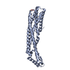


 PDBj
PDBj

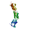
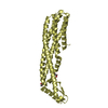
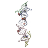
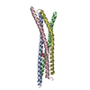
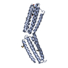
 Assembly
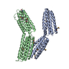


 Mass: 18.015 Da / Num. of mol.: 332 / Source method: isolated from a natural source / Formula: H2O
 Sample preparation
 / Beamline: ID14-4 / Wavelength: 0.9757
  Processing