 Movie
Movie Controller

[English] 日本語
 Yorodumi
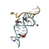
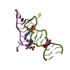
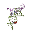
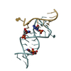
- PDB-6yml: Crystal structure of the SAM-SAH riboswitch with decarboxylated SAH -
+ Open data

- Basic information
| Entry | Database: PDB / ID: 6yml | |||||||||
|---|---|---|---|---|---|---|---|---|---|---|
| Title | Crystal structure of the SAM-SAH riboswitch with decarboxylated SAH | |||||||||
 Components Components |
| |||||||||
 Keywords Keywords | RNA / Pseudoknot / SAM / Riboswitch | |||||||||
| Function / homology | ADENOSINE MONOPHOSPHATE / 5'-S-(3-aminopropyl)-5'-thioadenosine / RNA / RNA (> 10) Function and homology information Function and homology information | |||||||||
| Biological species |  Roseobacter sp. (bacteria) Roseobacter sp. (bacteria) | |||||||||
| Method |  X-RAY DIFFRACTION / SYNCHROTRON / MOLECULAR REPLACEMENT / Resolution: 2.17 Å X-RAY DIFFRACTION / SYNCHROTRON / MOLECULAR REPLACEMENT / Resolution: 2.17 Å | |||||||||
 Authors Authors | Huang, L. / Lilley, D.M.J. | |||||||||
| Funding support |  United Kingdom, United Kingdom,  China, 2items China, 2items
| |||||||||
 Citation Citation | Journal: Nucleic Acids Res. / Year: 2020 Title: Crystal structure and ligand-induced folding of the SAM/SAH riboswitch. Authors: Huang, L. / Liao, T.W. / Wang, J. / Ha, T. / Lilley, D.M.J. | |||||||||
| History |
|
- Structure visualization
| Structure viewer | Molecule: MolmilJmol/JSmol |
|---|
- Downloads & links
-Download
| PDBx/mmCIF format | 6yml.cif.gz | 108.2 KB | Display | PDBx/mmCIF format |
|---|---|---|---|---|
| PDB format | pdb6yml.ent.gz | 70.6 KB | Display | PDB format |
| PDBx/mmJSON format | 6yml.json.gz | Tree view | PDBx/mmJSON format | |
| Others |  Other downloads Other downloads |
-Validation report
| Arichive directory | https://data.pdbj.org/pub/pdb/validation_reports/ym/6ymlftp://data.pdbj.org/pub/pdb/validation_reports/ym/6yml | HTTPS FTP |
|---|
-Related structure data
| Related structure data | 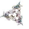 6yl5SC 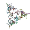 6ylbC 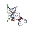 6ymiC 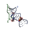 6ymjC 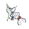 6ymkC 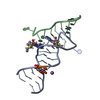 6ymmC S: Starting model for refinement C: citing same article ( |
|---|---|
| Similar structure data |


-Links
 PDBj
PDBj


- Assembly
| Deposited unit | 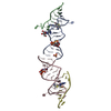
| ||||||||||||
|---|---|---|---|---|---|---|---|---|---|---|---|---|---|
| 1 | 
| ||||||||||||
| 2 | 
| ||||||||||||
| Unit cell |
|
-Components
-RNA chain , 2 types, 4 molecules ACBD
| #1: RNA chain | Mass: 8389.874 Da / Num. of mol.: 2 / Source method: obtained synthetically / Source: (synth.) Roseobacter sp. (bacteria)#2: RNA chain | Mass: 2895.791 Da / Num. of mol.: 2 / Source method: obtained synthetically / Source: (synth.) Roseobacter sp. (bacteria) |
|---|
-Non-polymers , 4 types, 9 molecules 
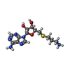


| #3: Chemical | ChemComp-AMP /  Mass: 347.221 Da / Num. of mol.: 1 / Source method: obtained synthetically / Formula: C10H14N5O7P / Feature type: SUBJECT OF INVESTIGATION / Comment: AMP*YM Mass: 347.221 Da / Num. of mol.: 1 / Source method: obtained synthetically / Formula: C10H14N5O7P / Feature type: SUBJECT OF INVESTIGATION / Comment: AMP*YM | ||||
|---|---|---|---|---|---|
| #4: Chemical |  Mass: 340.401 Da / Num. of mol.: 2 / Source method: obtained synthetically / Formula: C13H20N6O3S / Feature type: SUBJECT OF INVESTIGATION Mass: 340.401 Da / Num. of mol.: 2 / Source method: obtained synthetically / Formula: C13H20N6O3S / Feature type: SUBJECT OF INVESTIGATION#5: Chemical | ChemComp-SO4 / |  Mass: 96.063 Da / Num. of mol.: 1 / Source method: obtained synthetically / Formula: SO4 Mass: 96.063 Da / Num. of mol.: 1 / Source method: obtained synthetically / Formula: SO4#6: Water | ChemComp-HOH / | Mass: 18.015 Da / Num. of mol.: 5 / Source method: isolated from a natural source / Formula: H2O |
-Details
| Has ligand of interest | Y |
|---|
-Experimental details
-Experiment
| Experiment | Method: X-RAY DIFFRACTION / Number of used crystals: 1 |
|---|
- Sample preparation
| Crystal | Density Matthews: 3.72 Å3/Da / Density % sol: 66.9 % |
|---|---|
| Crystal grow | Temperature: 293 K / Method: vapor diffusion, sitting drop / pH: 6 Details: 0.01 M Magnesium Sulfate, 0.05 M Sodium Cacodylate pH 6.0, 1.8 M Lithium Sulfate monohydrate |
-Data collection
| Diffraction | Mean temperature: 100 K / Serial crystal experiment: N |
|---|---|
| Diffraction source | Source: SYNCHROTRON / Site: Diamond / Beamline: I24 / Wavelength: 0.9188 Å |
| Detector | Type: DECTRIS PILATUS3 6M / Detector: PIXEL / Date: Sep 15, 2019 |
| Radiation | Protocol: SINGLE WAVELENGTH / Monochromatic (M) / Laue (L): M / Scattering type: x-ray |
| Radiation wavelength | Wavelength: 0.9188 Å / Relative weight: 1 |
| Reflection | Resolution: 2.17→75.4 Å / Num. obs: 17121 / % possible obs: 100 % / Observed criterion σ(I): 0.6 / Redundancy: 9.7 % / Biso Wilson estimate: 69.42 Å2 / CC1/2: 0.997 / Rmerge(I) obs: 0.095 / Rpim(I) all: 0.033 / Net I/σ(I): 9.4 |
| Reflection shell | Resolution: 2.17→2.2 Å / Rmerge(I) obs: 1.5 / Mean I/σ(I) obs: 0.6 / Num. unique obs: 867 / CC1/2: 0.355 / Rpim(I) all: 1 / % possible all: 100 |
- Processing
| Software |
| ||||||||||||||||||||||||||||||||||||||||||
|---|---|---|---|---|---|---|---|---|---|---|---|---|---|---|---|---|---|---|---|---|---|---|---|---|---|---|---|---|---|---|---|---|---|---|---|---|---|---|---|---|---|---|---|
| Refinement | Method to determine structure: MOLECULAR REPLACEMENT Starting model: 6YL5 Resolution: 2.17→53.05 Å / SU ML: 0.3452 / Cross valid method: FREE R-VALUE / σ(F): 1.35 / Phase error: 32.1866
| ||||||||||||||||||||||||||||||||||||||||||
| Solvent computation | Shrinkage radii: 0.9 Å / VDW probe radii: 1.11 Å | ||||||||||||||||||||||||||||||||||||||||||
| Displacement parameters | Biso mean: 74.95 Å2 | ||||||||||||||||||||||||||||||||||||||||||
| Refinement step | Cycle: LAST / Resolution: 2.17→53.05 Å
| ||||||||||||||||||||||||||||||||||||||||||
| Refine LS restraints |
| ||||||||||||||||||||||||||||||||||||||||||
| LS refinement shell |
| ||||||||||||||||||||||||||||||||||||||||||
| Refinement TLS params. | Method: refined / Origin x: 20.5268773692 Å / Origin y: -28.5433780996 Å / Origin z: 17.9665400538 Å
| ||||||||||||||||||||||||||||||||||||||||||
| Refinement TLS group | Selection details: all |
