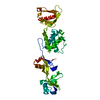
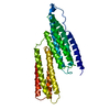
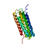
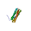
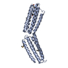
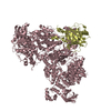
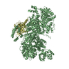
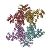
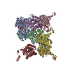
Journal: Cell / Year: 2019 Title: The Architecture of Talin1 Reveals an Autoinhibition Mechanism. Authors: Dirk Dedden / Stephanie Schumacher / Charlotte F Kelley / Martin Zacharias / Christian Biertümpfel / Reinhard Fässler / Naoko Mizuno / Abstract: Focal adhesions (FAs) are protein machineries essential for cell adhesion, migration, and differentiation. Talin is an integrin-activating and tension-sensing FA component directly connecting ...Focal adhesions (FAs) are protein machineries essential for cell adhesion, migration, and differentiation. Talin is an integrin-activating and tension-sensing FA component directly connecting integrins in the plasma membrane with the actomyosin cytoskeleton. To understand how talin function is regulated, we determined a cryoelectron microscopy (cryo-EM) structure of full-length talin1 revealing a two-way mode of autoinhibition. The actin-binding rod domains fold into a 15-nm globular arrangement that is interlocked by the integrin-binding FERM head. In turn, the rod domains R9 and R12 shield access of the FERM domain to integrin and the phospholipid PIP at the membrane. This mechanism likely ensures synchronous inhibition of integrin, membrane, and cytoskeleton binding. We also demonstrate that compacted talin1 reversibly unfolds to an ∼60-nm string-like conformation, revealing interaction sites for vinculin and actin. Our data explain how fast switching between active and inactive conformations of talin could regulate FA turnover, a process critical for cell adhesion and signaling.
Instrument: FEI VITROBOT MARK IV / Cryogen name: ETHANE-PROPANE / Humidity: 95 % / Chamber temperature: 277 K / Details: Blotted 4 seconds before plunging, blot force 4
-
Electron microscopy imaging
Experimental equipment
Model: Titan Krios / Image courtesy: FEI Company
Microscopy
Model: FEI TITAN KRIOS / Date: May 14, 2018
Electron gun
Electron source: FIELD EMISSION GUN / Accelerating voltage: 300 kV / Illumination mode: FLOOD BEAM
Electron lens
Mode: BRIGHT FIELD / Nominal magnification: 130000 X / Nominal defocus max: 3000 nm / Nominal defocus min: 1000 nm / Cs: 2.62 mm / C2 aperture diameter: 70 µm / Alignment procedure: COMA FREE
Average exposure time: 10 sec. / Electron dose: 76.8 e/Å2 / Detector mode: COUNTING / Film or detector model: GATAN K2 SUMMIT (4k x 4k) / Num. of grids imaged: 1 / Num. of real images: 11007
EM imaging optics
Energyfilter name: GIF Quantum LS / Energyfilter slit width: 20 eV
Image scans
Sampling size: 5 µm / Width: 4096 / Height: 4096 / Movie frames/image: 40 / Used frames/image: 1-40
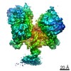
Resolution: 6.2 Å / Resolution method: FSC 0.143 CUT-OFF / Num. of particles: 30438 / Actual pixel size: 1.06 Å / Algorithm: FOURIER SPACE / Num. of class averages: 1 / Symmetry type: POINT
Atomic model building
B value: 377 / Protocol: FLEXIBLE FIT / Space: REAL
Atomic model building
3D fitting-ID: 1 / Pdb chain-ID: a / Source name: PDB / Type: experimental model
In the structure databanks used in Yorodumi, some data are registered as the other names, "COVID-19 virus" and "2019-nCoV". Here are the details of the virus and the list of structure data.
Jan 31, 2019. EMDB accession codes are about to change! (news from PDBe EMDB page)
EMDB accession codes are about to change! (news from PDBe EMDB page)
The allocation of 4 digits for EMDB accession codes will soon come to an end. Whilst these codes will remain in use, new EMDB accession codes will include an additional digit and will expand incrementally as the available range of codes is exhausted. The current 4-digit format prefixed with “EMD-” (i.e. EMD-XXXX) will advance to a 5-digit format (i.e. EMD-XXXXX), and so on. It is currently estimated that the 4-digit codes will be depleted around Spring 2019, at which point the 5-digit format will come into force.
The EM Navigator/Yorodumi systems omit the EMD- prefix.
Related info.:Q: What is EMD? / ID/Accession-code notation in Yorodumi/EM Navigator
Yorodumi is a browser for structure data from EMDB, PDB, SASBDB, etc.
This page is also the successor to EM Navigator detail page, and also detail information page/front-end page for Omokage search.
The word "yorodu" (or yorozu) is an old Japanese word meaning "ten thousand". "mi" (miru) is to see.
Related info.:EMDB / PDB / SASBDB / Comparison of 3 databanks / Yorodumi Search / Aug 31, 2016. New EM Navigator & Yorodumi / Yorodumi Papers / Jmol/JSmol / Function and homology information / Changes in new EM Navigator and Yorodumi
 Movie
Movie Controller


 Open data
 Basic information
 Components
 Keywords
 Function and homology information
 Homo sapiens (human)
 Authors
 Germany, 2items
  Citation
 Structure visualization
 Downloads & links
 Other downloads

 PDBj
PDBj


 Assembly
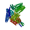

 Sample preparation
 Electron microscopy imaging
 FIELD EMISSION GUN / Accelerating voltage: 300 kV / Illumination mode: FLOOD BEAM
 Processing