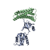
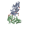
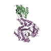
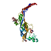
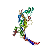
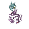
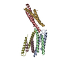
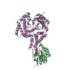
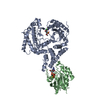
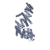
[English] 日本語
Yorodumi
- PDB-5ic1: Structural analysis of a talin triple domain module, E1794Y, E179... -

+
Open data


ID or keywords:

Loading...

-
Basic information

Entry
Database: PDB / ID: 5ic1
TitleStructural analysis of a talin triple domain module, E1794Y, E1797Y, Q1801Y mutant
ComponentsTalin-1
KeywordsPEPTIDE BINDING PROTEIN / Integrin / RIAM / autoinhibition / alternative
Function / homology
Function and homology information


GRB2:SOS provides linkage to MAPK signaling for Integrins / Integrin signaling / Smooth Muscle Contraction / p130Cas linkage to MAPK signaling for integrins / SEMA3A-Plexin repulsion signaling by inhibiting Integrin adhesion / MAP2K and MAPK activation / Platelet degranulation / LIM domain binding / vinculin binding / integrin activation ...GRB2:SOS provides linkage to MAPK signaling for Integrins / Integrin signaling / Smooth Muscle Contraction / p130Cas linkage to MAPK signaling for integrins / SEMA3A-Plexin repulsion signaling by inhibiting Integrin adhesion / MAP2K and MAPK activation / Platelet degranulation / LIM domain binding / vinculin binding / integrin activation / cell-substrate junction assembly / cortical actin cytoskeleton organization / phosphatidylserine binding / ruffle / phosphatidylinositol binding / integrin-mediated signaling pathway / adherens junction / structural constituent of cytoskeleton / cell-cell adhesion / ruffle membrane / actin filament binding / integrin binding / cytoskeleton / focal adhesion / cell surface / plasma membrane / cytoplasm
Similarity search - Function
: / Talin, R4 domain / Vinculin-binding site-containing domain / Talin, central / Talin, N-terminal F0 domain / Talin, central domain superfamily / Talin-1/2, rod-segment / Vinculin Binding Site / Talin, middle domain / N-terminal or F0 domain of Talin-head FERM ...: / Talin, R4 domain / Vinculin-binding site-containing domain / Talin, central / Talin, N-terminal F0 domain / Talin, central domain superfamily / Talin-1/2, rod-segment / Vinculin Binding Site / Talin, middle domain / N-terminal or F0 domain of Talin-head FERM / I/LWEQ domain / I/LWEQ domain superfamily / I/LWEQ domain / I/LWEQ domain profile. / I/LWEQ domain / Phosphotyrosine-binding domain / Alpha-catenin/vinculin-like superfamily / FERM domain signature 1. / FERM conserved site / FERM domain signature 2. / FERM central domain / FERM/acyl-CoA-binding protein superfamily / FERM central domain / FERM superfamily, second domain / FERM domain / FERM domain profile. / Band 4.1 domain / Band 4.1 homologues / PH-like domain superfamily / Ubiquitin-like domain superfamily
Similarity search - Domain/homology
Biological speciesMus musculus (house mouse)
MethodX-RAY DIFFRACTION / SYNCHROTRON / MOLECULAR REPLACEMENT / Resolution: 2.2 Å
AuthorsWu, J. / Chang, Y.-C.E. / Zhang, H. / Huang, Q.-Q.
Funding support United States, 3items
OrganizationGrant numberCountry
National Institutes of Health/National Cancer Institute (NIH/NCI)CA006927 United States
Pennsylvania Department of Health4100068716, ACS RSG-15-167-01-DMC United States
CCSG Supported Pilot Projects award5P30CA006927-51 United States
CitationJournal: Structure / Year: 2016
Title: Structural and Functional Analysis of a Talin Triple-Domain Module Suggests an Alternative Talin Autoinhibitory Configuration.
Authors: Zhang, H. / Chang, Y.C. / Huang, Q. / Brennan, M.L. / Wu, J.
History
DepositionFeb 22, 2016Deposition site: RCSB / Processing site: RCSB
Revision 1.0May 18, 2016Provider: repository / Type: Initial release
Revision 1.1Sep 20, 2017Group: Author supporting evidence / Database references / Derived calculations
Category: citation / pdbx_audit_support / pdbx_struct_oper_list
Item: _citation.journal_id_CSD / _pdbx_audit_support.funding_organization / _pdbx_struct_oper_list.symmetry_operation
Revision 1.2Dec 4, 2019Group: Author supporting evidence / Category: pdbx_audit_support / Item: _pdbx_audit_support.funding_organization
Revision 1.3Sep 27, 2023Group: Data collection / Database references / Refinement description
Category: chem_comp_atom / chem_comp_bond ...chem_comp_atom / chem_comp_bond / database_2 / diffrn_radiation_wavelength / pdbx_initial_refinement_model
Item: _database_2.pdbx_DOI / _database_2.pdbx_database_accession

-
Structure visualization

Structure viewerMolecule:
MolmilJmol/JSmol

Downloads & links

-
Assembly

Deposited unit
A: Talin-1
hetero molecules


Theoretical massNumber of molelcules
Total (without water)49,5934
Polymers49,4071
Non-polymers1863
Water2,432135
1


  • Idetical with deposited unit
  • defined by author&software
TypeNameSymmetry operationNumber
identity operation1_555x,y,z1
Buried area610 Å2
ΔGint9 kcal/mol
Surface area22740 Å2
MethodPISA
Unit cell
Length a, b, c (Å)60.633, 67.193, 65.955
Angle α, β, γ (deg.)90.00, 101.48, 90.00
Int Tables number4
Space group name H-MP1211

-
Components

#1: Protein Talin-1


Mass: 49406.570 Da / Num. of mol.: 1 / Mutation: E1794Y, E1797Y, Q1801Y
Source method: isolated from a genetically manipulated source
Source: (gene. exp.) Mus musculus (house mouse) / Gene: Tln1, Tln / Production host: Escherichia coli (E. coli) / References: UniProt: P26039
#2: Chemical ChemComp-EDO / 1,2-ETHANEDIOL / ETHYLENE GLYCOL / Ethylene glycol


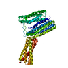
Mass: 62.068 Da / Num. of mol.: 3 / Source method: obtained synthetically / Formula: C2H6O2
#3: Water ChemComp-HOH / water / Water


Mass: 18.015 Da / Num. of mol.: 135 / Source method: isolated from a natural source / Formula: H2O

-
Experimental details

-
Experiment

ExperimentMethod: X-RAY DIFFRACTION / Number of used crystals: 1

-
Sample preparation

CrystalDensity Matthews: 2.66 Å3/Da / Density % sol: 53.85 %
Crystal growTemperature: 277 K / Method: vapor diffusion, hanging drop / Details: 0.2 M potassium phosphate, 20% (w/v) PEG 3350

-
Data collection

DiffractionMean temperature: 93 K
Diffraction sourceSource: SYNCHROTRON / Site: CHESS / Beamline: A1 / Wavelength: 0.6424 Å
DetectorType: ADSC QUANTUM 210 / Detector: CCD / Date: Jun 15, 2015
RadiationProtocol: SINGLE WAVELENGTH / Monochromatic (M) / Laue (L): M / Scattering type: x-ray
Radiation wavelengthWavelength: 0.6424 Å / Relative weight: 1
ReflectionResolution: 2.2→48.9 Å / Num. obs: 26339 / % possible obs: 99.8 % / Redundancy: 3.8 % / Net I/σ(I): 17.9

-
Processing

Software
NameVersionClassification
REFMAC5.6.0117refinement
HKL-2000data reduction
SCALEPACKdata scaling
PHASERphasing
RefinementMethod to determine structure: MOLECULAR REPLACEMENT
Starting model: 4W8P, 4F7G
Resolution: 2.2→48.86 Å / Cor.coef. Fo:Fc: 0.941 / Cor.coef. Fo:Fc free: 0.913 / SU B: 11.651 / SU ML: 0.147 / Cross valid method: THROUGHOUT / ESU R: 0.26 / ESU R Free: 0.208 / Details: HYDROGENS HAVE BEEN USED IF PRESENT IN THE INPUT
RfactorNum. reflection% reflectionSelection details
Rfree0.24893 1320 5 %RANDOM
Rwork0.2083 ---
obs0.21037 25139 99.75 %-
Solvent computationIon probe radii: 0.8 Å / Shrinkage radii: 0.8 Å / VDW probe radii: 1.2 Å
Displacement parametersBiso mean: 39.788 Å2
Baniso -1Baniso -2Baniso -3
1-0 Å20 Å20.01 Å2
2--0 Å20 Å2
3---0 Å2
Refinement stepCycle: 1 / Resolution: 2.2→48.86 Å
ProteinNucleic acidLigandSolventTotal
Num. atoms3308 0 12 135 3455
Refine LS restraints
Refine-IDTypeDev idealDev ideal targetNumber
X-RAY DIFFRACTIONr_bond_refined_d0.0070.023399
X-RAY DIFFRACTIONr_bond_other_d
X-RAY DIFFRACTIONr_angle_refined_deg1.0251.9694642
X-RAY DIFFRACTIONr_angle_other_deg
X-RAY DIFFRACTIONr_dihedral_angle_1_deg4.1435477
X-RAY DIFFRACTIONr_dihedral_angle_2_deg35.47326.452124
X-RAY DIFFRACTIONr_dihedral_angle_3_deg14.95815529
X-RAY DIFFRACTIONr_dihedral_angle_4_deg14.6891510
X-RAY DIFFRACTIONr_chiral_restr0.0710.2567
X-RAY DIFFRACTIONr_gen_planes_refined0.0040.0212553
X-RAY DIFFRACTIONr_gen_planes_other
X-RAY DIFFRACTIONr_nbd_refined
X-RAY DIFFRACTIONr_nbd_other
X-RAY DIFFRACTIONr_nbtor_refined
X-RAY DIFFRACTIONr_nbtor_other
X-RAY DIFFRACTIONr_xyhbond_nbd_refined
X-RAY DIFFRACTIONr_xyhbond_nbd_other
X-RAY DIFFRACTIONr_metal_ion_refined
X-RAY DIFFRACTIONr_metal_ion_other
X-RAY DIFFRACTIONr_symmetry_vdw_refined
X-RAY DIFFRACTIONr_symmetry_vdw_other
X-RAY DIFFRACTIONr_symmetry_hbond_refined
X-RAY DIFFRACTIONr_symmetry_hbond_other
X-RAY DIFFRACTIONr_symmetry_metal_ion_refined
X-RAY DIFFRACTIONr_symmetry_metal_ion_other
X-RAY DIFFRACTIONr_mcbond_it
X-RAY DIFFRACTIONr_mcbond_other
X-RAY DIFFRACTIONr_mcangle_it
X-RAY DIFFRACTIONr_mcangle_other
X-RAY DIFFRACTIONr_scbond_it
X-RAY DIFFRACTIONr_scbond_other
X-RAY DIFFRACTIONr_scangle_it
X-RAY DIFFRACTIONr_scangle_other
X-RAY DIFFRACTIONr_long_range_B_refined
X-RAY DIFFRACTIONr_long_range_B_other
X-RAY DIFFRACTIONr_rigid_bond_restr
X-RAY DIFFRACTIONr_sphericity_free
X-RAY DIFFRACTIONr_sphericity_bonded
LS refinement shellResolution: 2.2→2.257 Å / Total num. of bins used: 20
RfactorNum. reflection% reflection
Rfree0.299 87 -
Rwork0.262 1789 -
obs--100 %
Refinement TLS params.Method: refined / Origin x: -21.733 Å / Origin y: -11.5787 Å / Origin z: -13.1226 Å
111213212223313233
T0.0459 Å20.0142 Å20.014 Å2-0.1054 Å20.0128 Å2--0.0469 Å2
L0.6689 °20.6672 °20.3186 °2-0.7135 °20.2683 °2--0.3779 °2
S0.0108 Å °0.0479 Å °0.0559 Å °0.0339 Å °0.0262 Å °0.0757 Å °0.0409 Å °-0.0002 Å °-0.037 Å °

+
About Yorodumi

-
News

-
Feb 9, 2022. New format data for meta-information of EMDB entries

New format data for meta-information of EMDB entries

  • Version 3 of the EMDB header file is now the official format.
  • The previous official version 1.9 will be removed from the archive.

Related info.:EMDB header

External links:wwPDB to switch to version 3 of the EMDB data model

-
Aug 12, 2020. Covid-19 info

Covid-19 info

URL: https://pdbj.org/emnavi/covid19.php

New page: Covid-19 featured information page in EM Navigator.

Related info.:Covid-19 info / Mar 5, 2020. Novel coronavirus structure data

+
Mar 5, 2020. Novel coronavirus structure data

Novel coronavirus structure data

Related info.:Yorodumi Speices / Aug 12, 2020. Covid-19 info

External links:COVID-19 featured content - PDBj / Molecule of the Month (242):Coronavirus Proteases

+
Jan 31, 2019. EMDB accession codes are about to change! (news from PDBe EMDB page)

EMDB accession codes are about to change! (news from PDBe EMDB page)

  • The allocation of 4 digits for EMDB accession codes will soon come to an end. Whilst these codes will remain in use, new EMDB accession codes will include an additional digit and will expand incrementally as the available range of codes is exhausted. The current 4-digit format prefixed with “EMD-” (i.e. EMD-XXXX) will advance to a 5-digit format (i.e. EMD-XXXXX), and so on. It is currently estimated that the 4-digit codes will be depleted around Spring 2019, at which point the 5-digit format will come into force.
  • The EM Navigator/Yorodumi systems omit the EMD- prefix.

Related info.:Q: What is EMD? / ID/Accession-code notation in Yorodumi/EM Navigator

External links:EMDB Accession Codes are Changing Soon! / Contact to PDBj

+
Jul 12, 2017. Major update of PDB

Major update of PDB

  • wwPDB released updated PDB data conforming to the new PDBx/mmCIF dictionary.
  • This is a major update changing the version number from 4 to 5, and with Remediation, in which all the entries are updated.
  • In this update, many items about electron microscopy experimental information are reorganized (e.g. em_software).
  • Now, EM Navigator and Yorodumi are based on the updated data.

External links:wwPDB Remediation / Enriched Model Files Conforming to OneDep Data Standards Now Available in the PDB FTP Archive

-
Yorodumi

Thousand views of thousand structures

  • Yorodumi is a browser for structure data from EMDB, PDB, SASBDB, etc.
  • This page is also the successor to EM Navigator detail page, and also detail information page/front-end page for Omokage search.
  • The word "yorodu" (or yorozu) is an old Japanese word meaning "ten thousand". "mi" (miru) is to see.

Related info.:EMDB / PDB / SASBDB / Comparison of 3 databanks / Yorodumi Search / Aug 31, 2016. New EM Navigator & Yorodumi / Yorodumi Papers / Jmol/JSmol / Function and homology information / Changes in new EM Navigator and Yorodumi

Read more