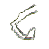
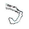
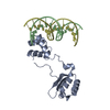
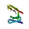
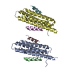
[English] 日本語
Yorodumi
- PDB-4w8p: Crystal structure of RIAM TBS1 in complex with talin R7R8 domains -

+
Open data


ID or keywords:

Loading...

-
Basic information

Entry
Database: PDB / ID: 4w8p
TitleCrystal structure of RIAM TBS1 in complex with talin R7R8 domains
Components
  • Amyloid beta A4 precursor protein-binding family B member 1-interacting protein
  • Talin-1
KeywordsPEPTIDE BINDING PROTEIN / Talin / rod domains / R7R8 / R8 / RIAM / TBS1
Function / homology
Function and homology information


GRB2:SOS provides linkage to MAPK signaling for Integrins / T cell activation via T cell receptor contact with antigen bound to MHC molecule on antigen presenting cell / Integrin signaling / Smooth Muscle Contraction / p130Cas linkage to MAPK signaling for integrins / SEMA3A-Plexin repulsion signaling by inhibiting Integrin adhesion / MAP2K and MAPK activation / LIM domain binding / Platelet degranulation / vinculin binding ...GRB2:SOS provides linkage to MAPK signaling for Integrins / T cell activation via T cell receptor contact with antigen bound to MHC molecule on antigen presenting cell / Integrin signaling / Smooth Muscle Contraction / p130Cas linkage to MAPK signaling for integrins / SEMA3A-Plexin repulsion signaling by inhibiting Integrin adhesion / MAP2K and MAPK activation / LIM domain binding / Platelet degranulation / vinculin binding / integrin activation / cell-substrate junction assembly / cortical actin cytoskeleton organization / T cell receptor complex / phosphatidylserine binding / positive regulation of cell adhesion / ruffle / phosphatidylinositol binding / integrin-mediated signaling pathway / adherens junction / structural constituent of cytoskeleton / platelet aggregation / ruffle membrane / cell-cell adhesion / actin filament binding / integrin binding / lamellipodium / cytoskeleton / focal adhesion / cell surface / signal transduction / plasma membrane / cytoplasm / cytosol
Similarity search - Function
Talin, central domain / GRB/APBB1IP / APBB1IP, PH domain / A middle domain of Talin 1 / : / Talin, R4 domain / Alpha-catenin/vinculin-like / Vinculin-binding site-containing domain / Talin, central / Talin, N-terminal F0 domain ...Talin, central domain / GRB/APBB1IP / APBB1IP, PH domain / A middle domain of Talin 1 / : / Talin, R4 domain / Alpha-catenin/vinculin-like / Vinculin-binding site-containing domain / Talin, central / Talin, N-terminal F0 domain / Talin, central domain superfamily / Talin-1/2, rod-segment / Vinculin Binding Site / Talin, middle domain / N-terminal or F0 domain of Talin-head FERM / I/LWEQ domain / I/LWEQ domain superfamily / I/LWEQ domain / I/LWEQ domain profile. / I/LWEQ domain / Ras association (RalGDS/AF-6) domain / Phosphotyrosine-binding domain / Ras-associating (RA) domain profile. / Ras association (RalGDS/AF-6) domain / Ras-associating (RA) domain / Alpha-catenin/vinculin-like superfamily / FERM domain signature 1. / FERM conserved site / FERM domain signature 2. / FERM central domain / FERM/acyl-CoA-binding protein superfamily / FERM central domain / FERM superfamily, second domain / FERM domain / FERM domain profile. / Band 4.1 domain / Band 4.1 homologues / PH domain / Four Helix Bundle (Hemerythrin (Met), subunit A) / PH domain profile. / Pleckstrin homology domain. / Pleckstrin homology domain / PH-like domain superfamily / Ubiquitin-like domain superfamily / Up-down Bundle / Mainly Alpha
Similarity search - Domain/homology
Talin-1 / Amyloid beta A4 precursor protein-binding family B member 1-interacting protein
Similarity search - Component
Biological speciesMus musculus (house mouse)
MethodX-RAY DIFFRACTION / SYNCHROTRON / MOLECULAR REPLACEMENT / Resolution: 1.5 Å
AuthorsChang, Y.C.E. / Zhang, H. / Wu, J.
CitationJournal: Structure / Year: 2014
Title: Structural and Mechanistic Insights into the Recruitment of Talin by RIAM in Integrin Signaling.
Authors: Chang, Y.C. / Zhang, H. / Franco-Barraza, J. / Brennan, M.L. / Patel, T. / Cukierman, E. / Wu, J.
History
DepositionAug 25, 2014Deposition site: RCSB / Processing site: RCSB
Revision 1.0Dec 3, 2014Provider: repository / Type: Initial release
Revision 1.1Dec 17, 2014Group: Database references
Revision 2.0Sep 27, 2023Group: Atomic model / Data collection ...Atomic model / Data collection / Database references / Derived calculations / Other / Refinement description / Source and taxonomy
Category: atom_site_anisotrop / chem_comp_atom ...atom_site_anisotrop / chem_comp_atom / chem_comp_bond / citation / database_2 / entity_src_gen / pdbx_database_status / pdbx_entity_src_syn / pdbx_initial_refinement_model / pdbx_struct_oper_list / refine_hist
Item: _atom_site_anisotrop.pdbx_PDB_model_num / _atom_site_anisotrop.pdbx_label_asym_id ..._atom_site_anisotrop.pdbx_PDB_model_num / _atom_site_anisotrop.pdbx_label_asym_id / _atom_site_anisotrop.pdbx_label_atom_id / _atom_site_anisotrop.pdbx_label_comp_id / _atom_site_anisotrop.pdbx_label_seq_id / _citation.journal_id_CSD / _database_2.pdbx_DOI / _database_2.pdbx_database_accession / _entity_src_gen.pdbx_alt_source_flag / _pdbx_database_status.pdb_format_compatible / _pdbx_entity_src_syn.pdbx_alt_source_flag / _pdbx_struct_oper_list.symmetry_operation / _refine_hist.number_atoms_solvent / _refine_hist.number_atoms_total / _refine_hist.pdbx_number_atoms_ligand / _refine_hist.pdbx_number_atoms_nucleic_acid / _refine_hist.pdbx_number_atoms_protein

-
Structure visualization

Structure viewerMolecule:
MolmilJmol/JSmol

Downloads & links

-
Assembly

Deposited unit
A: Talin-1
B: Amyloid beta A4 precursor protein-binding family B member 1-interacting protein
hetero molecules


Theoretical massNumber of molelcules
Total (without water)34,1284
Polymers34,0042
Non-polymers1242
Water5,495305
1


  • Idetical with deposited unit
  • defined by author&software
TypeNameSymmetry operationNumber
identity operation1_555x,y,z1
Buried area2030 Å2
ΔGint-6 kcal/mol
Surface area16280 Å2
MethodPISA
2
A: Talin-1
B: Amyloid beta A4 precursor protein-binding family B member 1-interacting protein
hetero molecules

A: Talin-1
B: Amyloid beta A4 precursor protein-binding family B member 1-interacting protein
hetero molecules


Theoretical massNumber of molelcules
Total (without water)68,2578
Polymers68,0094
Non-polymers2484
Water724
TypeNameSymmetry operationNumber
identity operation1_555x,y,z1
crystal symmetry operation2_555-x,-y,z1
Buried area7590 Å2
ΔGint-22 kcal/mol
Surface area29030 Å2
MethodPISA
Unit cell
Length a, b, c (Å)59.722, 105.884, 49.027
Angle α, β, γ (deg.)90.00, 90.00, 90.00
Int Tables number18
Space group name H-MP21212

-
Components

#1: Protein Talin-1


Mass: 31603.615 Da / Num. of mol.: 1
Source method: isolated from a genetically manipulated source
Source: (gene. exp.) Mus musculus (house mouse) / Gene: Tln1, Tln / Plasmid: pET28a / Production host: Escherichia coli (E. coli) / Strain (production host): BL21(DE3) / References: UniProt: P26039
#2: Protein/peptide Amyloid beta A4 precursor protein-binding family B member 1-interacting protein / APBB1-interacting protein 1 / Proline-rich EVH1 ligand 1 / PREL-1 / Proline-rich protein 48


Mass: 2400.636 Da / Num. of mol.: 1 / Source method: obtained synthetically / Details: synthetic peptide / Source: (synth.) Mus musculus (house mouse) / References: UniProt: Q8R5A3
#3: Chemical ChemComp-EDO / 1,2-ETHANEDIOL / ETHYLENE GLYCOL


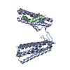
Mass: 62.068 Da / Num. of mol.: 2 / Source method: obtained synthetically / Formula: C2H6O2
#4: Water ChemComp-HOH / water


Mass: 18.015 Da / Num. of mol.: 305 / Source method: isolated from a natural source / Formula: H2O

-
Experimental details

-
Experiment

ExperimentMethod: X-RAY DIFFRACTION

-
Sample preparation

CrystalDensity Matthews: 2.2 Å3/Da / Density % sol: 44.19 %
Crystal growTemperature: 298 K / Method: vapor diffusion, hanging drop / pH: 7.5
Details: 100 mM NaCl, 20% (w/v) polyethylene glycol 3350 and 20% (v/v) ethylene glycol

-
Data collection

DiffractionMean temperature: 100 K
Diffraction sourceSource: SYNCHROTRON / Site: NSLS / Beamline: X29A / Wavelength: 1.075 Å
DetectorType: ADSC QUANTUM 315r / Detector: CCD / Date: Aug 8, 2013
RadiationProtocol: SINGLE WAVELENGTH / Monochromatic (M) / Laue (L): M / Scattering type: x-ray
Radiation wavelengthWavelength: 1.075 Å / Relative weight: 1
ReflectionResolution: 1.5→50 Å / Num. all: 285419 / Num. obs: 49933 / % possible obs: 98.6 % / Redundancy: 5.7 % / Rsym value: 0.051 / Net I/σ(I): 47.3
Reflection shellResolution: 1.5→1.53 Å / Redundancy: 4.5 % / Rmerge(I) obs: 0.558 / Mean I/σ(I) obs: 2.9 / % possible all: 96.9

-
Processing

SoftwareName: REFMAC / Version: 5.6.0117 / Classification: refinement
RefinementMethod to determine structure: MOLECULAR REPLACEMENT
Starting model: 2X0C
Resolution: 1.5→24.79 Å / Cor.coef. Fo:Fc: 0.951 / Cor.coef. Fo:Fc free: 0.943 / SU B: 3.21 / SU ML: 0.06 / Cross valid method: THROUGHOUT / ESU R: 0.093 / ESU R Free: 0.089 / Stereochemistry target values: MAXIMUM LIKELIHOOD / Details: HYDROGENS HAVE BEEN USED IF PRESENT IN THE INPUT
RfactorNum. reflection% reflectionSelection details
Rfree0.24192 2535 5.1 %RANDOM
Rwork0.22262 ---
obs0.22363 47327 98.32 %-
Solvent computationIon probe radii: 0.8 Å / Shrinkage radii: 0.8 Å / VDW probe radii: 1.2 Å / Solvent model: MASK
Displacement parametersBiso mean: 23.122 Å2
Baniso -1Baniso -2Baniso -3
1--0.01 Å20 Å20 Å2
2--0 Å20 Å2
3---0.01 Å2
Refinement stepCycle: 1 / Resolution: 1.5→24.79 Å
ProteinNucleic acidLigandSolventTotal
Num. atoms2364 0 8 305 2677
Refine LS restraints
Refine-IDTypeDev idealDev ideal targetNumber
X-RAY DIFFRACTIONr_bond_refined_d0.0050.0192451
X-RAY DIFFRACTIONr_bond_other_d
X-RAY DIFFRACTIONr_angle_refined_deg0.9341.9743335
X-RAY DIFFRACTIONr_angle_other_deg
X-RAY DIFFRACTIONr_dihedral_angle_1_deg4.0165338
X-RAY DIFFRACTIONr_dihedral_angle_2_deg36.28725.882102
X-RAY DIFFRACTIONr_dihedral_angle_3_deg12.51615417
X-RAY DIFFRACTIONr_dihedral_angle_4_deg8.4081514
X-RAY DIFFRACTIONr_chiral_restr0.0680.2393
X-RAY DIFFRACTIONr_gen_planes_refined0.0050.0211859
X-RAY DIFFRACTIONr_gen_planes_other
X-RAY DIFFRACTIONr_nbd_refined
X-RAY DIFFRACTIONr_nbd_other
X-RAY DIFFRACTIONr_nbtor_refined
X-RAY DIFFRACTIONr_nbtor_other
X-RAY DIFFRACTIONr_xyhbond_nbd_refined
X-RAY DIFFRACTIONr_xyhbond_nbd_other
X-RAY DIFFRACTIONr_metal_ion_refined
X-RAY DIFFRACTIONr_metal_ion_other
X-RAY DIFFRACTIONr_symmetry_vdw_refined
X-RAY DIFFRACTIONr_symmetry_vdw_other
X-RAY DIFFRACTIONr_symmetry_hbond_refined
X-RAY DIFFRACTIONr_symmetry_hbond_other
X-RAY DIFFRACTIONr_symmetry_metal_ion_refined
X-RAY DIFFRACTIONr_symmetry_metal_ion_other
X-RAY DIFFRACTIONr_mcbond_it
X-RAY DIFFRACTIONr_mcbond_other
X-RAY DIFFRACTIONr_mcangle_it
X-RAY DIFFRACTIONr_mcangle_other
X-RAY DIFFRACTIONr_scbond_it
X-RAY DIFFRACTIONr_scbond_other
X-RAY DIFFRACTIONr_scangle_it
X-RAY DIFFRACTIONr_scangle_other
X-RAY DIFFRACTIONr_long_range_B_refined
X-RAY DIFFRACTIONr_long_range_B_other
X-RAY DIFFRACTIONr_rigid_bond_restr
X-RAY DIFFRACTIONr_sphericity_free
X-RAY DIFFRACTIONr_sphericity_bonded
LS refinement shellResolution: 1.499→1.538 Å
RfactorNum. reflection% reflection
Rfree0.303 161 5 %
Rwork0.28 3113 -
obs--94.16 %
Refinement TLS params.

Method: refined / Refine-ID: X-RAY DIFFRACTION

IDL112)L122)L132)L222)L232)L332)S11 (Å °)S12 (Å °)S13 (Å °)S21 (Å °)S22 (Å °)S23 (Å °)S31 (Å °)S32 (Å °)S33 (Å °)T112)T122)T132)T222)T232)T332)Origin x (Å)Origin y (Å)Origin z (Å)
10.08230.2375-0.01750.7862-0.17250.1940.0094-0.00560.01670.05150.03860.0645-0.0061-0.0462-0.0480.01660.0212-0.00590.0415-0.00640.05066.0665-7.2118-15.0829
26.5191-1.86064.23412.2857-1.2852.94610.12410.1546-0.2469-0.1602-0.01080.1460.10540.0957-0.11330.02970.01920.00590.05050.00840.03293.8259-31.9773-19.9889
Refinement TLS group
IDRefine-IDRefine TLS-IDAuth asym-IDAuth seq-ID
1X-RAY DIFFRACTION1A1357 - 1657
2X-RAY DIFFRACTION2B5 - 25

+
About Yorodumi

-
News

-
Feb 9, 2022. New format data for meta-information of EMDB entries

New format data for meta-information of EMDB entries

  • Version 3 of the EMDB header file is now the official format.
  • The previous official version 1.9 will be removed from the archive.

Related info.:EMDB header

External links:wwPDB to switch to version 3 of the EMDB data model

-
Aug 12, 2020. Covid-19 info

Covid-19 info

URL: https://pdbjlvh1.pdbj.org/emnavi/covid19.php

New page: Covid-19 featured information page in EM Navigator.

Related info.:Covid-19 info / Mar 5, 2020. Novel coronavirus structure data

+
Mar 5, 2020. Novel coronavirus structure data

Novel coronavirus structure data

Related info.:Yorodumi Speices / Aug 12, 2020. Covid-19 info

External links:COVID-19 featured content - PDBj / Molecule of the Month (242):Coronavirus Proteases

+
Jan 31, 2019. EMDB accession codes are about to change! (news from PDBe EMDB page)

EMDB accession codes are about to change! (news from PDBe EMDB page)

  • The allocation of 4 digits for EMDB accession codes will soon come to an end. Whilst these codes will remain in use, new EMDB accession codes will include an additional digit and will expand incrementally as the available range of codes is exhausted. The current 4-digit format prefixed with “EMD-” (i.e. EMD-XXXX) will advance to a 5-digit format (i.e. EMD-XXXXX), and so on. It is currently estimated that the 4-digit codes will be depleted around Spring 2019, at which point the 5-digit format will come into force.
  • The EM Navigator/Yorodumi systems omit the EMD- prefix.

Related info.:Q: What is EMD? / ID/Accession-code notation in Yorodumi/EM Navigator

External links:EMDB Accession Codes are Changing Soon! / Contact to PDBj

+
Jul 12, 2017. Major update of PDB

Major update of PDB

  • wwPDB released updated PDB data conforming to the new PDBx/mmCIF dictionary.
  • This is a major update changing the version number from 4 to 5, and with Remediation, in which all the entries are updated.
  • In this update, many items about electron microscopy experimental information are reorganized (e.g. em_software).
  • Now, EM Navigator and Yorodumi are based on the updated data.

External links:wwPDB Remediation / Enriched Model Files Conforming to OneDep Data Standards Now Available in the PDB FTP Archive

-
Yorodumi

Thousand views of thousand structures

  • Yorodumi is a browser for structure data from EMDB, PDB, SASBDB, etc.
  • This page is also the successor to EM Navigator detail page, and also detail information page/front-end page for Omokage search.
  • The word "yorodu" (or yorozu) is an old Japanese word meaning "ten thousand". "mi" (miru) is to see.

Related info.:EMDB / PDB / SASBDB / Comparison of 3 databanks / Yorodumi Search / Aug 31, 2016. New EM Navigator & Yorodumi / Yorodumi Papers / Jmol/JSmol / Function and homology information / Changes in new EM Navigator and Yorodumi

Read more