| Entry | Database: PDB / ID: 2x0c
|
|---|
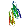
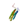
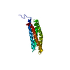
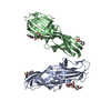
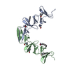
| Title | Crystal Structure of the R7R8 domains of Talin |
|---|
 Components Components | TALIN-1 |
|---|
 Keywords Keywords | CELL ADHESION / CYTOSKELETON / CELL MEMBRANE / ACTIN / SYNEMIN / INTEGRIN / VINCULIN / CELL PROJECTION |
|---|
| Function / homology |  Function and homology information Function and homology information
GRB2:SOS provides linkage to MAPK signaling for Integrins / Integrin signaling / p130Cas linkage to MAPK signaling for integrins / MAP2K and MAPK activation / SEMA3A-Plexin repulsion signaling by inhibiting Integrin adhesion / LIM domain binding / Smooth Muscle Contraction / Platelet degranulation / vinculin binding / cortical microtubule organization ...GRB2:SOS provides linkage to MAPK signaling for Integrins / Integrin signaling / p130Cas linkage to MAPK signaling for integrins / MAP2K and MAPK activation / SEMA3A-Plexin repulsion signaling by inhibiting Integrin adhesion / LIM domain binding / Smooth Muscle Contraction / Platelet degranulation / vinculin binding / cortical microtubule organization / integrin activation / cell-substrate junction assembly / cortical actin cytoskeleton organization / phosphatidylserine binding / ruffle / phosphatidylinositol binding / integrin-mediated signaling pathway / adherens junction / cell-cell adhesion / structural constituent of cytoskeleton / integrin binding / ruffle membrane / actin filament binding / actin cytoskeleton organization / cytoskeleton / focal adhesion / cell surface / plasma membrane / cytoplasm / cytosolSimilarity search - Function Talin, central domain / A middle domain of Talin 1 / Alpha-catenin/vinculin-like / : / Talin VBS2 domain / Vinculin-binding site-containing domain / Talin, central / Talin, central domain superfamily / Talin-1/2, rod-segment / : ...Talin, central domain / A middle domain of Talin 1 / Alpha-catenin/vinculin-like / : / Talin VBS2 domain / Vinculin-binding site-containing domain / Talin, central / Talin, central domain superfamily / Talin-1/2, rod-segment / : / : / Vinculin Binding Site / Talin, middle domain / Talin, R4 domain / Talin 1-like, rod segment domain / Talin, N-terminal F0 domain / : / N-terminal or F0 domain of Talin-head FERM / Talin IBS2B domain / I/LWEQ domain / I/LWEQ domain superfamily / I/LWEQ domain / I/LWEQ domain profile. / I/LWEQ domain / Phosphotyrosine-binding domain / IRS-type PTB domain / PTB domain (IRS-1 type) / Alpha-catenin/vinculin-like superfamily / FERM domain signature 1. / FERM conserved site / FERM domain signature 2. / FERM/acyl-CoA-binding protein superfamily / FERM central domain / FERM superfamily, second domain / FERM domain / FERM domain profile. / Band 4.1 domain / Band 4.1 homologues / Four Helix Bundle (Hemerythrin (Met), subunit A) / PH-like domain superfamily / Ubiquitin-like domain superfamily / Up-down Bundle / Mainly AlphaSimilarity search - Domain/homology |
|---|
| Biological species |   MUS MUSCULUS (house mouse) MUS MUSCULUS (house mouse) |
|---|
| Method |  X-RAY DIFFRACTION / SYNCHROTRON / MAD / Resolution: 2 Å X-RAY DIFFRACTION / SYNCHROTRON / MAD / Resolution: 2 Å |
|---|
 Authors Authors | Gingras, A.R. / Goult, B.T. / Bate, N. / Barsukov, I.L. / Emsley, J. / Critchely, D.R. |
|---|
 Citation Citation | Journal: J.Biol.Chem. / Year: 2010
Title: Central Region of Talin Has a Unique Fold that Binds Vinculin and Actin.
Authors: Gingras, A.R. / Bate, N. / Goult, B.T. / Patel, B. / Kopp, P.M. / Emsley, J. / Barsukov, I.L. / Roberts, G.C.K. / Critchley, D.R. |
|---|
| History | | Deposition | Dec 8, 2009 | Deposition site: PDBE / Processing site: PDBE |
|---|
| Revision 1.0 | Jul 7, 2010 | Provider: repository / Type: Initial release |
|---|
| Revision 1.1 | Jul 13, 2011 | Group: Advisory / Version format compliance |
|---|
| Revision 1.2 | Mar 1, 2023 | Group: Database references / Other / Structure summary / Category: database_2 / pdbx_database_status / struct
Item: _database_2.pdbx_DOI / _database_2.pdbx_database_accession ..._database_2.pdbx_DOI / _database_2.pdbx_database_accession / _pdbx_database_status.status_code_sf / _struct.title |
|---|
| Revision 1.3 | Jun 19, 2024 | Group: Data collection / Category: chem_comp_atom / chem_comp_bond |
|---|
|
|---|
 Movie
Movie Controller


 Open data
 Basic information
 Structure visualization
 Downloads & links
 Other downloads
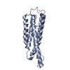


 PDBj
PDBj


 Assembly

 Mass: 18.015 Da / Num. of mol.: 185 / Source method: isolated from a natural source / Formula: H2O
 Sample preparation
 / Beamline: ID23-1 / Wavelength: 0.97905
  Processing