 Movie
Movie Controller

[English] 日本語
 Yorodumi
- PDB-1y19: Structural basis for phosphatidylinositol phosphate kinase type I... -
+ Open data

- Basic information
| Entry | Database: PDB / ID: 1y19 | ||||||
|---|---|---|---|---|---|---|---|
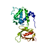
| Title | Structural basis for phosphatidylinositol phosphate kinase type I-gamma binding to talin at focal adhesions | ||||||
 Components Components |
| ||||||
 Keywords Keywords | STRUCTURAL PROTEIN / SIGNALING PROTEIN / FOCAL ADHESION / FERM DOMAIN / CYTOSKELETON / NPXY MOTIF / PTB DOMAIN | ||||||
| Function / homology |  Function and homology information Function and homology informationtalin binding / 1-phosphatidylinositol-4-phosphate 5-kinase / presynaptic endocytic zone membrane / Regulation of CDH1 posttranslational processing and trafficking to plasma membrane / 1-phosphatidylinositol-4-phosphate 5-kinase activity / GRB2:SOS provides linkage to MAPK signaling for Integrins / Integrin signaling / p130Cas linkage to MAPK signaling for integrins / MAP2K and MAPK activation / Synthesis of PIPs at the plasma membrane ...talin binding / 1-phosphatidylinositol-4-phosphate 5-kinase / presynaptic endocytic zone membrane / Regulation of CDH1 posttranslational processing and trafficking to plasma membrane / 1-phosphatidylinositol-4-phosphate 5-kinase activity / GRB2:SOS provides linkage to MAPK signaling for Integrins / Integrin signaling / p130Cas linkage to MAPK signaling for integrins / MAP2K and MAPK activation / Synthesis of PIPs at the plasma membrane / SEMA3A-Plexin repulsion signaling by inhibiting Integrin adhesion / uropod / LIM domain binding / PI5P, PP2A and IER3 Regulate PI3K/AKT Signaling / Smooth Muscle Contraction / phosphatidylinositol metabolic process / Platelet degranulation / vinculin binding / cortical microtubule organization / Clathrin-mediated endocytosis / integrin activation / phosphatidylinositol biosynthetic process / cell-substrate junction assembly / cortical actin cytoskeleton organization / phosphatidylserine binding / regulation of postsynaptic neurotransmitter receptor internalization / phosphatidylinositol phosphate biosynthetic process / exocytosis / regulation of synaptic vesicle endocytosis / phagocytosis / phagocytic cup / ruffle / axonogenesis / phosphatidylinositol binding / integrin-mediated signaling pathway / adherens junction / cell-cell adhesion / structural constituent of cytoskeleton / platelet aggregation / integrin binding / ruffle membrane / chemotaxis / actin filament binding / actin cytoskeleton organization / cytoskeleton / endosome membrane / postsynaptic density / focal adhesion / glutamatergic synapse / cell surface / nucleoplasm / ATP binding / plasma membrane / cytoplasm / cytosol Similarity search - Function | ||||||
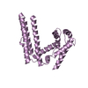
| Biological species |  | ||||||
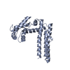
| Method |  X-RAY DIFFRACTION / SYNCHROTRON / MOLECULAR REPLACEMENT / Resolution: 2.6 Å X-RAY DIFFRACTION / SYNCHROTRON / MOLECULAR REPLACEMENT / Resolution: 2.6 Å | ||||||
 Authors Authors | de Pereda, J.M. / Wegener, K. / Santelli, E. / Bate, N. / Ginsberg, M.H. / Critchley, D.R. / Campbell, I.D. / Liddington, R.C. | ||||||
 Citation Citation | Journal: J.Biol.Chem. / Year: 2005 Title: Structural bases for phosphatidylinositol phosphate kinase type I-gamma binding to talin at focal adhesions Authors: de Pereda, J.M. / Wegener, K. / Santelli, E. / Bate, N. / Ginsberg, M.H. / Critchley, D.R. / Campbell, I.D. / Liddington, R.C. | ||||||
| History |
|
- Structure visualization
| Structure viewer | Molecule: MolmilJmol/JSmol |
|---|
- Downloads & links
-Download
| PDBx/mmCIF format | 1y19.cif.gz | 257.2 KB | Display | PDBx/mmCIF format |
|---|---|---|---|---|
| PDB format | pdb1y19.ent.gz | 210.4 KB | Display | PDB format |
| PDBx/mmJSON format | 1y19.json.gz | Tree view | PDBx/mmJSON format | |
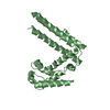
| Others |  Other downloads Other downloads |
-Validation report
| Arichive directory | https://data.pdbj.org/pub/pdb/validation_reports/y1/1y19ftp://data.pdbj.org/pub/pdb/validation_reports/y1/1y19 | HTTPS FTP |
|---|
-Related structure data
| Related structure data | |
|---|---|
| Similar structure data |


-Links
 PDBj
PDBj


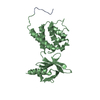
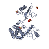
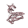
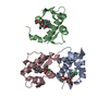
- Assembly
| Deposited unit | 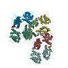
| ||||||||||
|---|---|---|---|---|---|---|---|---|---|---|---|
| 1 | 
| ||||||||||
| 2 | 
| ||||||||||
| 3 | 
| ||||||||||
| 4 | 
| ||||||||||
| 5 | 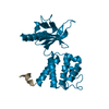
| ||||||||||
| 6 | 
| ||||||||||
| Unit cell |
|
-Components
| #1: Protein/peptide | Mass: 1711.829 Da / Num. of mol.: 6 / Fragment: C-TERMINAL REGION Source method: isolated from a genetically manipulated source Details: chimera of chain A/B, C/D, E/F, G/H, I/J, K/L / Source: (gene. exp.)  #2: Protein | Mass: 23281.965 Da / Num. of mol.: 6 / Fragment: F2 AND F3 SUBDOMAINS OF THE FERM DOMAIN Source method: isolated from a genetically manipulated source Details: chimera of chain A/B, C/D, E/F, G/H, I/J, K/L / Source: (gene. exp.) Description: FORMS CHIMERA WITH PHOSPHATIDYL INOSITOL KINASE TYPE 1 GAMMA AT THE C-TERMINUS Gene: Tln1, Tln / Plasmid: pET15b / Species (production host): Escherichia coli / Production host: #3: Water | ChemComp-HOH / |  Mass: 18.015 Da / Num. of mol.: 466 / Source method: isolated from a natural source / Formula: H2O Mass: 18.015 Da / Num. of mol.: 466 / Source method: isolated from a natural source / Formula: H2OHas protein modification | Y | |
|---|
-Experimental details
-Experiment
| Experiment | Method: X-RAY DIFFRACTION / Number of used crystals: 1 |
|---|
- Sample preparation
| Crystal | Density Matthews: 2.51 Å3/Da / Density % sol: 52.2 % |
|---|---|
| Crystal grow | Temperature: 277 K / Method: vapor diffusion / pH: 6.2 Details: 0.1 M Na/K phosphte buffer, 35% MPD, pH 6.2, VAPOR DIFFUSION, temperature 277K |
-Data collection
| Diffraction | Mean temperature: 100 K |
|---|---|
| Diffraction source | Source: SYNCHROTRON / Site: SSRL  / Beamline: BL1-5 / Wavelength: 1.008 Å / Beamline: BL1-5 / Wavelength: 1.008 Å |
| Detector | Type: ADSC QUANTUM 4 / Detector: CCD / Date: Feb 27, 2003 |
| Radiation | Protocol: SINGLE WAVELENGTH / Monochromatic (M) / Laue (L): M / Scattering type: x-ray |
| Radiation wavelength | Wavelength: 1.008 Å / Relative weight: 1 |
| Reflection | Resolution: 2.6→50 Å / Num. obs: 43284 / % possible obs: 96.7 % / Observed criterion σ(I): -4 / Redundancy: 2.4 % / Biso Wilson estimate: 57 Å2 / Rmerge(I) obs: 0.058 / Net I/σ(I): 15.2 |
| Reflection shell | Resolution: 2.6→2.69 Å / Rmerge(I) obs: 0.459 / Mean I/σ(I) obs: 2.4 / % possible all: 84.4 |
- Processing
| Software |
| ||||||||||||||||||||||||||||
|---|---|---|---|---|---|---|---|---|---|---|---|---|---|---|---|---|---|---|---|---|---|---|---|---|---|---|---|---|---|
| Refinement | Method to determine structure: MOLECULAR REPLACEMENT / Resolution: 2.6→30 Å / Cross valid method: THROUGHOUT / σ(F): 0 / Stereochemistry target values: ENGH & HUBER
| ||||||||||||||||||||||||||||
| Displacement parameters |
| ||||||||||||||||||||||||||||
| Refinement step | Cycle: LAST / Resolution: 2.6→30 Å
| ||||||||||||||||||||||||||||
| Refine LS restraints |
| ||||||||||||||||||||||||||||
| LS refinement shell | Resolution: 2.6→2.69 Å /
|
