 Movie
Movie Controller


+ Open data

- Basic information
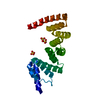
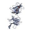
| Entry | Database: PDB / ID: 1mk9 | ||||||
|---|---|---|---|---|---|---|---|
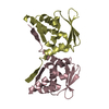
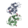
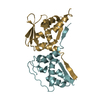
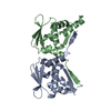
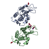
| Title | CRYSTAL STRUCTURE OF AN INTEGRIN BETA3-TALIN CHIMERA | ||||||
 Components Components |
| ||||||
 Keywords Keywords | STRUCTURAL PROTEIN / FOCAL ADHESION / INTEGRIN BINDING / FERM DOMAIN / CYTOSKELETON NPXY MOTIF / PTB DOMAIN | ||||||
| Function / homology |  Function and homology information Function and homology informationregulation of serotonin uptake / positive regulation of adenylate cyclase-inhibiting opioid receptor signaling pathway / tube development / alpha9-beta1 integrin-ADAM8 complex / regulation of trophoblast cell migration / integrin alphaIIb-beta3 complex / regulation of postsynaptic neurotransmitter receptor diffusion trapping / maintenance of postsynaptic specialization structure / alphav-beta3 integrin-vitronectin complex / regulation of extracellular matrix organization ...regulation of serotonin uptake / positive regulation of adenylate cyclase-inhibiting opioid receptor signaling pathway / tube development / alpha9-beta1 integrin-ADAM8 complex / regulation of trophoblast cell migration / integrin alphaIIb-beta3 complex / regulation of postsynaptic neurotransmitter receptor diffusion trapping / maintenance of postsynaptic specialization structure / alphav-beta3 integrin-vitronectin complex / regulation of extracellular matrix organization / platelet alpha granule membrane / positive regulation of glomerular mesangial cell proliferation / integrin alphav-beta3 complex / negative regulation of lipoprotein metabolic process / alphav-beta3 integrin-PKCalpha complex / fibrinogen binding / alphav-beta3 integrin-HMGB1 complex / negative regulation of lipid transport / vascular endothelial growth factor receptor 2 binding / positive regulation of vascular endothelial growth factor signaling pathway / Elastic fibre formation / cell-substrate junction assembly / alphav-beta3 integrin-IGF-1-IGF1R complex / positive regulation of bone resorption / platelet-derived growth factor receptor binding / mesodermal cell differentiation / angiogenesis involved in wound healing / filopodium membrane / regulation of release of sequestered calcium ion into cytosol / glycinergic synapse / extracellular matrix binding / positive regulation of vascular endothelial growth factor receptor signaling pathway / positive regulation of cell adhesion mediated by integrin / apolipoprotein A-I-mediated signaling pathway / regulation of bone resorption / negative regulation of low-density lipoprotein particle clearance / wound healing, spreading of epidermal cells / apoptotic cell clearance / positive regulation of fibroblast migration / integrin complex / cell adhesion mediated by integrin / Molecules associated with elastic fibres / smooth muscle cell migration / heterotypic cell-cell adhesion / positive regulation of smooth muscle cell migration / negative chemotaxis / Mechanical load activates signaling by PIEZO1 and integrins in osteocytes / positive regulation of cell-matrix adhesion / Syndecan interactions / p130Cas linkage to MAPK signaling for integrins / regulation of postsynaptic neurotransmitter receptor internalization / cellular response to insulin-like growth factor stimulus / positive regulation of osteoblast proliferation / protein disulfide isomerase activity / microvillus membrane / platelet-derived growth factor receptor signaling pathway / cell-substrate adhesion / PECAM1 interactions / GRB2:SOS provides linkage to MAPK signaling for Integrins / TGF-beta receptor signaling activates SMADs / lamellipodium membrane / fibronectin binding / negative regulation of macrophage derived foam cell differentiation / negative regulation of lipid storage / blood coagulation, fibrin clot formation / ECM proteoglycans / Integrin cell surface interactions / negative regulation of endothelial cell apoptotic process / positive regulation of T cell migration / coreceptor activity / cellular response to platelet-derived growth factor stimulus / ruffle / Integrin signaling / positive regulation of endothelial cell proliferation / positive regulation of substrate adhesion-dependent cell spreading / substrate adhesion-dependent cell spreading / embryo implantation / positive regulation of endothelial cell migration / cell adhesion molecule binding / positive regulation of smooth muscle cell proliferation / Turbulent (oscillatory, disturbed) flow shear stress activates signaling by PIEZO1 and integrins in endothelial cells / cell-matrix adhesion / protein kinase C binding / response to activity / Signal transduction by L1 / integrin-mediated signaling pathway / regulation of actin cytoskeleton organization / wound healing / cellular response to mechanical stimulus / Signaling by high-kinase activity BRAF mutants / cell-cell adhesion / platelet activation / MAP2K and MAPK activation / structural constituent of cytoskeleton / VEGFA-VEGFR2 Pathway / platelet aggregation / integrin binding / cellular response to xenobiotic stimulus / positive regulation of fibroblast proliferation / ruffle membrane Similarity search - Function | ||||||
| Biological species |  Homo sapiens (human) Homo sapiens (human) | ||||||
| Method |  X-RAY DIFFRACTION / MOLECULAR REPLACEMENT / Resolution: 2.8 Å X-RAY DIFFRACTION / MOLECULAR REPLACEMENT / Resolution: 2.8 Å | ||||||
 Authors Authors | Garcia-Alvarez, B. / De Pereda, J.M. / Calderwood, D.A. / Ulmer, T.S. / Critchley, D. / Campbell, I.D. / Ginsberg, M.H. / Liddington, R.C. | ||||||
 Citation Citation | Journal: Mol.Cell / Year: 2003 Title: Structural Determinants of Integrin Recognition by Talin Authors: Garcia-Alvarez, B. / de Pereda, J.M. / Calderwood, D.A. / Ulmer, T.S. / Critchley, D. / Campbell, I.D. / Ginsberg, M.H. / Liddington, R.C. | ||||||
| History |
| ||||||
| Remark 999 | SEQUENCE AUTHORS INFORMED THAT THE SEQUENCE OF CHICKEN TALIN IS NOT AVAILABLE IN ANY REFERENCE ...SEQUENCE AUTHORS INFORMED THAT THE SEQUENCE OF CHICKEN TALIN IS NOT AVAILABLE IN ANY REFERENCE DATABASE. THE SEQUENCE HAS BEEN DESCRIBED IN THE PUBLICATION: HEMMINGS, L., REES, D.J.G., OHANIAN, V., BOLTON, S.J., GILMORE, A.P., PATEL, N., PRIDDLE, H., TREVITHICK, J.E., HYNES, R.O., & CRITCHLEY, D.R. (1996). TALIN CONTAINS THREE ACTIN-BINDING SITES EACH OF WHICH IS ADJACENT TO A VINCULIN-BINDING SITE. J. CELL SCI., 109, 2715-2726. |
- Structure visualization
| Structure viewer | Molecule: MolmilJmol/JSmol |
|---|
- Downloads & links
-Download
| PDBx/mmCIF format | 1mk9.cif.gz | 157 KB | Display | PDBx/mmCIF format |
|---|---|---|---|---|
| PDB format | pdb1mk9.ent.gz | 127.3 KB | Display | PDB format |
| PDBx/mmJSON format | 1mk9.json.gz | Tree view | PDBx/mmJSON format | |
| Others |  Other downloads Other downloads |
-Validation report
| Arichive directory | https://data.pdbj.org/pub/pdb/validation_reports/mk/1mk9ftp://data.pdbj.org/pub/pdb/validation_reports/mk/1mk9 | HTTPS FTP |
|---|
-Related structure data
| Related structure data | 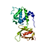 1mixSC 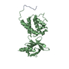 1mizC  1mk7C S: Starting model for refinement C: citing same article ( |
|---|---|
| Similar structure data |


-Links
 PDBj
PDBj


- Assembly
| Deposited unit | 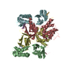
| ||||||||
|---|---|---|---|---|---|---|---|---|---|
| 1 | 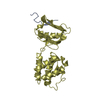
| ||||||||
| 2 | 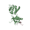
| ||||||||
| 3 | 
| ||||||||
| 4 | 
| ||||||||
| Unit cell |
|
-Components
| #1: Protein/peptide | Mass: 1835.990 Da / Num. of mol.: 4 / Fragment: PARTIAL CYTOPLASMIC TAIL (Residues 739-750) Source method: isolated from a genetically manipulated source Details: Forms chimera with talin at the C-terminus / Source: (gene. exp.) Homo sapiens (human) / Plasmid: pET15B / Species (production host): Escherichia coli / Production host:  #2: Protein | Mass: 22150.656 Da / Num. of mol.: 4 Fragment: F2 AND F3 SUBDOMAINS OF FERM DOMAIN (Residues 209-400) Source method: isolated from a genetically manipulated source Details: Forms chimera with Integrin Beta3 at the N-terminus Source: (gene. exp.) #3: Water | ChemComp-HOH / |  Mass: 18.015 Da / Num. of mol.: 31 / Source method: isolated from a natural source / Formula: H2O Mass: 18.015 Da / Num. of mol.: 31 / Source method: isolated from a natural source / Formula: H2OHas protein modification | Y | |
|---|
-Experimental details
-Experiment
| Experiment | Method: X-RAY DIFFRACTION / Number of used crystals: 1 |
|---|
- Sample preparation
| Crystal | Density Matthews: 2.21 Å3/Da / Density % sol: 44.39 % | ||||||||||||||||||||||||||||||||||||||||||||||||||||||||
|---|---|---|---|---|---|---|---|---|---|---|---|---|---|---|---|---|---|---|---|---|---|---|---|---|---|---|---|---|---|---|---|---|---|---|---|---|---|---|---|---|---|---|---|---|---|---|---|---|---|---|---|---|---|---|---|---|---|
| Crystal grow | Temperature: 277 K / Method: vapor diffusion, hanging drop / pH: 8.6 Details: PEG 8000, MAGNESIUM CHLORIDE, TRIS-HCL, pH 8.6, VAPOR DIFFUSION, HANGING DROP, temperature 277K | ||||||||||||||||||||||||||||||||||||||||||||||||||||||||
| Crystal grow | *PLUS Temperature: 4 ℃ / pH: 7 | ||||||||||||||||||||||||||||||||||||||||||||||||||||||||
| Components of the solutions | *PLUS
|
-Data collection
| Diffraction | Mean temperature: 100 K |
|---|---|
| Diffraction source | Source: ROTATING ANODE / Type: RIGAKU FR-D / Wavelength: 1.5418 |
| Detector | Type: RIGAKU RAXIS IV / Detector: IMAGE PLATE / Date: May 5, 2002 |
| Radiation | Protocol: SINGLE WAVELENGTH / Monochromatic (M) / Laue (L): M / Scattering type: x-ray |
| Radiation wavelength | Wavelength: 1.5418 Å / Relative weight: 1 |
| Reflection | Resolution: 2.8→29.83 Å / Num. obs: 18904 / % possible obs: 91.8 % / Observed criterion σ(I): -3 / Redundancy: 3.11 % / Biso Wilson estimate: 28.3 Å2 / Rsym value: 0.09 / Net I/σ(I): 13 |
| Reflection shell | Resolution: 2.8→2.9 Å / Mean I/σ(I) obs: 4 / % possible all: 94.5 |
| Reflection | *PLUS Highest resolution: 2.8 Å / Num. measured all: 57904 / Rmerge(I) obs: 0.09 |
| Reflection shell | *PLUS % possible obs: 94.5 % / Rmerge(I) obs: 0.307 |
- Processing
| Software |
| ||||||||||||||||||||
|---|---|---|---|---|---|---|---|---|---|---|---|---|---|---|---|---|---|---|---|---|---|
| Refinement | Method to determine structure: MOLECULAR REPLACEMENT Starting model: PDB ENTRY 1MIX Resolution: 2.8→29.83 Å / Rfactor Rfree error: 0.009 / Isotropic thermal model: RESTRAINED / Cross valid method: THROUGHOUT / σ(F): 0 / Stereochemistry target values: Engh & Huber
| ||||||||||||||||||||
| Solvent computation | Solvent model: FLAT MODEL / Bsol: 27.9626 Å2 / ksol: 0.315075 e/Å3 | ||||||||||||||||||||
| Displacement parameters | Biso mean: 26.7 Å2
| ||||||||||||||||||||
| Refine analyze |
| ||||||||||||||||||||
| Refinement step | Cycle: LAST / Resolution: 2.8→29.83 Å
| ||||||||||||||||||||
| Refine LS restraints |
| ||||||||||||||||||||
| LS refinement shell | Resolution: 2.8→2.98 Å / Rfactor Rfree error: 0.027 / Total num. of bins used: 6
| ||||||||||||||||||||
| Xplor file |
| ||||||||||||||||||||
| Refinement | *PLUS Highest resolution: 2.8 Å / Lowest resolution: 30 Å / Rfactor Rfree: 0.324 / Rfactor Rwork: 0.239 | ||||||||||||||||||||
| Solvent computation | *PLUS | ||||||||||||||||||||
| Displacement parameters | *PLUS | ||||||||||||||||||||
| Refine LS restraints | *PLUS
|
