 Movie
Movie Controller

[English] 日本語
 Yorodumi
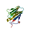
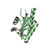
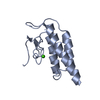
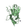
- PDB-2jik: Crystal structure of PDZ domain of Synaptojanin-2 binding protein -
+ Open data

- Basic information
| Entry | Database: PDB / ID: 2jik | ||||||
|---|---|---|---|---|---|---|---|
| Title | Crystal structure of PDZ domain of Synaptojanin-2 binding protein | ||||||
 Components Components | SYNAPTOJANIN-2 BINDING PROTEIN | ||||||
 Keywords Keywords | MEMBRANE PROTEIN / TRANSMEMBRANE / OUTER MEMBRANE / MITOCHONDRIA DISTRIBUTION / PDZ / MEMBRANE / SCAFFOLD / MITOCHONDRION | ||||||
| Function / homology |  Function and homology information Function and homology informationnegative regulation of sprouting angiogenesis / regulation of Notch signaling pathway / negative regulation of endothelial cell migration / negative regulation of endothelial cell proliferation / regulation of endocytosis / protein targeting / Rho protein signal transduction / negative regulation of angiogenesis / negative regulation of ERK1 and ERK2 cascade / mitochondrial outer membrane / mitochondrion Similarity search - Function | ||||||
| Biological species |  HOMO SAPIENS (human) HOMO SAPIENS (human) | ||||||
| Method |  X-RAY DIFFRACTION / SYNCHROTRON / MOLECULAR REPLACEMENT / Resolution: 1.35 Å X-RAY DIFFRACTION / SYNCHROTRON / MOLECULAR REPLACEMENT / Resolution: 1.35 Å | ||||||
 Authors Authors | Tickle, J. / Phillips, C. / Pike, A.C.W. / Cooper, C. / Salah, E. / Elkins, J. / Turnbull, A.P. / Edwards, A. / Arrowsmith, C.H. / Weigelt, J. ...Tickle, J. / Phillips, C. / Pike, A.C.W. / Cooper, C. / Salah, E. / Elkins, J. / Turnbull, A.P. / Edwards, A. / Arrowsmith, C.H. / Weigelt, J. / Sundstrom, M. / Doyle, D. | ||||||
 Citation Citation | Journal: To be Published Title: Crystal Structure of Pdz Domain of Synaptojanin-2 Binding Protein Authors: Tickle, J. / Phillips, C. / Pike, A.C.W. / Cooper, C. / Salah, E. / Elkins, J. / Turnbull, A.P. / Edwards, A. / Arrowsmith, C.H. / Weigelt, J. / Sundstrom, M. / Doyle, D. | ||||||
| History |
| ||||||
| Remark 700 | SHEET THE SHEET STRUCTURE OF THIS MOLECULE IS BIFURCATED. IN ORDER TO REPRESENT THIS FEATURE IN ... SHEET THE SHEET STRUCTURE OF THIS MOLECULE IS BIFURCATED. IN ORDER TO REPRESENT THIS FEATURE IN THE SHEET RECORDS BELOW, TWO SHEETS ARE DEFINED. |
- Structure visualization
| Structure viewer | Molecule: MolmilJmol/JSmol |
|---|
- Downloads & links
-Download
| PDBx/mmCIF format | 2jik.cif.gz | 100.1 KB | Display | PDBx/mmCIF format |
|---|---|---|---|---|
| PDB format | pdb2jik.ent.gz | 77.1 KB | Display | PDB format |
| PDBx/mmJSON format | 2jik.json.gz | Tree view | PDBx/mmJSON format | |
| Others |  Other downloads Other downloads |
-Validation report
| Arichive directory | https://data.pdbj.org/pub/pdb/validation_reports/ji/2jikftp://data.pdbj.org/pub/pdb/validation_reports/ji/2jik | HTTPS FTP |
|---|
-Related structure data
| Related structure data |  2jinC  2fe5S C: citing same article ( S: Starting model for refinement |
|---|---|
| Similar structure data |


-Links
 PDBj
PDBj- Assembly
| Deposited unit | 
| ||||||||
|---|---|---|---|---|---|---|---|---|---|
| 1 | 
| ||||||||
| 2 | 
| ||||||||
| Unit cell |
| ||||||||
| Noncrystallographic symmetry (NCS) | NCS oper: (Code: given Matrix: (-0.99955, -0.03004, -0.00035), Vector: |
-Components
| #1: Protein | Mass: 11034.173 Da / Num. of mol.: 2 / Fragment: PDZ DOMAIN, RESIDUES 6-100 Source method: isolated from a genetically manipulated source Source: (gene. exp.) HOMO SAPIENS (human) / Plasmid: PNIC28-BSA4 / Production host:  #2: Water | ChemComp-HOH / |  Mass: 18.015 Da / Num. of mol.: 240 / Source method: isolated from a natural source / Formula: H2O Mass: 18.015 Da / Num. of mol.: 240 / Source method: isolated from a natural source / Formula: H2O |
|---|
-Experimental details
-Experiment
| Experiment | Method: X-RAY DIFFRACTION / Number of used crystals: 1 |
|---|
- Sample preparation
| Crystal | Density Matthews: 1.88 Å3/Da / Density % sol: 34.75 % |
|---|---|
| Crystal grow | pH: 4 Details: 20% PEG6K, 1M LITHIUM CHLORIDE, 0.1M CITRATE PH4.0, pH 4.00 |
-Data collection
| Diffraction | Mean temperature: 100 K |
|---|---|
| Diffraction source | Source: SYNCHROTRON / Site: SLS  / Beamline: X10SA / Wavelength: 1.03315 / Beamline: X10SA / Wavelength: 1.03315 |
| Detector | Type: MARRESEARCH / Detector: CCD / Date: Apr 16, 2007 |
| Radiation | Protocol: SINGLE WAVELENGTH / Monochromatic (M) / Laue (L): M / Scattering type: x-ray |
| Radiation wavelength | Wavelength: 1.03315 Å / Relative weight: 1 |
| Reflection | Resolution: 1.35→38.35 Å / Num. obs: 35482 / % possible obs: 97.7 % / Observed criterion σ(I): 0 / Redundancy: 6.7 % / Biso Wilson estimate: 11.8 Å2 / Rmerge(I) obs: 0.08 / Net I/σ(I): 17.3 |
| Reflection shell | Resolution: 1.35→1.42 Å / Redundancy: 6.3 % / Rmerge(I) obs: 0.34 / Mean I/σ(I) obs: 5.3 / % possible all: 97.4 |
- Processing
| Software |
| ||||||||||||||||||||||||||||||||||||||||||||||||||||||||||||||||||||||||||||||||||||||||||||||||||||||||||||||||||||||||||||||||||||||||||||||||||||||||||||||||||||||||||||||||||||||
|---|---|---|---|---|---|---|---|---|---|---|---|---|---|---|---|---|---|---|---|---|---|---|---|---|---|---|---|---|---|---|---|---|---|---|---|---|---|---|---|---|---|---|---|---|---|---|---|---|---|---|---|---|---|---|---|---|---|---|---|---|---|---|---|---|---|---|---|---|---|---|---|---|---|---|---|---|---|---|---|---|---|---|---|---|---|---|---|---|---|---|---|---|---|---|---|---|---|---|---|---|---|---|---|---|---|---|---|---|---|---|---|---|---|---|---|---|---|---|---|---|---|---|---|---|---|---|---|---|---|---|---|---|---|---|---|---|---|---|---|---|---|---|---|---|---|---|---|---|---|---|---|---|---|---|---|---|---|---|---|---|---|---|---|---|---|---|---|---|---|---|---|---|---|---|---|---|---|---|---|---|---|---|---|
| Refinement | Method to determine structure: MOLECULAR REPLACEMENT Starting model: PDB ENTRY 2FE5 Resolution: 1.35→30 Å / Cor.coef. Fo:Fc: 0.963 / Cor.coef. Fo:Fc free: 0.941 / SU B: 2.4 / SU ML: 0.047 / Cross valid method: THROUGHOUT / ESU R: 0.077 / ESU R Free: 0.077 / Stereochemistry target values: MAXIMUM LIKELIHOOD / Details: HYDROGENS HAVE BEEN ADDED IN THE RIDING POSITIONS.
| ||||||||||||||||||||||||||||||||||||||||||||||||||||||||||||||||||||||||||||||||||||||||||||||||||||||||||||||||||||||||||||||||||||||||||||||||||||||||||||||||||||||||||||||||||||||
| Solvent computation | Ion probe radii: 0.8 Å / Shrinkage radii: 0.8 Å / VDW probe radii: 1.2 Å / Solvent model: MASK | ||||||||||||||||||||||||||||||||||||||||||||||||||||||||||||||||||||||||||||||||||||||||||||||||||||||||||||||||||||||||||||||||||||||||||||||||||||||||||||||||||||||||||||||||||||||
| Displacement parameters | Biso mean: 9.62 Å2
| ||||||||||||||||||||||||||||||||||||||||||||||||||||||||||||||||||||||||||||||||||||||||||||||||||||||||||||||||||||||||||||||||||||||||||||||||||||||||||||||||||||||||||||||||||||||
| Refinement step | Cycle: LAST / Resolution: 1.35→30 Å
| ||||||||||||||||||||||||||||||||||||||||||||||||||||||||||||||||||||||||||||||||||||||||||||||||||||||||||||||||||||||||||||||||||||||||||||||||||||||||||||||||||||||||||||||||||||||
| Refine LS restraints |
|