 Movie
Movie Controller

[English] 日本語
 Yorodumi
- PDB-3jqx: Crystal structure of Clostridium histolyticum colH collagenase co... -
+ Open data

- Basic information
| Entry | Database: PDB / ID: 3jqx | ||||||
|---|---|---|---|---|---|---|---|
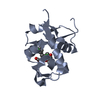
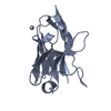
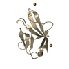
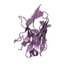
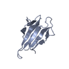
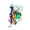
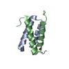
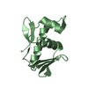
| Title | Crystal structure of Clostridium histolyticum colH collagenase collagen binding domain 3 at 2.2 Angstrom resolution in the presence of calcium and cadmium | ||||||
 Components Components | ColH protein | ||||||
 Keywords Keywords | CELL ADHESION / beta barrel / dual calcium-binding motif / Collagen | ||||||
| Function / homology |  Function and homology information Function and homology informationtripeptidase activity / microbial collagenase / collagen metabolic process / collagen binding / metalloendopeptidase activity / endopeptidase activity / calcium ion binding / proteolysis / extracellular region / zinc ion binding Similarity search - Function | ||||||
| Biological species |  Clostridium histolyticum (bacteria) Clostridium histolyticum (bacteria) | ||||||
| Method |  X-RAY DIFFRACTION / SYNCHROTRON / MOLECULAR REPLACEMENT / Resolution: 2.2 Å X-RAY DIFFRACTION / SYNCHROTRON / MOLECULAR REPLACEMENT / Resolution: 2.2 Å | ||||||
 Authors Authors | Sakon, J. / Philominathan, S.T.L. / Bauer, R. / Matsushita, O. | ||||||
 Citation Citation | Journal: J.Bacteriol. / Year: 2013 Title: Structural Comparison of ColH and ColG Collagen-Binding Domains from Clostridium histolyticum. Authors: Bauer, R. / Wilson, J.J. / Philominathan, S.T. / Davis, D. / Matsushita, O. / Sakon, J. | ||||||
| History |
|
- Structure visualization
| Structure viewer | Molecule: MolmilJmol/JSmol |
|---|
- Downloads & links
-Download
| PDBx/mmCIF format | 3jqx.cif.gz | 89.3 KB | Display | PDBx/mmCIF format |
|---|---|---|---|---|
| PDB format | pdb3jqx.ent.gz | 66.7 KB | Display | PDB format |
| PDBx/mmJSON format | 3jqx.json.gz | Tree view | PDBx/mmJSON format | |
| Others |  Other downloads Other downloads |
-Validation report
| Arichive directory | https://data.pdbj.org/pub/pdb/validation_reports/jq/3jqxftp://data.pdbj.org/pub/pdb/validation_reports/jq/3jqx | HTTPS FTP |
|---|
-Related structure data
| Related structure data |  3jqwC  4hpkC 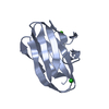 2o8oS S: Starting model for refinement C: citing same article ( |
|---|---|
| Similar structure data |


-Links
 PDBj
PDBj
- Assembly
| Deposited unit | 
| ||||||||
|---|---|---|---|---|---|---|---|---|---|
| 1 | 
| ||||||||
| 2 | 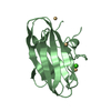
| ||||||||
| 3 | 
| ||||||||
| Unit cell |
|
-Components
| #1: Protein | Mass: 13412.737 Da / Num. of mol.: 3 / Fragment: collagen binding domain Source method: isolated from a genetically manipulated source Source: (gene. exp.) Clostridium histolyticum (bacteria) / Gene: colH / Production host: #2: Chemical |   Mass: 40.078 Da / Num. of mol.: 3 / Source method: obtained synthetically / Formula: Ca Mass: 40.078 Da / Num. of mol.: 3 / Source method: obtained synthetically / Formula: Ca#3: Chemical | ChemComp-CD /   Mass: 112.411 Da / Num. of mol.: 9 / Source method: obtained synthetically / Formula: Cd Mass: 112.411 Da / Num. of mol.: 9 / Source method: obtained synthetically / Formula: Cd#4: Water | ChemComp-HOH / |  Mass: 18.015 Da / Num. of mol.: 197 / Source method: isolated from a natural source / Formula: H2O Mass: 18.015 Da / Num. of mol.: 197 / Source method: isolated from a natural source / Formula: H2O |
|---|
-Experimental details
-Experiment
| Experiment | Method: X-RAY DIFFRACTION / Number of used crystals: 1 |
|---|
- Sample preparation
| Crystal | Density Matthews: 2.37 Å3/Da / Density % sol: 48.15 % |
|---|---|
| Crystal grow | Temperature: 298 K / Method: vapor diffusion, hanging drop / pH: 7.5 Details: sodium acetate, CdSO4,CaCl2, pH 7.5, VAPOR DIFFUSION, HANGING DROP, temperature 298K |
-Data collection
| Diffraction | Mean temperature: 100 K |
|---|---|
| Diffraction source | Source: SYNCHROTRON / Site: APS  / Beamline: 19-ID / Wavelength: 0.97937 Å / Beamline: 19-ID / Wavelength: 0.97937 Å |
| Detector | Type: ADSC QUANTUM 315 / Detector: CCD |
| Radiation | Protocol: SINGLE WAVELENGTH / Monochromatic (M) / Laue (L): M / Scattering type: x-ray |
| Radiation wavelength | Wavelength: 0.97937 Å / Relative weight: 1 |
| Reflection | Resolution: 2.2→30 Å / Num. obs: 18873 / % possible obs: 98.7 % / Observed criterion σ(F): 2 / Observed criterion σ(I): 2 / Redundancy: 13.9 % / Rsym value: 0.12 / Net I/σ(I): 9.6 |
| Reflection shell | Resolution: 2.2→2.5 Å / Redundancy: 3.4 % / Mean I/σ(I) obs: 2.4 / Num. unique all: 1431 / Rsym value: 0.54 / % possible all: 97.7 |
- Processing
| Software |
| |||||||||||||||||||||||||||||||||||||||||||||||||||||||||||||||||||||||||||||||||||||||||||||||
|---|---|---|---|---|---|---|---|---|---|---|---|---|---|---|---|---|---|---|---|---|---|---|---|---|---|---|---|---|---|---|---|---|---|---|---|---|---|---|---|---|---|---|---|---|---|---|---|---|---|---|---|---|---|---|---|---|---|---|---|---|---|---|---|---|---|---|---|---|---|---|---|---|---|---|---|---|---|---|---|---|---|---|---|---|---|---|---|---|---|---|---|---|---|---|---|---|
| Refinement | Method to determine structure: MOLECULAR REPLACEMENT Starting model: 2o8o ColG-3b collagen binding domain Resolution: 2.2→28.68 Å / Cor.coef. Fo:Fc: 0.949 / Cor.coef. Fo:Fc free: 0.908 / SU B: 5.905 / SU ML: 0.15 / Cross valid method: THROUGHOUT / ESU R: 0.262 / ESU R Free: 0.224 / Stereochemistry target values: MAXIMUM LIKELIHOOD
| |||||||||||||||||||||||||||||||||||||||||||||||||||||||||||||||||||||||||||||||||||||||||||||||
| Solvent computation | Ion probe radii: 0.8 Å / Shrinkage radii: 0.8 Å / VDW probe radii: 1.2 Å / Solvent model: MASK | |||||||||||||||||||||||||||||||||||||||||||||||||||||||||||||||||||||||||||||||||||||||||||||||
| Displacement parameters | Biso mean: 20.309 Å2
| |||||||||||||||||||||||||||||||||||||||||||||||||||||||||||||||||||||||||||||||||||||||||||||||
| Refinement step | Cycle: LAST / Resolution: 2.2→28.68 Å
| |||||||||||||||||||||||||||||||||||||||||||||||||||||||||||||||||||||||||||||||||||||||||||||||
| Refine LS restraints |
| |||||||||||||||||||||||||||||||||||||||||||||||||||||||||||||||||||||||||||||||||||||||||||||||
| LS refinement shell | Resolution: 2.196→2.253 Å / Total num. of bins used: 20
|
